A tracheotomy is a surgical opening created in the windpipe via the neck. It is primarily considered when it is unlikely that a patient will be weaned off invasive ventilation. A cannula keeps the opening open.
A tracheotomy can facilitate respiratory support and simplify airway management.
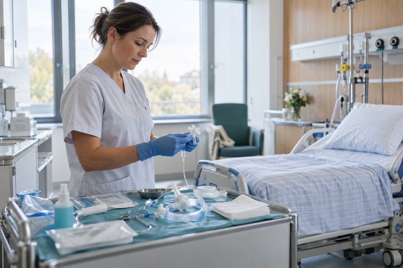
Image generated by AI
What is a tracheotomy?
A tracheotomy involves creating an opening to the trachea through the skin and tissue. This opening is called a tracheostoma. A tracheal cannula prevents the opening from closing up.
Medical records may also use the term ‘tracheostomy’ as well as the English terms ‘tracheostomy’ and ‘tracheotomy’. ‘Trachea’ is the medical term for the windpipe. In each case, the procedure refers to an airway access point in the neck.
Thoracic surgery, by contrast, is performed via an incision into the chest. A tracheotomy, on the other hand, creates an airway opening exclusively in the neck.
When is a tracheotomy indicated?
The indication – that is, the medical reason for the treatment – applies in particular when a person is unlikely to be weaned off invasive ventilation.
- Prolonged weaning: It is foreseeable that invasive ventilation will be required for an extended period.
- Obstruction: A tumour of the larynx or pharynx causes a narrowing of the upper airways.
- Mucus retention: In cases of advanced neurological disease, mucus cannot be adequately cleared.
In adults on invasive ventilation, a routine early tracheotomy shortly after intubation is not recommended. Initially, it is advisable to wait and see how weaning from mechanical ventilation progresses.
| Procedure | Who is it suitable for? | Differential diagnosis |
|---|---|---|
| Tracheotomy | In cases of prolonged ventilatory support, airway obstruction or inadequate secretion clearance | Direct access via the neck; can also be performed as an elective procedure |
| Non-invasive mask ventilation | For suitable patients, where support via a mask is sufficient | No surgically created access |
| Further observation | If, following intubation, it is still unclear whether prolonged ventilation will remain necessary | Avoiding routine early surgery |
| Coniotomy | Particularly in emergency medicine in an extremely life-threatening situation | Incision at a different site |
How is a tracheotomy performed in intensive care?
The two main methods are open surgical tracheotomy and percutaneous dilatational tracheotomy. In the percutaneous procedure, the access site is created through the skin and then dilated.
In English specialist texts, the second method is referred to as a percutaneous dilatational tracheostomy, or PDT for short. A variant of the dilatational method developed by Ciaglia uses a conical dilator, which widens the access in a single step. The English term ‘intensive care’ refers to care provided in an intensive care unit.
- Preparation: Prior to a percutaneous tracheostomy, the patient’s blood clotting status and any medication they are taking are assessed.
- Guidance: An ultrasound scan can visualise blood vessels in the intended access route.
- Anaesthesia: If the patient is breathing spontaneously, the planned operation can be carried out under local anaesthesia.
- Access: The medical team surgically opens the trachea or dilates the punctured access site.
- Monitoring: In adults, the percutaneous puncture should be monitored under bronchoscopy.
- Securing: Finally, the cannula is inserted to ensure that the tracheostoma remains open.
A percutaneous tracheostomy can be performed at the patient’s bedside in the intensive care unit, whilst open surgery can be carried out in the operating theatre. In a study on tracheostomies performed at the intensive care bedside, the procedure took an average of 29 minutes; times ranging from 15 to 60 minutes were observed.
The length of the hospital stay is not determined solely by the procedure itself. The decisive factors are the condition that led to the need for airway management and the subsequent course of intensive care.
What are the effects of the treatment?
A tracheotomy provides a stable access point for mechanical ventilation and can facilitate the removal of mucus. However, it does not treat the underlying cause of the breathing difficulties. The tracheotomy therefore does not determine whether the underlying condition will resolve.
If the artificial airway is no longer required, the cannula can be removed at a later date. In early neurological and neurosurgical rehabilitation, this was successful in around 55 to 60 per cent of patients who were initially on ventilator support. This result cannot be generalised to every condition.
What complications can arise?
Possible complications may arise during the procedure, at the tracheostoma site or in relation to the cannula. The individual risk depends, amongst other things, on the underlying condition and the procedure chosen.
- Immediate problems: In an older study conducted at the intensive care bedside, these occurred in around 5.5 per cent of the patients examined.
- Wound infection: According to a Cochrane review, this was less common following percutaneous placement than following open surgery.
- Technical difficulties: These occurred more frequently with percutaneous procedures in a meta-analysis.
- Tracheal stenosis: Scarring leading to a narrowing of the trachea can occur following either procedure.
- Obstruction: Dried mucus can block the cannula and impair breathing.
A systematic review documented tracheotomy-related deaths following both open surgical and percutaneous procedures. Due to incomplete documentation, the data do not allow for accurate predictions for individual patients.
How should the tracheostomy tube be cared for?
Following the procedure, the focus is on caring for the access site, managing mucus and checking swallowing function. Following a percutaneous dilation tracheotomy, the first scheduled cannula change should take place no earlier than 7 to 10 days later.
Patients who are discharged home with a cannula following cancer surgery are taught in hospital how to clean and care for it, change the cannula and carry out the necessary suctioning. Having a defined procedure in place for dealing with blockages or other cannula-related problems is an important quality indicator.
With a suitable speaking cannula, speaking may be possible. The ability to swallow must be assessed before eating or drinking, as food or liquid may enter the airways.
After the cannula has been removed, the opening should visibly shrink. If this does not happen within about 14 days, a tracheobronchoscopy – an examination of the airways – should be carried out to determine the cause.
What does health insurance cover?
A medically necessary tracheotomy carried out as part of essential hospital treatment is generally covered by statutory health insurance.
People over the age of 18 with statutory health insurance usually pay 10 euros per day for full inpatient treatment, limited to 28 days per calendar year. This hospital co-payment is not a charge for the tracheotomy itself.
Those with private health insurance should check, before a planned admission, which hospital costs their policy covers. Depending on the policy, it may be necessary to pay an invoice yourself initially.
Which doctors perform a tracheotomy?
A tracheotomy is carried out by a medical team with experience in this procedure. Depending on the clinical situation, specialists in thoracic surgery or anaesthesiology may be involved. Specialist training in thoracic surgery covers both surgical and dilational tracheotomies.
When choosing a clinic, factors to consider include experience with the procedure, trained nursing staff and a defined protocol for dealing with cannula-related problems. During the preliminary consultation, you can ask the following questions:
- Which procedure is planned and why?
- How will the access site be monitored during insertion?
- Who will carry out the first cannula change?
- How will I be trained to deal with a blocked cannula?
You will find tracheotomy specialists listed at the end of this text.
Frequently asked questions about tracheostomies
Why is a tracheostomy performed?
A tracheostomy is primarily considered when prolonged mechanical ventilation is anticipated, a tumour is narrowing the airway, or mucus cannot be cleared adequately. Routine early placement is not recommended in adults on invasive ventilation.
Can a tracheostomy be performed under local anaesthesia?
Yes, in the case of a planned operation where the patient is still breathing independently, local anaesthesia may be possible. The type of anaesthesia suitable depends on the patient’s respiratory status and the planned procedure.
Is it possible to speak again after a tracheotomy?
Yes, a suitable speaking valve can enable speech. Whether this is an option depends on the individual situation.
Can a tracheostomy be reversed?
Yes, the cannula can be removed at a later date. The tracheostoma should then visibly shrink.