The trachea carries inhaled air from the throat and nasal cavity to the bronchi. It is also responsible for exhaling “used” air. A narrowing of the trachea (tracheal stenosis) manifests as difficulty breathing, which can progress to acute respiratory distress. The good news: For each of the various causes of this condition, an ear, nose, and throat (ENT) specialist has appropriate treatment options available.
Learn more below about the causes, symptoms, and treatment of tracheal stenosis, and find suitable specialists.
Causes of Tracheal Stenosis
The most common causes of tracheal narrowing are:
- Inflammation of the trachea caused by a bacterial or viral infection or by inhaling irritant gases
- Tumors in the trachea (benign or malignant), such as tracheal cancer
- Growths or tumors in the immediate vicinity that press on the trachea and narrow its diameter (often associated with an enlarged thyroid gland)
- Scarring in the trachea following inflammation, injury, long-term mechanical ventilation, or a tracheotomy
- Obstruction caused by the accidental inhalation of a foreign object
- Congenital narrowing of the trachea (genetic causes)
The listed causes show that there are hardly any preventive measures. However, frequent inhalation of harmful aerosols, such as those found in tobacco smoke, promotes inflammation in the trachea. Tracheal narrowing due to genetic predisposition is extremely rare and typically manifests in childhood.
 The trachea is an elastic tube that begins at the larynx @ Paweł Kacperek /AdobeStock
Symptoms and Diagnostic Methods for Tracheal Stenosis
Depending on the cause, tracheal stenosis may develop slowly or suddenly, as is the case, for example, with a foreign body. Initially, patients do not notice the tracheal stenosis. It is only during physical exertion that breathing difficulties and severe shortness of breath occur.
Those affected find the shortness of breath very unpleasant and, in some cases, even frightening. A severe narrowing of the trachea typically leads to breathing sounds that manifest as a kind of humming or whistling.
To make a diagnosis, the ENT specialist primarily uses the following three methods:
- Ultrasound
- Computed tomography (CT)
- Tracheal endoscopy
Ultrasound and CT are noninvasive and completely painless. Tracheal endoscopy, on the other hand, is performed under local anesthesia or brief general anesthesia.
During an ultrasound, the ENT specialist can assess the nature of the narrowing and thus determine which treatment is appropriate. A CT scan, as an X-ray imaging procedure, can provide information about tissue changes or abnormalities in the trachea.
The endoscopic examination provides the doctor with a direct view of the trachea. A small camera at the tip of the endoscope captures the images. During the examination, the doctor can remove tissue using a laser or take tissue samples for histological examination.
Treatment Options for Tracheal Stenosis
Depending on the cause and severity of the condition, the doctor can choose between non-invasive and invasive treatment options.
Mild tracheal stenosis that does not impair oxygen supply is treated by an internist or ENT specialist with anti-inflammatory medications. It is also possible that the body’s own immune system will resolve the tracheal stenosis on its own.
Tracheal stenosis that impairs breathing usually also results in an inadequate supply of oxygen to the body. This leads to accelerated cellular aging and damage to the central nervous system.
The following minimally invasive and surgical treatment options are used to correct the narrowing in the trachea:
- Laser therapy for the endoscopic removal of tissue (scar tissue)
- Endoscopic administration of locally acting anti-inflammatory medications into the trachea
- Tracheal surgery, in which the surgeon removes the affected section and sutures the slightly shortened trachea back together
- Insertion of a stent into the trachea for stabilization
- Tracheotomy to create an artificial opening into the trachea (tracheostoma)
Your treating physician will discuss the advantages and disadvantages of all possible treatment options with you. Together, you will then decide on the most appropriate form of therapy.
Course and Prognosis of Tracheal Stenosis
The course and prognosis following treatment depend heavily on the cause. If the doctor was able to eliminate the cause, the patient has a good prognosis.
Possible causes that need to be addressed:
- Removal of growths outside the trachea
- Surgical removal of the narrowing in the trachea followed by restoration of patency
Overall, the prognosis is good for all forms of treatment, particularly for surgical procedures on the trachea. If the narrowing in the trachea is high up and close to the larynx, the procedure should be performed by specially trained physicians. The goal is to minimize risks such as vocal cord damage and similar complications.
Tracheotomy in Emergency Situations
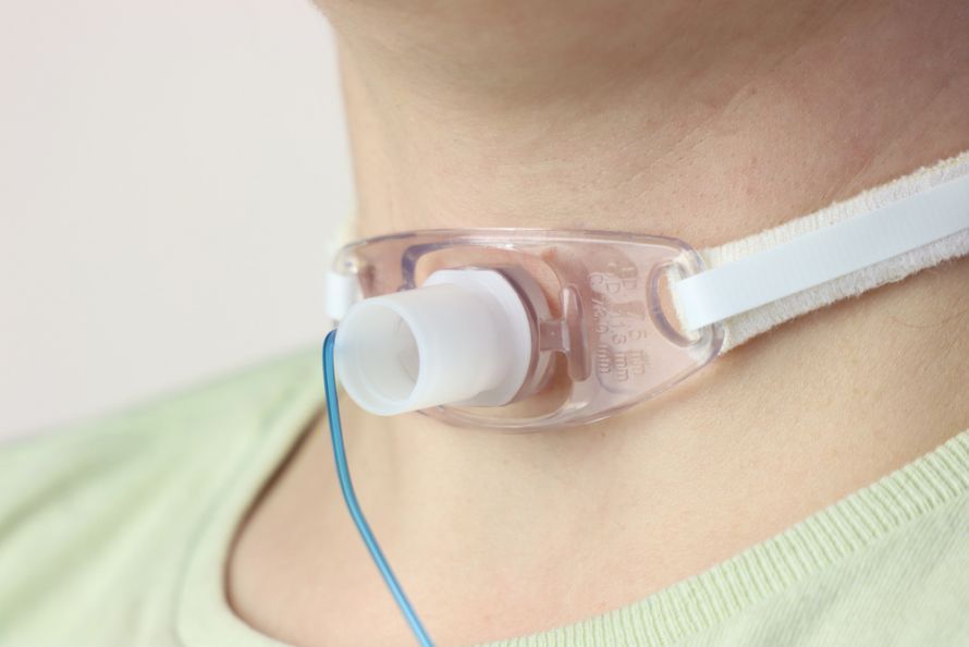 A tracheotomy is an artificial opening into the trachea that ensures breathing @ Jeniffer /AdobeStock
Although TV media often portray it differently, tracheotomies are actually quite rare. If the entrance to the trachea becomes obstructed, this is an extreme case that can lead to death by suffocation.
If a foreign object is causing the blockage, doctors can remove it with their fingers or tweezers. It’s also possible that a wasp sting in the throat could trigger an allergic reaction, causing the entrance to the trachea to swell shut.
In an emergency—that is, if the obstruction cannot be cleared within a few minutes—a tracheotomy is a life-saving procedure. A tracheotomy performed using a tracheal cannula is part of the treatment for certain pre-existing conditions. This procedure is performed by experienced physicians in specialized clinics.
Share this article