MRCP stands for magnetic resonance cholangiopancreatography. It is a specialized imaging technique used in magnetic resonance imaging (MRI). It provides a highly detailed image of the bile ducts on a monitor. MRCP is a gentle, non-invasive method for diagnosing conditions of the bile ducts, liver, and pancreas.
Here you will find further information as well as a selection of MRCP specialists and centers.
How does an MRCP work?
During an MRCP, the patient lies inside the tube of a large magnetic resonance imaging (MRI) scanner. In most MRI machines, an electric current flowing through several wire coils generates the magnetic field. Inside the MRI scanner are additional wire coils that transmit and receive radiofrequency waves.
This produces signals that pass through the body and are picked up again by the wire coils through which current flows.
A computer processes this data and uses it to generate highly detailed images of
- organs,
- soft tissues,
- bones, and
- all other internal body structures.
Unlike conventional X-rays, the body is not exposed to harmful radiation during an MRI. During an MRCP, the imaging is limited strictly to the upper abdomen, including
- gallbladder,
- liver, and
- pancreas.
What does an MRI look like?
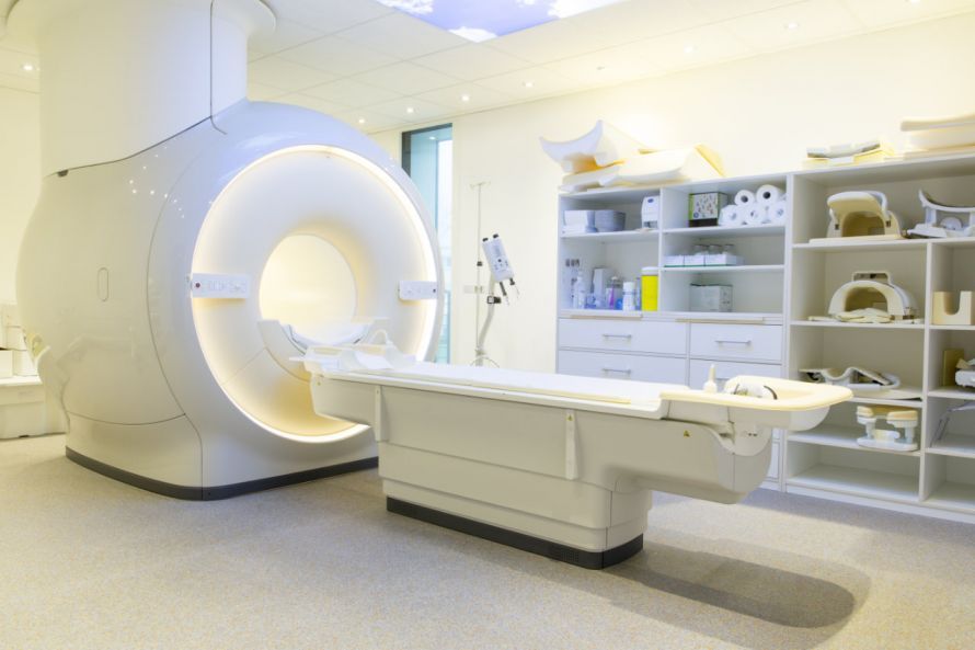
Today, there are various types of magnetic resonance imaging (MRI) machines. The traditional MRI is relatively common. It consists of a large cylindrical tube that is completely surrounded by a circular magnet. Inside the tube is a movable examination table that can be moved directly into the interior of the magnet.
A newer type is the so-called short MRI system, in which the magnetic tube does not extend the full length of the body.
With an open MRI, the sides of the machine are open, so the patient does not lie inside a closed tube. The open MRI offers a good alternative, especially for patients who suffer from claustrophobia or are severely overweight.
How can you prepare for an MRCP?
If you have an MRCP scheduled, it’s best to fast. This means you should not eat or drink anything for eight hours before the MRCP.
The gallbladder is visualized using a contrast agent. In rare cases, people may have an allergic reaction to it. Therefore, the doctor will check with you in advance to see if you have any such allergies.
The MRI contrast agent itself, however, is relatively harmless. Unlike other contrast agents, it is not based on iodine compounds but consists of gadolinium.
An MRCP may be contraindicated in cases of certain pre-existing medical conditions. This means that an MRI cannot be performed if you have a contrast agent intolerance. In particular, an MRCP must be avoided if you have existing kidney disease.
Every patient should also be aware in advance that they will have to lie in a narrow tube for an extended period of time during an MRI. It can also get very loud at times.
If you find such situations uncomfortable or suffer from claustrophobia, you should definitely inform your doctor. In most cases, you will then be given a mild sedative before the exam.
Since metal objects of any kind can interfere with the magnetic resonance field, you must remove all metallic objects before the exam. This includes all types of
- jewelry such as watches, necklaces, and earrings, as well as
- body piercings in the nose, navel, etc.
Often overlooked in this context are implants such as
- dental implants,
- an artificial hip, or
- an artificial knee.
Also,
- pacemakers,
- shunts,
- clips,
- screws, and metal plates
are also affected. If you have any such implants in your body, you must be sure to inform your doctor!
Performing an MRCP
The patient lies down on the MRI scanner’s extendable table and is secured in the correct position. This is necessary to ensure that the patient remains as still as possible during the exam. Only then can the machine produce sharp, clear images.
The radiologists insert an intravenous line through which the contrast agent for the exam will later be administered. This may cause a brief, slight pain. To prevent the line from clotting, a saline solution is infused until the contrast agent is actually ready to be injected. The patient is then moved into the MRI scanner.
First, the technologist takes a few images for calibration. Only then does he or she slowly inject the contrast agent through the IV line. This may cause a brief burning sensation, but it usually goes away quickly. Some patients also report a metallic taste in their mouth after the injection, but this also disappears within a very short time.
The upper abdomen feels slightly warm during the exam. You must lie completely still during the imaging phases. Depending on the area currently being imaged by the MRI, these phases can last from a few seconds to several minutes. It is therefore important to always follow the doctor’s instructions exactly.
During the scan, the wire coils used to transmit radiofrequency waves in the MRI machine are activated. This can be heard as a loud knocking and humming noise. Patients who are sensitive to noise should wear earplugs before the MRCP to minimize the noise level.
The actual MRCP usually takes only about 10 minutes. However, it is most often performed as part of a standard MRI of the entire upper abdomen. This takes about 30 minutes, so the entire examination lasts about 45 minutes.
After the catheter is removed, redness may occur around the puncture site in rare cases. This usually does not require further treatment.
If you did not receive a sedative before the MRCP, you can resume your normal daily activities immediately after the exam. However, some patients may experience mild side effects from the contrast agent, such as nausea and occasional pain.
In very rare cases, allergic reactions may occur, including
- rash,
- watery eyes, or
- other general allergy symptoms.
In such cases, you should notify a doctor immediately.
Who interprets the MRCP results?
The MRCP is performed by a radiologist who is specially trained to interpret the imaging results of the MRI exam.
They analyze the images and write a report. This report includes the examination results, the diagnosis, and, if applicable, a recommendation for specific treatment.
What are the benefits and risks of MRCP?
Advantages of MRCP:
- No radiation harmful to the body
- More detail than other imaging methods (e.g., X-ray or CT)
- Provides information not only on the location of gallstones but also on whether the flow of bile is obstructed
- The contrast agent used in MRCP causes far fewer allergic reactions than the iodine-based contrast agents used in conventional X-rays and CT scans
- MRCP is far less invasive than ERCP and, in comparison, has virtually no side effects, complications, or long-term damage.
Risks of MRCP:
- If sedatives are administered, the normal risks associated with sedation apply
- Metal implants can cause problems or malfunction during the procedure (e.g., pacemakers)
- There is a low risk of an allergic reaction to the contrast agent
In general, the benefits of MRCP outweigh the risks. It is, especially compared to ERCP, the first-line choice for diagnosing gallstones and other diseases
- of the bile ducts,
- the pancreas, and
- the liver.