The term “prenatal diagnostics” refers to tests performed during pregnancy. These tests are designed to identify risks to the pregnant woman and her unborn child at an early stage. Diagnostics are primarily performed using ultrasound. In specific cases where there is cause for concern, a diagnostic procedure (e.g., amniocentesis) may be helpful for further evaluation.
Below you will find additional information as well as a selection of specialists in prenatal diagnostics.
Prenatal Diagnostics—What Is It?
Prenatal diagnostics involves testing an unborn child in the womb. It is used to detect various risks at an early stage.
Various tests are available, such as
- testing for abnormalities in the implantation of the placenta,
- fetal growth restriction due to inadequate nutrient supply or diabetic metabolic conditions, or
- the presence of fetal diseases or malformations.
Three ultrasound examinations are an integral part of prenatal care. In addition, there are other useful tests.
First-Trimester Screening
One diagnostic test that many pregnant women undergo is first-trimester screening.
This screening has several objectives:
- Early detailed assessment of fetal anatomy using high-resolution ultrasound.
- To determine the risk of chromosomal abnormalities
- based on the analysis of blood markers in the mother’s blood
- using measurements of fetal nuchal translucency and other surrogate parameters (e.g., nasal bone)
- based on the mother’s age
- Assessment of the risk of preeclampsia and/or intrauterine growth restriction (= insufficient nutrient supply to the fetus via the placenta)
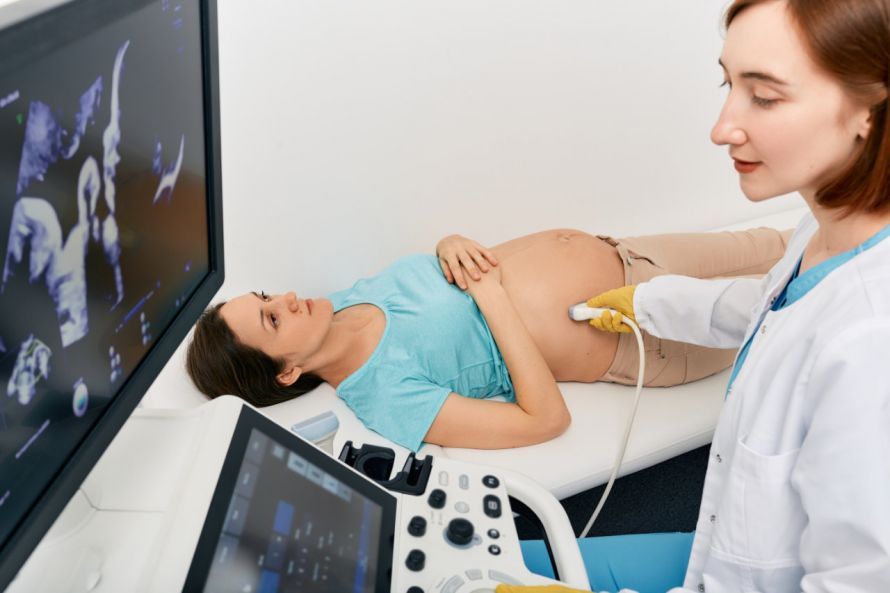
During the ultrasound, the gynecologist measures the fetus from crown to rump. This allows the doctor to monitor the growth of the unborn child and determine the gestational age. The doctor checks the fetal heart rate and examines whether organs such as the limbs or the heart are properly formed and developing.
Even at this early stage, more than half of all structural abnormalities can be detected by an experienced practitioner.
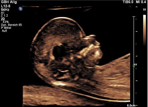
Measurement of Fetal Nuchal Translucency
The measurement of fetal nuchal translucency (NT) takes place between 11+0 and 13+6 weeks of gestation. It plays a central role in first-trimester screening.
Essentially, it refers to a collection of fluid under the skin in the nuchal region of the fetus. It is present in all fetuses and is not considered an abnormality. However, if the fluid layer is thickened, the risk of a fetal abnormality increases.
An increased nuchal translucency (NT) is associated with a higher probability of
- malformations, such as heart defects, or
- chromosomal abnormalities, such as trisomy 21 (Down syndrome).
However, it is never definitive proof of a problem and requires further testing. In combination with the mother’s age and hormone levels from her blood, a statistical risk assessment can be performed. When performed carefully, this risk assessment can indicate the presence of trisomy with 95 percent accuracy.
Blood test
In recent years, blood tests using cell-free DNA have also become established. These tests also allow for screening for trisomies 21, 13, and 18, as well as for sex chromosomes.
The detection rates for trisomies are slightly higher than with conventional first-trimester screening (97–99%).
Here, too, there are false-positive and false-negative test results. Furthermore, the test does not cover the full spectrum of possible chromosomal abnormalities. For this reason, the test alone should not be considered a definitive diagnosis. When combined with a qualified ultrasound, this test is a good diagnostic option.
The blood test currently involves additional costs for expectant parents. Furthermore, in 2–3% of cases, the test yields no result.
Chorionic Villus Sampling
Chorionic villus sampling (CVS), a diagnostic procedure involving the aspiration of early placental tissue, is used for the early detection of chromosomal abnormalities. The test can also detect single-gene disorders, such as hereditary metabolic or muscular disorders known to run in the family. It is performed starting in the 11th week of pregnancy.
In this diagnostic procedure, the doctor inserts a fine hollow needle through the abdominal wall and the wall of the pregnant woman’s uterus. Using this, the doctor extracts cellular tissue from the developing placenta. From this sample, fetal cells are isolated in the laboratory and examined for their chromosomal makeup.
The results of chorionic villus sampling are reliable. In very rare cases (<1%), findings (e.g., mosaicism) may occur that require a repeat procedure for definitive clarification.
In exceptional cases, the removal of fetal cells can lead to contractions or rupture of the amniotic sac, potentially causing a miscarriage. When performed by an experienced practitioner, this risk is 0.2%, which is within the range of the natural miscarriage rate.
Amniocentesis (amniotic fluid testing)
Amniocentesis can be performed starting in the 16th week of pregnancy. Here, too, the doctor uses a hollow needle to collect a sample.
Under ultrasound guidance, the doctor inserts the hollow needle into the amniotic sac and withdraws a small amount of amniotic fluid. The amniotic fluid contains cells from the unborn child, which are examined in the laboratory for abnormalities.
When performed by an experienced practitioner, the risk of miscarriage is 0.1%.
Umbilical Cord Puncture
Umbilical cord puncture (chordocentesis) allows doctors to examine the blood of the unborn child.
The doctor inserts a fine hollow needle into the pregnant woman’s abdomen and draws a small amount of blood from the umbilical vein. This procedure is painless for the baby. The exact composition of the blood is analyzed in the laboratory.
This diagnostic procedure is primarily used to detect:
- Anemia
- Blood group incompatibilities (Rhesus factor)
- Infections
Furthermore, unclear chromosomal findings following an amniocentesis can be verified using umbilical cord puncture.
In one out of every hundred women, umbilical cord puncture can trigger a miscarriage. For this reason, this diagnostic procedure should only be performed when there is concrete suspicion and by an experienced practitioner.

About the medical author
Sabine Schneider
Editor-in-Chief
Sabine Schneider – medical author: Explore expert articles and medical expertise in the Leading Medicine Guide.
More about the medical author →