A stress ECG is an electrocardiogram recorded while the patient is engaging in physical activity. It is used to diagnose cardiac conditions such as coronary artery disease or arrhythmias.
Further information and a list of doctors who perform stress ECGs can be found below.
What is an ECG (electrocardiogram)?
An ECG is a very common and important diagnostic test used to record the electrical activity of the heart muscle cells. “Electro” describes the electrical excitation, “cardio” refers to the heart, and “gram” means “writing.” That is why the ECG is also called a “heart tracing.”
Every heartbeat is controlled by an electrical impulse that originates in the so-called sinus node (pacemaker) and spreads throughout the entire heart and all heart muscle cells. Only when this electrical activity proceeds regularly and in a coordinated manner can the heart beat effectively and strongly.
The ECG, as the so-called “heart recording,” can detect excitation disturbances and thus reveal damage to the heart muscle and cardiac arrhythmias. This is very important in cases such as a heart attack and can be lifesaving.
Which doctors can perform an ECG?
The ECG is particularly important for specialists who focus on heart diseases. These include, in particular, cardiologists and cardiac surgeons. However, because it is simple to perform and can be done anywhere, the ECG is used very frequently across all medical specialties, both in hospitals and in private practices. Interpreting an ECG and recognizing important findings is part of medical training. Rare and complex findings, on the other hand, fall within the specialty of cardiology.
How is an ECG performed?
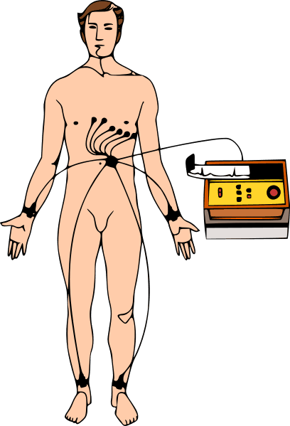
Even though it involves measuring electrical activity, the ECG is a completely side-effect-free and painless test. No electric shocks are administered, as one might assume upon first reading. During an ECG, measurement electrodes are placed externally on the chest and extremities. These are either taped in place or secured with special suction electrodes.
This test is simple to perform and can be done anywhere. It is supplemented by measurements of the pulse and blood pressure, but it is much more specific and accurate in diagnosing heart rhythm disorders and conduction abnormalities.
Is an ECG better than a “heart catheterization”?
An ECG is primarily used to rule out heart disease. For this reason, it is used, for example, during preventive medical checkups. An ECG is also frequently performed before major surgeries to detect previously undetected and asymptomatic heart conditions. Much more often, however, the ECG is normal, allowing serious heart conditions to be ruled out. In such cases, further testing is no longer necessary, and the anesthesiologist has no objections to the surgery. This spares the patient from having to undergo a cardiac catheterization.
However, if abnormalities are present on the ECG, further tests are necessary for a more detailed evaluation and a complete understanding of the clinical picture. In addition to cardiac catheterization, these include an echocardiogram and, in some cases, an MRI scan of the heart muscle and coronary arteries (cardiac MRI).
What types of ECGs are there?
There are various types of ECGs, all of which are based on the same principle. A resting ECG is performed once on a patient who is in a normal physical state. The rhythm, frequency, and intensity of the heart’s pumping function are not affected by external factors, particularly physical exertion.
A stress ECG is performed while the patient is engaging in physical exertion. The patient’s performance is compared to ergometric standards to identify deviations from the norm.
To obtain more meaningful results and monitor the body in specific situations, a long-term ECG is used. In this procedure, the patient wears a device on their body—usually for 24 hours—that measures their heart rate around the clock. The ECG device’s electrodes are attached to the skin, and the device itself is housed in a small pouch that the patient can wear around their neck. A long-term ECG is usually performed in a hospital, though in some cases it can also be done on an outpatient basis at home. The results are then evaluated by a cardiologist.
When is a resting ECG performed?
Resting ECGs are used when a medical history review and general physical examination—particularly of blood pressure and pulse—raise suspicion of heart disease.
For example,
- a pulse that is too fast or irregular, as well as
- fluctuations in blood pressure (both high and low) should always be considered suspicious. However, even in cases of
- known heart conditions
- for monitoring after a heart attack, or
- before major surgical procedures.
The ECG is then part of a series of follow-up routine examinations designed to identify the cause of any disturbances in the functioning of the cardiovascular system.
A resting ECG is performed, so to speak, to get an initial impression of the heart’s function. In most cases, the resting ECG is sufficient to rule out serious heart conditions. If the patient’s medical history is unremarkable and the patient reports no chest pain at rest or during exertion, a normal resting ECG is sufficient, and further testing is not necessary.
What suspected diagnoses can be made with an ECG?
An ECG can provide clues to a wide range of conditions; here are some important examples:
- High blood pressure
- Coronary heart disease
- Heart attacks
- Heart valve disease
- Heart failure
- Arrhythmias
- Tachycardia (rapid heart rate)
- Bradycardia (slow heart rate)
- Atrial fibrillation
- Myocarditis
- Cardiomyopathy (diseases of the heart muscle)
- Sleep apnea
The conditions listed above can be detected by an ECG before the patient experiences significant symptoms. Furthermore, after treatment for a condition—such as heart valve surgery—an ECG can monitor the patient’s condition and the success of the treatment, and provide early warning signs if problems recur.
When is a stress ECG performed?
A stress ECG is ordered when
- symptoms are reported during physical exertion
- following coronary artery bypass surgery, or
- after the placement of stents
This allows for the early detection of narrowings in the stents or in the bypass through changes in the ECG during exercise. However, it is important that the exercise be moderate so as not to trigger a cardiac complication during exercise. In the first few weeks—and in some cases, months—following coronary artery bypass surgery, physical exertion and the performance of a stress ECG are contraindicated, meaning they should not be performed.
Anyone planning to participate in a major athletic competition should also have a stress ECG performed. A sports medicine check-up, for example, is recommended for all athletes who are planning to run a marathon or who regularly participate in one. In addition to a thorough physical examination and blood tests, a sports medicine check-up also includes a stress ECG. If the results are normal, a certificate of fitness for sports is issued. For some competitions, this sports medicine certificate is already a prerequisite for being allowed to participate in such a sporting event.
When are further tests necessary?
In most cases, the ECG is not an indicator of the severity of a heart condition. It merely shows that there is a disturbance in the electrical activity of the heart muscle cells. An ECG can be used, so to speak, to make a preliminary diagnosis, which is then confirmed by further tests such as an ultrasound or an MRI.
The gold standard for confirming a diagnosis of coronary heart disease—which refers to narrowing of the coronary arteries as the cause of a heart attack—is coronary angiography. However, this is an invasive procedure that requires inserting a catheter into the body. The ECG is non-invasive, which means that the integrity of the body is guaranteed.
To make more accurate assessments of heart disease in cases of abnormal ECG findings, further tests are necessary. These primarily include an echocardiogram (“heart echo”), which carries no risks or side effects and can be repeated as often as needed. An echocardiogram can be used, for example, to assess the heart valves, the heart chambers, and blood flow.
In summary, the ECG represents a non-invasive, painless method for assessing the heart’s function. It provides comprehensive and precise information about the heart’s health, which can be evaluated by all medical professionals—and especially by cardiologists—to generate detailed findings. It is one of the most important tests in medicine and has become an indispensable part of everyday clinical practice.

About the medical author
Sabine Schneider
Editor-in-Chief
Sabine Schneider – medical author: Explore expert articles and medical expertise in the Leading Medicine Guide.
More about the medical author →