Hypertrophic obstructive cardiomyopathy (HOCM) is a disease of the heart muscle. The main characteristic of HOCM is thickening of the wall of the left ventricle. The condition is incurable and is occasionally accompanied by life-threatening symptoms. However, with appropriate treatment, those affected can lead a largely symptom-free life. Here you will find further information as well as a list of selected HCM specialists.
What is hypertrophic obstructive cardiomyopathy (HOCM)?
HCM is a disease of the heart muscle. Along with hypertrophic non-obstructive cardiomyopathy (HNCM), it is classified as a type of hypertrophic cardiomyopathy (HCM). Accounting for about 70% of cases, the obstructive form is significantly more common than the non-obstructive form.
Definition: Hypertrophic cardiomyopathy is an umbrella term for all diseases of the heart muscle (cardio = heart and myopathy = muscle disease) that are associated with an excessive increase (hypertrophy) in muscle mass. In the obstructive forms, there is a narrowing at the heart’s outflow tract, causing the heart muscle to enlarge in order to overcome the increased resistance.
What causes hypertrophic obstructive cardiomyopathy (HOCM)?
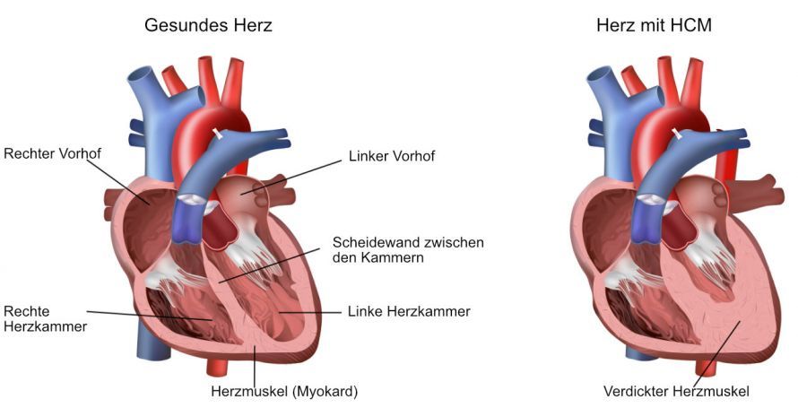
In HCM, a narrowing (obstruction) occurs in the outflow tract of the left ventricle. From here, blood flows from the ventricle through the aortic valve into the aorta and thus into the body.
The obstruction may be caused by
- an enlargement and bulging of the septum (the wall separating the heart chambers) into the left ventricle, as well as
- a pathological (abnormal) movement of the mitral valve (the valve between the left atrium and the left ventricle)
.
In all forms of hypertrophic cardiomyopathy, the heart wall of the left ventricle—and occasionally that of the right ventricle—is enlarged.
In the obstructive form, the septum (the wall separating the heart chambers) is also enlarged (a condition known as septal hypertrophy). The septum then bulges into the left ventricle, causing the ventricle to become smaller.
In addition, the mitral valve may also be affected. Due to the narrowing, a suction force pulls the mitral valve further into the ventricle. This results in mitral valve dysfunction (known as mitral valve insufficiency, or mitral insufficiency for short).
As a result, with every heartbeat, blood may flow backward from the left ventricle into the left atrium, leading to a buildup of blood. In addition, the ejection fraction from the left ventricle is reduced, meaning that less blood is pumped into the body with each heartbeat. This leads to heart failure, which can manifest as reduced physical performance, exhaustion, fatigue, or even circulatory collapse.
What are the symptoms of HCM?
The degree and severity of symptoms depend on the duration and severity of the disease. Generally, a heart affected by HOCM is unable to pump enough blood into the circulatory system. The symptoms are therefore often comparable to the typical symptoms of chronic heart failure (cardiac insufficiency).
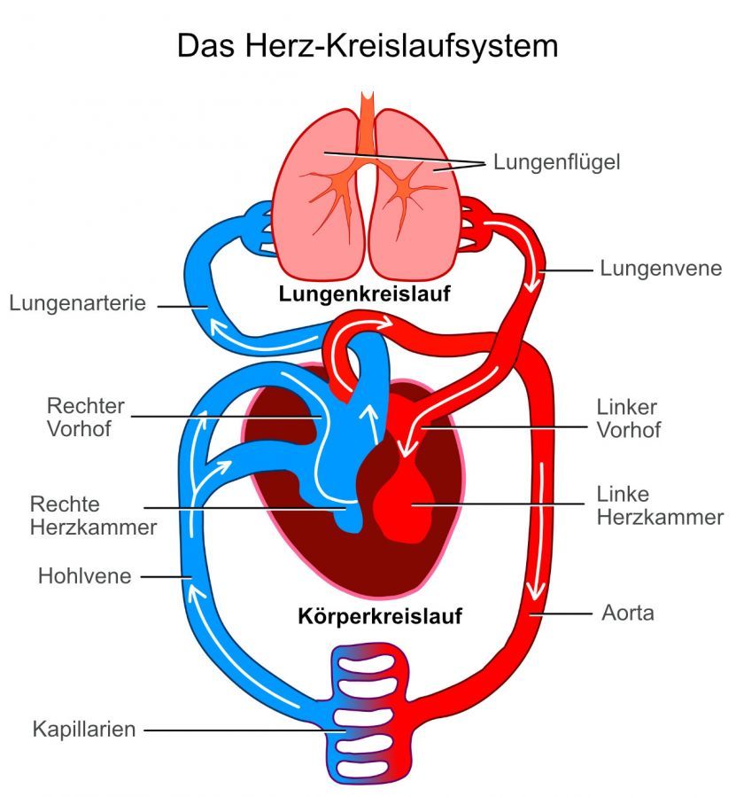
If the mitral valve no longer closes properly, blood can be forced back into the pulmonary circulation. This manifests itself, for example, in
- rapid fatigue,
- reduced physical performance, and
- shortness of breath on exertion.
If this backflow into the left atrium persists for a long time, the atrium enlarges, leading to functional impairment. This can result in various problems, including, in many cases, chronic atrial fibrillation. Those affected may be asymptomatic, especially in the early stages, so that the atrial fibrillation remains undetected for a long time. More severe symptoms only occur during maximum physical exertion.
This may be the case if the septal hypertrophy is only mild and the atrium and mitral valve are not affected. This is also one reason why athletes (and those who aspire to become athletes) are advised to undergo stress tests and fitness evaluations with a heart specialist (cardiologist). This is particularly important before major physical exertion—such as training for or competing in a marathon—to detect previously undiagnosed heart conditions and thereby prevent complications during peak exertion.
Angina pectoris (chest pain) and fluid buildup in the legs and lungs may also occur. If the brain is not supplied with enough oxygen, dizziness and syncope (loss of consciousness) may occur.

HOCM leads to a thickening of the heart muscles. For this reason, they require more oxygen than a healthy heart. At the same time, the heart’s pumping capacity is reduced, which in turn leads to insufficient oxygen supply to the heart muscle.
This imbalance can cause a feeling of tightness and pressure in the chest (angina pectoris) in those affected. This chest pain can occur both during physical exertion and at rest.
In addition, cardiac arrhythmias may occur. Those affected sometimes describe these as a “skipping heartbeat.” Combined with general heart failure, they can lead to fainting spells or dizziness.
What are the causes of HOCM?
Genetic factors are among the main causes of hypertrophic obstructive cardiomyopathy. Most cases can be explained by defects in the genetic material. These defects lead to disruptions in the formation of specific proteins that are necessary for the development of certain muscle units.
Affected individuals can pass these genetic defects directly on to their offspring. For this reason, HOCM often runs in families. However, the disease can manifest differently in each offspring. It is also possible for an individual to carry the defective gene but have no symptoms or signs of the disease. Nevertheless, the disease may reappear in the next generation. Therefore, genetic counseling and testing are strongly recommended in cases of a family history of heart disease.
What tests are necessary to diagnose HOCM?
During the consultation, the patient’s symptoms and medical history are first reviewed. If HOCM is suspected, the patient’s family medical history is also relevant. If the condition has already occurred within the family, the risk of developing the disease is also increased for other family members. The closer the degree of kinship, the higher the risk.
During the physical examination, the doctor looks for symptoms of an irregular heart rhythm and heart failure. The heart is also auscultated, as a flow murmur is typical of HOCM. This murmur often becomes more pronounced during physical exertion.
Special diagnostic methods are available to confirm the diagnosis:
An ultrasound of the heart (echocardiography, also called a heart echo) is a radiation-free and risk-free ultrasound examination. High-frequency waves are emitted, making the structures of the heart visible. This allows for the detection of a thickened heart wall or a thickened heart muscle.
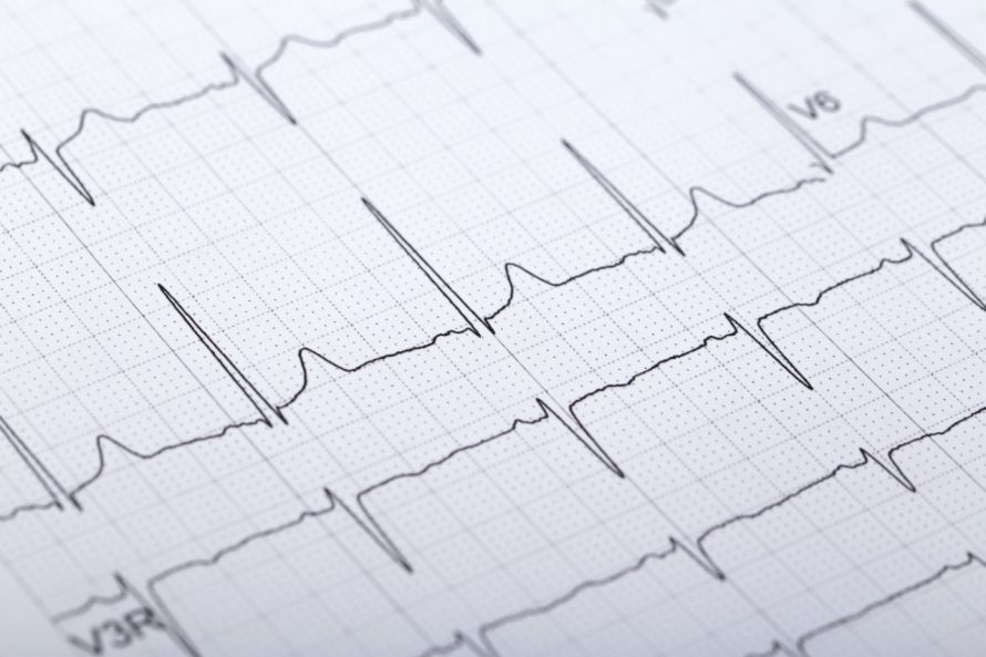
An electrocardiogram (ECG) measures and records the heart’s electrical activity. To obtain a complete picture,
- an ECG of the heart “at rest”—that is, without exertion—is performed,
- a long-term ECG using a portable device over 24 or even 48 hours, as well as
- a stress ECG, for which the patient sits on an ergometer.
The ECG trend chart in HOCM patients shows a specific arrhythmia or a persistently elevated heart rate.
During a cardiac catheterization, a thin tube is inserted into the heart. Instruments for examination and treatment can be guided through this tube. This allows for the assessment of the coronary arteries and the collection of tissue samples from the heart muscle. The tissue is then examined under a microscope to confirm the diagnosis.
An MRI (magnetic resonance imaging) uses a magnetic field to produce high-resolution images of the heart. The examination is painless and does not involve harmful radiation.
What does treatment for HCM involve?
To date, hypertrophic obstructive cardiomyopathy is incurable. Doctors can only manage the symptoms; they cannot treat the underlying cause.
In general, it is recommended that those affected avoid putting excessive strain on the heart and take it easy physically. If symptoms are limiting daily life, various treatment options may be considered for relief.
The first point of contact for HOCM patients is a specialist in heart disease—that is, a cardiologist. The German Society of Cardiology has published guidelines for the diagnosis and treatment of HOCM, which you can access here.
The chosen treatment depends on the symptoms and their cause; for example,
- medication (beta-blockers or antiarrhythmic drugs) to regulate the heartbeat,
- the implantation of a defibrillator (ICD, “implantable cardioverter defibrillator”) in cases of arrhythmias or when there is a risk of sudden cardiac death, or
- the removal of muscle tissue in cases of severe septal hypertrophy.
An important interventional procedure for treating HOCM is transcoronary ablation of septal hypertrophy (TASH). The underlying principle is to reduce blood flow to the thickened tissue so that it shrinks again.
This goal is achieved during a cardiac catheterization procedure: The injection of ethanol into the affected coronary artery creates an artificial infarction in the area of hypertrophy (thickening). This causes the tissue to die, allowing the thickening to regress. The treatment success rate is nearly 90%.
What does drug therapy for HOCM involve?
Drug therapy for hypertrophic obstructive cardiomyopathy (HOCM) aims to relieve symptoms, reduce the risk of arrhythmias, and support heart function. Here are some medications commonly used to treat HOCM:
- Beta-blockers: They reduce the heart rate and lessen the strain on the heart muscle. They also help reduce the risk of arrhythmias.
- Calcium channel blockers: These medications can help lower the heart rate and reduce the force of the heart’s contractions. They can also be helpful in controlling arrhythmias.
- ACE inhibitors or angiotensin II receptor blockers (ARBs): These medications can help lower blood pressure and reduce the strain on the heart by dilating blood vessels.
- Diuretics: These can be used to treat heart failure and reduce fluid buildup in the body, which can alleviate the symptoms of HOCM.
- Antiarrhythmic drugs: These are medications used to treat heart rhythm disorders; they are used to control irregular heartbeats and reduce the risk of life-threatening arrhythmias.
The choice of medications and their dosages depends on
- the patient’s individual situation
- the severity of the symptoms, and
- the presence of comorbidities
. Treatment is often determined in close collaboration with a cardiologist and monitored regularly to achieve the best possible outcomes.
Can children also have HOCM?
HOCM is a congenital condition, so it can manifest as early as childhood. However, it is rarely diagnosed in infancy or early childhood; it is usually first diagnosed during adolescence.
Genetic testing can be performed on asymptomatic children in families with a history of HOCM. This test determines whether the child is a carrier of the genetic mutation responsible for the condition. If so, the child should be monitored more closely.
In children with HOCM, physical activity (for example, during school and recreational sports) can increase obstruction of the left ventricular outflow tract. This can lead to rapid exhaustion and shortness of breath, as well as fainting spells.

HOCM carries a risk of sudden cardiac death. Therefore, children with hypertrophic obstructive cardiomyopathy must not participate in competitive sports or engage in isometric muscle exertion. Isometric muscle exertion refers to muscle exertion without movement—that is, the simple tensing of muscles.
Sudden cardiac death, in particular—which, as described, can also occur during physical activity—is a serious complication. That is why it is very important to make the correct diagnosis early on, even in children, and to assess the risk of sudden cardiac death.
The risk is higher if children
- a history of arrhythmias and fainting episodes, as well as
- septal hypertrophy
- an enlarged atrium and/or
- an enlarged left ventricular wall
are present.
In such cases, the implantation of a defibrillator (known as an ICD) can reduce the risk of sudden cardiac death.
What is the prognosis for HOCM?
Hypertrophic obstructive cardiomyopathy does not necessarily cause noticeable symptoms. On the other hand, life-threatening arrhythmias can occur as a complication. If HOCM goes undetected, the condition is one of the most common causes of sudden cardiac death.
If left untreated, this disease claims the lives of
- about six percent of affected children and adolescents, as well as
- about one percent of adults die from this disease each year.
Athletes are particularly frequently affected.
Early detection and treatment of HCM can mitigate many of the risks and symptoms of the disease.
With optimal treatment, there is nothing to prevent a normal life expectancy. However, the affected person’s ability to work depends on the severity of the condition and their personal physical condition.
Share this article

Sources
- Amboss (2020) Hypertrophe Kardiomyopathie (HCM). https://next.amboss.com/de/article/_S05Xf?q=hypertrophe%20kardiomyopathie
- Batzner A et al. (2019) Hypertrophe obstruktive Kardiomyopathie. Dtsch Arztebl Int 116: 47-53; DOI: 10.3238/arztebl.2019.0047
- S2k-Leitlinie „Primäre Kardiomyopathien im Kindes- und Jugendalter“ (AWMF-Register-Nr. 023-029), Stand 23.05.2024: register.awmf.org/de/leitlinien/detail/023-029
- Miron A et al. A Validated Model for Sudden Cardiac Death Risk Prediction in Pediatric Hypertrophic Cardiomyopathy. Circulation 2020; DOI: 10.1161/CIRCULATIONAHA.120.047235
- Sieverding L. (2015) Kardiomyopathien bei Kindern und Jugendlichen. In: Hoffmann G., Lentze M., Spranger J., Zepp F. (eds) Pädiatrie. Springer Reference Medizin. Springer, Berlin, Heidelberg. link.springer.com/rwe/10.1007/978-3-642-54671-6_206-1?error=cookies_not_supported&code=48b9ba87-fd53-4738-bc96-f202c6bbbabe