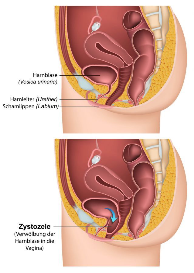
A cystocele, also known as bladder prolapse, occurs when the bladder shifts downward toward the vagina. This is usually caused by a weakening of the pelvic floor, which can be exacerbated by pregnancies, childbirth, hormonal changes during menopause, or a congenital weakness of the connective tissue. As a result of the prolapse, the bladder can no longer be held in its normal position, which can cause discomfort during urination, a sensation of a foreign body in the vagina, or urinary incontinence.
Cystocele is one of the most common forms of pelvic organ prolapse and primarily affects women. Depending on the severity, the spectrum ranges from mild prolapse without symptoms to a pronounced bulging of the vaginal wall that significantly impairs daily life. Thanks to modern diagnostic methods and individualized treatment options—ranging from targeted pelvic floor exercises to cystocele surgery—bladder prolapse can be treated very effectively today.
How does a cystocele (bladder prolapse) develop?
A cystocele develops when the bladder is no longer adequately supported by the pelvic floor muscles and the surrounding connective tissue. As a result, the bladder shifts downward toward the vagina and partially bulges into the anterior vaginal wall. This descent—also known as a bladder prolapse—is the most common form of pelvic organ prolapse in women.
The causes are varied. Often, weakening of the pelvic floor muscles due to childbirth, overstretching during pregnancy, or birth injuries causes the bladder and vaginal wall to lose their stable position. Especially after multiple births or when pregnancies are closely spaced, the supporting tissue cannot fully regenerate.
A congenital weakness of the connective tissue can also increase the risk of a cystocele. The tissue that normally holds the pelvic organs in place is then too elastic to provide lasting support for the bladder. In such cases, a cystocele can even occur at a young age.
With advancing age and hormonal changes during menopause, the elasticity of the tissue continues to decline. Falling estrogen levels weaken the vaginal walls and the supporting tissue around the bladder, making it easier for the bladder to descend.
Other contributing factors include obesity, chronic coughing (e.g., due to COPD or smoking), constipation, constant heavy lifting, or working in a standing position for many years. All of these factors increase abdominal pressure and can accelerate bladder prolapse.
In rare cases, a cystocele can even cause a kink in the urethra or ureters, which can lead to urinary retention, incomplete bladder emptying, and, in the long term, kidney damage. If the bladder is not completely emptied, the risk of bladder infections and urinary incontinence also increases.
A severe cystocele may occur in combination with other forms of pelvic organ prolapse, such as a rectocele (prolapse of the rectum) or uterine descent (prolapse of the uterus). In such cases, specialists refer to a combined pelvic organ prolapse, which requires a comprehensive gynecological examination and individualized treatment options.
What are the symptoms of a cystocele?
A cystocele may be barely noticeable at first. Many patients do not notice the bladder prolapse until they experience a sensation of a foreign body in the vagina or pressure in the lower abdomen. A typical sensation is that “something is pulling downward” or bulging in the vagina.
As the condition progresses, problems with urination often arise. Affected women complain of urinary retention, frequent urge to urinate, urinary incontinence, or a feeling of incomplete bladder emptying. Recurrent urinary tract infections can also indicate a cystocele.
Many women also experience pain during sexual intercourse (dyspareunia) or a feeling of fullness in the pelvic area. In severe cases, the bulge in the vaginal wall may be partially visible.
If a cystocele remains untreated, symptoms can worsen—potentially leading to impaired urine flow or a kink in the urethra, which increases the risk of complications.
How is a cystocele diagnosed?
A cystocele is typically diagnosed through a gynecological examination. The initial consultation with the patient already provides initial clues, such as complaints related to urination, urinary incontinence, or a sensation of a foreign body in the vagina.
During the examination, the doctor assesses the vaginal wall, the anterior vaginal wall, and the bladder to determine the extent of the bladder prolapse. Uroflowmetry can be used to evaluate bladder function—this procedure measures urine flow to detect possible urinary retention.
This is usually supplemented by an ultrasound examination of the pelvic organs. It shows whether the bladder empties completely and whether other organs, such as the uterus or rectum, are also affected. In some cases, a cystoscopy is performed for a more detailed assessment.
How is a cystocele treated?
Treatment for a cystocele depends on the severity of the bladder prolapse and the individual’s symptoms. In mild cases, non-surgical measures—such as targeted pelvic floor exercises to strengthen the pelvic floor muscles and keep the bladder in place—are usually effective. Regular exercise can alleviate symptoms and often make surgery unnecessary.
If hormonal changes during menopause are the cause, local estrogen therapy using creams or vaginal suppositories can strengthen the connective tissue. Alternatively, a pessary inserted into the vagina can stabilize the bladder and the anterior vaginal wall. It is fitted by a doctor and checked regularly.
In cases of severe cystocele or when conservative methods are insufficient, cystocele surgery is necessary. The surgical procedure is usually performed vaginally under general or spinal anesthesia. During the procedure, the supporting tissue around the bladder is tightened or reinforced to return the bladder to its normal position. In rare cases, the body’s own tissue or artificial material is used to secure the bladder in place.
Physical exertion should be avoided for several weeks after surgery. Follow-up physical therapy with exercises to strengthen the pelvic floor muscles supports healing and helps prevent a recurrence.
Progression, Complications, and Follow-Up Care
The course of a cystocele depends heavily on its severity and the chosen treatment. In early stages, regular pelvic floor muscle exercises can prevent further bladder prolapse. However, if the condition remains untreated, the bladder prolapse may worsen over time.
Possible complications of a cystocele include urinary incontinence, urinary retention, urine leakage, or recurrent urinary tract infections because the bladder cannot be completely emptied. In rare cases, a severe cystocele can cause a kink in the ureters and, in the long term, even lead to kidney damage.
Consistent follow-up care is important after cystocele surgery. Gentle exercise, avoiding heavy lifting, and regular pelvic floor exercises support the healing process. Medical follow-up visits ensure that the bladder remains in its normal position and that the vaginal wall does not bulge again.
FAQ: Common Questions About Cystocele (Bladder Prolapse)
Is a cystocele dangerous, or can it become serious?
In most cases, a cystocele is easily treatable. However, if it goes undiagnosed for an extended period, the severity of the cystocele may increase. In rare cases, a cystocele can be so severe that the bladder descends into the vagina, causing the vagina to narrow completely or partially. This makes it more difficult to empty the bladder and can lead to urinary retention or kidney damage. Therefore, a cystocele should always be evaluated by a doctor.
How is the severity of a cystocele determined?
Doctors generally distinguish between three grades of cystocele:
In Grade 1, the bladder prolapse is only mild and causes few symptoms. In Grade 2, the bladder partially descends into the vagina, while in Grade 3, a noticeable prolapse may be visible. The severity is determined through a gynecological examination or imaging to determine the appropriate treatment.
When is surgery necessary for a cystocele?
Surgery is recommended if non-surgical measures such as pelvic floor exercises or a pessary do not provide sufficient relief. In severe cases, the bladder is repositioned to its normal position, and the bladder and vaginal walls are supported to maintain function long-term. The procedure is usually performed vaginally; some specialists prefer to perform the surgery after menstruation to reduce the risk of infection.
Is a cystocele the most common type of pelvic organ prolapse?
Yes, a cystocele is the most common type of pelvic organ prolapse in women. It results from a weakening of the muscles and connective tissue that normally hold the bladder in place. Especially after multiple births or during menopause, the tissue can lose elasticity, causing the bladder to shift downward.
What are the symptoms of a cystocele, and when should you see a doctor?
Typical symptoms of a cystocele include a sensation of a foreign body, pressure in the pelvis, frequent urge to urinate, and urinary leakage. If such symptoms occur, a medical evaluation should be sought promptly. An untreated cystocele can become serious and, in rare cases, lead to a kink in the ureters, which can impair kidney function.
Share this article
Sources
- Lamblin G, Delorme E, Cosson M, Rubod C. Cystocele and functional anatomy of the pelvic floor: review and update of the various theories. Int Urogynecol J. 2016 Sep;27(9):1297-305. doi: 10.1007/s00192-015-2832-4. Epub 2015 Sep 4. PMID: 26337427.
- Iglesia CB, Smithling KR. Pelvic Organ Prolapse. Am Fam Physician. 2017 Aug 1;96(3):179-185. PMID: 28762694.
- Bu L, Yang D, Nie F, Li Q, Wang YF. Correlation of the type and degree of cystocele with stress urinary incontinence by transperineal ultrasound. J Med Ultrason (2001).
2020 Jan;47(1):123-130. doi: 10.1007/s10396-019-00972-0. Epub 2019 Sep 6. PMID: 31493276. - Stewart JR, Hamner JJ, Heit MH. Thirty Years of Cystocele/Rectocele Repair in the United States. Female Pelvic Med Reconstr Surg. 2016 Jul-Aug;22(4):243-7. doi: 10.1097/SPV.0000000000000240. PMID: 26825407.