Vesicoureteral reflux (VUR) refers to the pathological backflow of urine through the ureter into the kidney. The increased pressure in the kidney can cause lasting damage. Urinary reflux can be present from birth.
Here you will find further information as well as a selection of specialists and centers for vesicorenal reflux.
What is vesicorenal reflux?
Vesicorenal reflux (VRR) is also known in medicine as vesicoureteral reflux (VUR). This refers to the backward flow of urine from the bladder into the ureter. The urine can flow all the way back into the renal pelvis.
Over a prolonged period, the urine can exert excessive pressure at these sites. This poses a risk of kidney damage. In addition, the entry of bacteria can lead to painful kidney inflammation. This can result in scarring of the kidney tissue.
What are the different types of vesicorenal reflux?
Doctors distinguish between primary and secondary forms of vesicorenal reflux. While the primary form is congenital, the secondary form is acquired over the course of a person’s life.
Furthermore, a distinction is made between low-pressure VRR and high-pressure VRR:
- Low-pressure VRR: Urine reflux begins as early as the bladder’s filling phase.
- High-pressure VRR: Reflux occurs during the bladder’s emptying phase.
Vesicorenal reflux presents with varying degrees of severity. An important factor in this is the extent to which the anatomical structures are altered by the pressure of the urine. A typical pressure-induced change is the enlargement (technically: dilatation)
- of the ureter,
- the renal pelvis, and
- the renal calyces.
Since 1985, vesicorenal reflux has been classified into five different severity grades (according to Parkkulainen/Heikel):
- Grade I: Reflux into the ureter, but not into the renal pelvis.
- Grade II: Reflux extending into the renal pelvis without dilatation (= without changes to the renal calyces).
- Grade III: Reflux into the renal pelvis with mild dilatation (enlargement) of the calyx system and mild blunting of the renal calyces.
- Grade IV: Reflux with dilatation of the calyceal system, accompanied by mild ureteral kinking and blunting of the calyces, with papillae still visible.
- Grade V: Reflux with severe dilation of the urinary tract and ureteral kinking, as well as blunted renal calyces with papillae that are largely no longer visible.
Causes of Vesicorenal Reflux
Normally, urine flows from the kidney through the two ureters to the bladder. The end of the ureter at the bladder acts as a valve, ensuring that urine does not flow back to the kidneys.
However, a disruption of this function is sometimes congenital. This is caused by malformations of the ureteral orifice in the wall of the bladder. In most cases, the intramural course of the ureter is too short. Consequently, when pressure rises in the bladder, the upper urinary tract cannot be properly sealed off.
Secondary vesicorenal reflux results from direct impairment of the ureteral orifice. Possible triggers include cystitis or neurogenic bladder dysfunction.
In some cases, overdistension of the bladder wall is also responsible for the development of vesicorenal reflux.
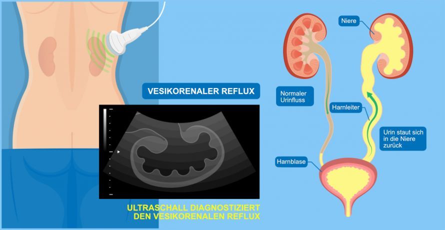
In vesicorenal reflux, urine backs up all the way to the kidneys and damages them © rumruay | AdobeStock
What are the symptoms of vesicorenal reflux?
Babies and toddlers with vesicorenal reflux typically suffer from:
- failure to thrive
- pallor
- low weight
- recurrent bedwetting
- Abdominal pain
- Diarrhea
- Vomiting
In older children and adult patients, the following symptoms may occur:
- Constant urge to urinate
- Burning sensation when urinating
- Pain in the kidney area
- a foul odor in the urine
- Pain in the flank while urinating or when the bladder is full
The backup of urine increases the risk of urinary tract infections. Harmful bacteria can reach as far as the renal pelvis. The resulting kidney infections can lead to scarring of the kidney tissue. This condition is called reflux nephropathy.
As vesicorenal reflux progresses, patients may experience complications such as
- high blood pressure or
- kidney insufficiency (kidney failure).
Incontinence can develop as early as childhood. However, in most cases—which are generally less severe—vesicorenal reflux in children often resolves spontaneously.
Diagnosis of Vesicorenal Reflux
If vesicorenal reflux is suspected, the examination should be performed by a specialist in urology or pediatric urology. The first step in the examination is to review the patient’s medical history (anamnesis).
The doctor will ask about the patient’s symptoms and any previous medical conditions.
General urological examination
Further clues may be provided by
- a physical examination,
- a urine test, and
- a blood test
. This allows for the detection of potential damage.
Sonography: Ultrasound Examination at VRR
Another diagnostic tool is sonography (ultrasound examination). Using this method, the urologist can determine whether the ureters or renal pelvis are dilated.
MCU: Voiding Cystourethrogram
A voiding cystourethrogram is used to assess the severity of vesicorenal reflux. During this examination, contrast medium is injected into the bladder via a thin catheter.
Under fluoroscopic guidance (and, if necessary, with ultrasound imaging), reflux of the contrast medium into the ureter or renal pelvis during filling or urination can be visualized if reflux is present.
In addition,
- bladder size,
- shape, and
- changes in the urethra
can be assessed.
Cystoscopy
Following an MCU, the doctor may perform a cystoscopy in some cases. This allows the shape, size, and location of the ureteral orifices to be determined.
At the same time, reflux can be treated during the same procedure (reflux injection).
DMSA scintigraphy
In some cases, a renal scintigraphy is also considered useful. It is a type of nuclear medicine examination. This test detects scarring of the kidney tissue resulting from previous inflammation.
How is vesicorenal reflux treated?
The method of treatment for vesicorenal reflux depends on the severity of the condition.
Treatment with Medications (Antibiotics)
If the condition is only mild, antibiotic treatment is sufficient.
This effectively combats urinary tract infections and prevents new infections. There is a high likelihood that the reflux will, so to speak, resolve on its own as the child gets older.
Endoscopic injection at the ureteral orifice
Endoscopic injection causes
- the end of the ureter is lifted,
- the ureteral orifice is narrowed, and
- ideally, the reflux is eliminated.
During the procedure, dextranomer/hyaluronic acid (Deflux) is injected into the bladder beneath the ureteral orifice via cystoscopy. The injection is performed under general anesthesia.
The milder the reflux, the higher the chance of long-term success.
Surgical Treatment
Open surgery is the most successful treatment for reflux, with a success rate of approximately 95 percent. It is primarily used for severe reflux or when the injection method has failed.
The most commonly used technique is the Lich-Gregoir antireflux procedure. It extends the course of the ureter through the bladder wall by creating a muscular tunnel. The bladder does not need to be opened—the procedure is performed through a small incision in the lower abdomen.
Surgery should generally be performed if
- recurrent pyelonephritis or febrile cystitis (“breakthrough infections”) occur despite antibiotic therapy,
- scarring of the renal tissue is increasing,
- there are high degrees of reflux (IV–V) with no prospect of spontaneous resolution,
- the parents refuse to have their child take medication for years, or
- medication therapy is not being followed reliably.
It is important to perform an annual ultrasound examination of the kidney, both after endoscopic injection and after open reflux correction. This allows for monitoring kidney growth through puberty and ruling out a recurrence of urinary reflux.
Immediately following the procedures, follow-up examinations (urinalysis, ultrasound, blood pressure checks) should be conducted more frequently.
Prognosis, Prevention, and Course
If vesicorenal reflux is congenital, there are no preventive measures. For the secondary form, urologists recommend emptying the bladder in two stages. This means waiting a few minutes after the first emptying before the next one.
Even without treatment, vesicorenal reflux often resolves on its own. In some cases, however, serious complications are possible, so it is generally advisable to consult a doctor.
Share this article
Sources
- Vesikorenaler Reflux: https://flexikon.doccheck.com/
- Blasenspiegelung (Zytoskopie): https://flexikon.doccheck.com/
- Ärzteblatt: https://www.aerzteblatt.de/
- S2k-Leitlinie: Diagnostik und Therapie der neurogenen Blasenfunktionsstörungen bei Kindern und Jugendlichen mit spinaler Dysplasie: https://www.awmf.org/ (PDF)