Fibromatosis refers to a group of benign connective tissue disorders characterized by excessive tissue growth. Among the best-known forms of fibromatosis are Ledderhose disease, which affects the sole of the foot, and Dupuytren’s contracture, which affects the palm of the hand. Although fibromatosis is benign, it can grow aggressively in localized areas and affect surrounding structures. The condition often manifests as lumps, hardened areas, or a palpable cord within the tissue.
Depending on the form, a distinction is made between superficial fibromatosis and deep fibromatosis. Treatment options include surgical procedures, radiation therapy, or other therapeutic approaches. An early diagnosis can help alleviate symptoms and slow the progression of the disease. Individualized treatment planning is particularly important in cases of aggressive fibromatosis.
What is fibromatosis?
Fibromatosis (ICD code: M72) is an overgrowth of collagenous connective tissue. Connective tissue is loose to firm tissue consisting of
- intercellular matrix
- adipose tissue and
- collagen
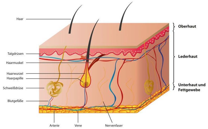
It also includes the supportive and protective sheaths surrounding muscles and organs. Fibromatoses can occur in a wide variety of locations throughout the body. Like cancers, they are classified as neoplastic diseases. Neoplastic means that disordered, autonomous, and excessive new tissue growth occurs.
Although the growth associated with fibromatosis is benign—meaning there are no malignant cancer cells present— Malignant cells can form metastases that spread to other organs. Malignant transformation—that is, the change from a benign growth to a malignant tumor—is also very rare.
However, fibromatosis can sometimes grow very aggressively and in an ulcerative manner. It can invade the surrounding tissue and damage important structures there. Affected structures may include, for example,
- blood vessels
- the lymphatic system, and
- nerves.
In many cases, myofibroblasts are involved in the development of fibromatosis. Myofibroblasts are cells that are classified as intermediate between muscle cells (myoblasts) and connective tissue cells (fibroblasts).
What are the causes of fibromatosis?
There are various possible causes for the development of fibromatosis. Possible causes include inflammation or injury. The connective tissue attempts to repair the local tissue damage but produces too many cells, leading to abnormal growths.
In many cases, however, there is no clear explanation for the development of fibromatosis. Some forms of fibromatosis are congenital or can be inherited.
What types of fibromatosis are there?
Fibromatoses are generally classified based on the part of the body where they occur. For example, they can be superficial or deep fibromatoses. There are a wide variety of fibromatoses; a comprehensive discussion of all subtypes is not possible within the scope of this article and would exceed its length. This is due to the fact that connective tissue can be found in nearly every part of the body, resulting in an extraordinarily large number of fibromatoses and different forms of manifestation.
Are all types of fibromatosis visible from the outside?
Fibromatoses are only visible from the outside if they have formed in the superficial tissues. These include, in particular, the skin, but also the layers directly beneath the skin (subcutaneous tissue), as well as mucous membranes and body orifices. Fibromatoses that have formed in deeper structures are not visible from the outside and are diagnosed only based on corresponding clinical signs and symptoms. Fibromatoses are therefore classified into superficial and deep forms.
What are the types of superficial fibromatoses?
The following conditions are classified as superficial fibromatoses:
- Ledderhose disease (plantar fibromatosis of the sole of the foot)
- Dupuytren’s disease (fibromatosis of the palm)
- Peyronie’s disease (induratio penis plastica, a connective tissue disorder of the penis)
- Desmoid tumor (aggressive fibromatosis affecting the muscle fascia)
- Fibromatosis colli (commonly occurs on the neck in young children and can lead to torticollis)
- Nodular fasciitis (formation of nodules in the upper extremities)
- Jones syndrome (fibromatosis of the gums)
What are deep fibromatoses?
Deep fibromatoses include, among others, the following conditions:
- Sclerosing mediastinitis (a hardening inflammation of the connective tissue in the mediastinum of the chest)
- Sclerosing mesenteritis (inflammation of the small intestine affecting the connective tissue interlaced with fatty tissue)
- Ormond’s disease (retroperitoneal fibromatosis, a connective tissue proliferation in the posterior abdominal cavity and lower back)
What are the symptoms of fibromatosis?
Symptoms vary depending on the location of the fibromatosis.
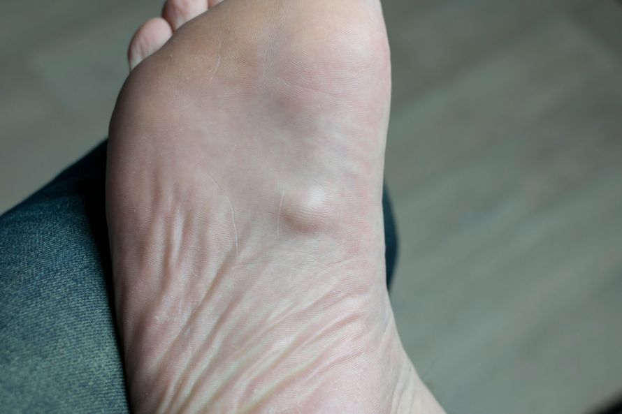
Superficial fibromatoses often develop on the soles of the feet or in the palms of the hands. The associated symptoms can then be felt directly and are often visible as well. The main symptoms include
- a sensation of pressure or tightness
- pain, as well as
- skin irritation
. Small or larger lumps may also form. The tissue changes cannot be clearly distinguished from the surrounding tissue but are clearly visible.
A condition known as nodular fasciitis is very commonly diagnosed. This is a form of superficial fibromatosis and usually manifests as the formation of individual lumps (= nodules) in the area of the muscle sheaths (fascia). This often results in small, tumor-like nodules on the upper extremities.
Symptoms of deep fibromatosis, depending on the location in the body, include:
- Digestive problems with fibromatosis in the abdominal cavity
- Back pain and kidney damage in cases of fibromatosis in the lower posterior abdomen (Ormond’s disease).
- Shortness of breath in cases of fibromatosis in the chest
How is fibromatosis diagnosed?
Diagnosing fibromatosis can be difficult in some cases. Some of the symptoms may also indicate other conditions, so further testing is necessary even if there are abnormal skin findings.
In addition to blood tests, this includes examining the suspicious tissue structures under a microscope. This requires a tissue sample (biopsy), which is then used to perform a histological analysis of the growths. During the examination, the structure and composition of the tissue are assessed. This usually makes it possible to determine whether fibromatosis is present and, if so, which type of the condition is involved.
If a strong suspicion of fibromatosis is raised or confirmed at this stage, the tissue growth is then surgically removed. This removed tissue is also prepared for histological examination by being cut into small pieces and thin sections. Specialists (pathologists) then determine whether the tumor is benign or malignant and assess the growth’s grade of malignancy.
In medical terminology, “malignancy” refers to the degree to which a disease is harmful. A finding with a higher degree of malignancy means that the disease has a greater potential to destroy the body. Rapid treatment is therefore absolutely essential for the patient. For example, fibroblastic-myofibroblastic tumors or malignant ulcers may occur. However, in the case of a malignant tumor, the doctor does not refer to it as fibromatosis but, depending on the findings, as a fibrosarcoma.
If malignant cells are detected, further tests are usually necessary. These involve checking the blood and the function of the internal organs, in part to identify any pre-existing conditions. Imaging techniques, such as
- MRI (magnetic resonance imaging),
- CT (computed tomography), and
- bone scintigraphy
, it is almost always necessary to determine whether the tumor has spread—that is, whether metastases have formed.
How is fibromatosis treated?
The treatment of fibromatosis depends on the specific type involved. In many cases, the connective tissue growth can initially be monitored. Of particular interest here is whether and how quickly the growth is expanding, as well as the severity of pain.
Surgical removal may be advisable if pain increases or for cosmetic reasons, and is strongly recommended if the growth increases rapidly in size. The affected tissue is excised relatively generously to ensure its safe and complete removal.
The affected tissue must be completely removed. If individual cells or margins of the excision remain, the condition often recurs.
If it is not possible to remove all of the diseased tissue, radiation therapy may be helpful. Radiation therapy is administered locally—that is, only at the site of the tissue growth—and aims to destroy the diseased cells. However, as with surgical removal, this can often result in additional damage to healthy tissue. Nevertheless, it is often necessary to prevent a recurrence.
FAQ
What is fibromatosis?
Fibromatosis is a benign connective tissue disorder characterized by excessive proliferation of connective tissue. Fibromatosis is a benign condition that, unlike a malignant tumor, does not form metastases. Nevertheless, some forms of fibromatosis can grow aggressively locally and infiltrate surrounding structures. The best-known forms include Ledderhose disease, Dupuytren’s disease, and aggressive desmoid-type fibromatosis.
What are the symptoms of Ledderhose disease?
Ledderhose disease affects the sole of the foot and often leads to palpable nodules, plaques, or a cord-like mass in the connective tissue. The overgrowth of connective tissue can cause pain, hardening, and discomfort when walking. The condition is classified as a superficial fibromatosis and may be triggered by repeated irritation or genetic factors.
How is fibromatosis diagnosed?
The diagnosis is initially made clinically through examination of the affected region. MRI, histological examinations, and, if necessary, a biopsy may also be used. In cases of deep fibromatosis, imaging helps assess the extent of the involvement as well as any potential compression of adjacent structures. It is particularly important to differentiate this condition from fibrosarcoma or other malignant changes.
What treatment options are available for fibromatosis?
Treatment depends on the type, location, and growth pattern of the fibromatosis. In addition to surgical resection, radiation therapy may be used to slow the growth of the lesions. In cases of aggressive fibromatosis, drug therapies such as tamoxifen are sometimes used. The goal of treatment is to reduce symptoms and prevent recurrence as much as possible.
Can fibromatosis recur after treatment?
Yes, there is an increased risk of recurrence, particularly with aggressive fibromatosis and desmoid tumors. New growths can develop even after complete resection. That is why regular follow-up examinations are important. For some patients, a combination of surgical and medical interventions may be appropriate for long-term management of the condition.
Share this article