When bones ache and muscles weaken, many people think of osteoporosis. However, osteomalacia may also be the cause of these symptoms. This painful softening of the bones is usually caused by a deficiency in vitamin D, calcium, or phosphate, which disrupts the mineralization of bone tissue. While the same condition in children is known as rickets and leads to deformities, adults with osteomalacia often suffer from diffuse bone pain and an increased risk of fractures. In addition to insufficient sunlight, causes can include kidney or liver disease, malabsorption in the intestine, or, rarely, oncogenic osteomalacia caused by a tumor. Early diagnosis through laboratory tests and X-rays is crucial for restoring bone strength.
What is osteomalacia?
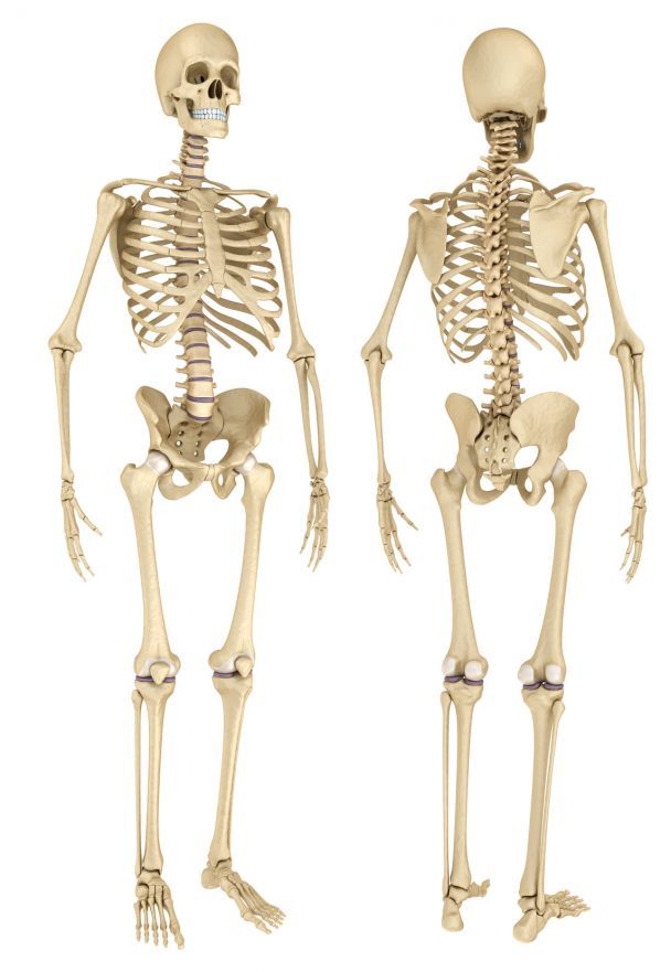
The human skeleton consists of approximately 206 bones of varying thickness and shape. Strong bones are essential for a stable body structure. This requires an adequate supply of nutrients and a healthy metabolism.
Osteomalacia is a disorder of bone mineralization. It is most commonly caused by a vitamin D or calcium deficiency. As a result, insufficient calcium and phosphorus are incorporated into the bones. The consequences are demineralization and softening of the bones, accompanied by various skeletal changes. These include knee misalignments such as bowlegs or knock-knees, as well as scoliosis.
At first glance, osteomalacia resembles osteoporosis. The difference between osteoporosis and osteomalacia is that in osteomalacia, the mineral content of the bone decreases, but not its volume. The bone matrix thus remains intact. In osteoporosis, bone density decreases.
Osteomalacia affects only adults. In children, doctors refer to the same condition as rickets.
Causes and Risk Factors for Osteomalacia
The main cause of bone softening, or osteomalacia, is vitamin D deficiency. Vitamin D plays a crucial role in the human body: it promotes the absorption of calcium from the intestines. This calcium is then lacking in the body and is not stored in sufficient quantities in the bone matrix.
Vitamin D deficiency arises for two reasons:
- insufficient intake of vitamin D through the diet
- insufficient exposure of the skin to sunlight
When sunlight hits the skin, the following occurs: The body converts the precursors of vitamin D (7-dehydrocholesterol) into active vitamin D in several steps. In technical terms, vitamin D is also called calcitriol or vitamin D3. Sunlight can thus meet 80% of the body’s vitamin D requirements.
Spending time outdoors therefore actively contributes to vitamin D levels and, consequently, to bone health.
In the absence of sufficient sun exposure, for example,
- in winter,
- for older or bedridden individuals, or
- due to the constant use of sunscreen
can lead to a deficiency in active vitamin D. This can sometimes result in osteomalacia due to reduced calcium absorption in the intestines.

In addition, the following conditions can also lead to bone softening:
- intestinal disorders such as celiac disease, ulcerative colitis, and short bowel syndrome,
- kidney dysfunction,
- congenital enzyme defects, and
- medications such as antiepileptics, fluoride, aluminum, and lithium.
What symptoms occur with osteomalacia?
Some of the symptoms associated with osteomalacia result from mineral deficiency. Other symptoms are consequences of the demineralized bone structure.
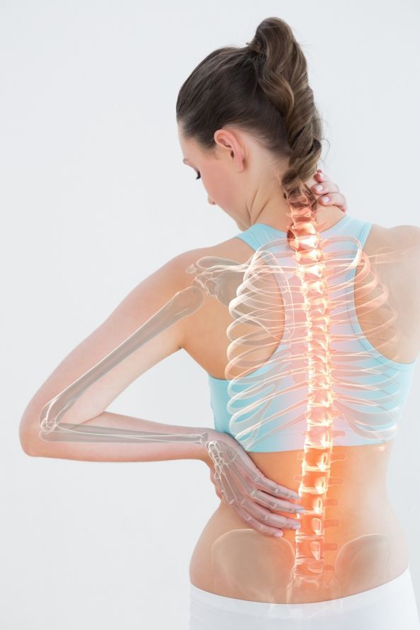
A major symptom of osteomalacia is dull, persistent pain, particularly
are particularly affected. These parts of the body bear a large portion of the body’s weight. Therefore, osteomalacia usually first becomes noticeable in these areas.
Other symptoms include:
- deformities due to reduced bone density (including curvature of the spine, bowlegs, and knock-knees)
- muscle weakness
- rapid fatigue
- increased susceptibility to bone fractures, particularly femoral neck fractures.
Bone pain is often mistaken for that caused by rheumatic diseases. Therefore, an accurate and comprehensive diagnosis is particularly important.
How to Make the Correct Diagnosis
Initial indications of bone softening emerge from a general physical examination. During this examination, the doctor assesses the patient’s physical condition as well as the symptoms described by the patient. The doctor also asks whether there have been any cases of osteomalacia in the patient’s family.
Important laboratory parameters are checked as part of a blood test. In cases of osteomalacia, the levels of the enzyme alkaline phosphatase and parathyroid hormone in the blood serum are consistently elevated. In addition, patients often have low levels of vitamin D and calcium. If the osteomalacia was caused by kidney disease, reduced phosphate levels are also observed.
An X-ray examination provides further evidence of whether bone softening is present. Here, the already deformed bones appear with a blurred structure.
A skeletal scintigraphy can detect increased bone metabolism. A bone density measurement confirms the diagnosis by revealing a low mineral content in the bone matrix.
If the diagnosis remains unclear, the doctor may then perform a bone biopsy. This allows the doctor to more clearly distinguish osteomalacia from other bone diseases, such as osteoporosis.
Treatment and Prognosis for Osteomalacia: Vitamin D and Calcium
The exact prognosis for osteomalacia depends on the stage of the disease at the time of diagnosis. In general, osteomalacia is easily treatable and curable. The earlier deficiencies or early-stage osteomalacia are detected and corrected, the less severe the consequences of demineralization will be. Osteomalacia is often completely cured within four to six months.
In rare cases, doctors may perform orthopedic or surgical procedures for osteomalacia. These are considered when the bones are severely deformed and the patient is suffering as a result.
If the bones are already severely affected, orthoses are one option for relief. These are medical devices that support, relieve pressure on, and correct the limbs. Physical therapy and exercise therapy are also helpful as supportive measures. They can help the patient develop stable and healthy posture.

The optimal treatment for osteomalacia promotes the remineralization of bone structure. The following options are available:
- Taking vitamin D supplements or receiving injections. A total daily intake of 5 µg of vitamin D is recommended; if a deficiency is present, a higher dose may be necessary after consulting a doctor.
- Regular exposure to sunlight: At least 15 minutes daily, ideally allowing the face, arms, and legs to be exposed without sunscreen.
- Phosphate supplementation and alkalizing substances in cases of kidney dysfunction.
- Take calcium supplements and follow a calcium-rich diet.
- Foods rich in vitamin D.
Foods high in vitamin D
- Fish (such as salmon, herring, trout, or sardines),
- dairy products (cheese, butter, milk),
- eggs,
- various mushrooms (button mushrooms, porcini mushrooms, chanterelles),
- avocados,
- legumes, and
- whole grains.
Animal products provide vitamin D in the form of D3, while plant-based products contain vitamin D as D2. The chemical form of vitamin D3 corresponds to the type of vitamin D produced in the skin through exposure to sunlight. This is why the body can utilize it effectively.
In general, while the diet can provide a consistent supply of vitamin D, many foods contain relatively little of it.
Depending on an individual’s constitution and symptoms, it may be advisable to have vitamin D levels checked by a doctor. Many people experience vitamin D deficiency, especially in the spring after the sun-deprived winter months.
FAQ: The 8 Most Important Questions About Osteomalacia
What is the difference between osteomalacia and osteoporosis?
Osteomalacia is a disorder of mineralization, meaning that the bones become soft because calcium and phosphate are not properly incorporated. Bone volume is often preserved, but bone quality suffers. In osteoporosis, on the other hand, mineralization is intact, but the overall bone mass and structure break down, making the bones porous. Both conditions increase the risk of fractures but have different underlying causes related to bone metabolism.
What are the symptoms of osteomalacia?
Typical symptoms of osteomalacia include persistent, dull bone pain, which often occurs in the lumbar spine, pelvis, and legs. This is often accompanied by pronounced muscle weakness, particularly in the thighs, which makes walking and climbing stairs difficult. In advanced stages of the disease, bones can become deformed, and fractures can occur easily, even with minimal stress.
How is osteomalacia diagnosed?
The doctor usually confirms the suspicion through laboratory tests. Typical findings include low levels of calcium and phosphate in the blood, elevated alkaline phosphatase levels, and low vitamin D levels. Parathyroid hormone levels are often secondarily elevated. X-rays show blurred bone structures or typical remodeling zones (Looser remodeling zones). To confirm the diagnosis in unclear cases, a bone biopsy may be necessary, in which bone tissue is removed and examined.
What is oncogenic osteomalacia?
Oncogenic osteomalacia is a rare form of osteomalacia caused by a tumor. This tumor produces hormone-like substances, most commonly fibroblast growth factor 23 (FGF23). This factor causes the kidneys to excrete more phosphate, leading to hypophosphatemia (low phosphate levels in the blood). Since phosphate is essential for bone hardness, the bones become soft. After the tumor is removed, this form usually resolves.
What role does vitamin D play?
Vitamin D—more specifically, the hormone calcitriol—is crucial for the absorption of calcium from the intestines. Without sufficient vitamin D, the body cannot provide enough calcium and phosphate for bone formation. A vitamin D deficiency—whether due to insufficient sun exposure, poor diet, or disorders of vitamin D metabolism in the liver or kidneys—is the main cause of osteomalacia and rickets.
How is osteomalacia treated?
Treatment depends on the cause. In cases of classic vitamin D deficiency, high-dose vitamin D (e.g., 1,000 to 2,000 units of vitamin D per day) is administered together with calcium. In cases of malabsorption, these nutrients often must be administered by injection. If hypophosphatemic osteomalacia is present, phosphate must be replaced. Adequate UV exposure is also important to stimulate the body’s own synthesis of vitamin D in the skin.
What is hypophosphatemic rickets?
This is a mostly hereditary form of rickets and osteomalacia in which the kidneys excrete too much phosphate due to a genetic defect (often on the X chromosome) (phosphate diabetes). Vitamin D alone is not effective in this case, as phosphate levels remain low regardless of vitamin D levels. Treatment requires the administration of phosphate and active vitamin D (calcitriol).
Who is particularly at risk?
Older adults who rarely go outdoors (residents of nursing homes) are at increased risk, as are people who completely cover their skin for cultural reasons. Patients with chronic gastrointestinal diseases (celiac disease), liver, or kidney dysfunction are also at risk. Vegans who do not consume vitamin D through fish or cod liver oil should also ensure they are getting enough, as recommended by the German Nutrition Society.
Share this article