Vasa praevia is a rare but dangerous pregnancy complication. In this condition, blood vessels from the fetal umbilical cord are located in the amniotic membranes at the internal os of the cervix. Around the time of birth, these vessels can rupture and, in the worst-case scenario, lead to the death of the unborn child from blood loss.
Here you’ll find information on symptoms and signs, treatment, and selected specialists for vasa praevia!
What is vasa praevia?
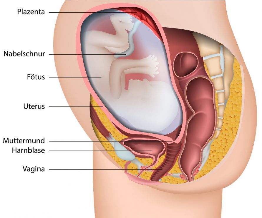
The umbilical cord is one to two centimeters thick and 50 to 60 centimeters long. It connects the unborn child to the mother’s body via the placenta, supplying the child with blood and nutrients. The fetal umbilical cord vessels normally do not run through the amniotic membranes but extend directly from the placenta to the fetus.
However, if fetal umbilical vessels are located within the amniotic membranes, this is referred to as insertio velamentosa. If they are also located within the amniotic membranes in front of the internal os, this is known as vasa praevia.
If the membranes rupture, these blood vessels can burst. As a result, the fetus may lose a critical amount of blood. Severe bleeding can be fatal for the fetus.
What are the symptoms of vasa praevia?
First, a slow fetal heart rate is observed in cases of vasa previa. Second, affected women experience vaginal bleeding when the membranes rupture.
This bleeding is often painless and usually begins shortly after labor begins.
Which doctors are experts in vasa praevia?
Experts and points of contact for vasa praevia are gynecologists—specialists in women’s health—and, in particular, obstetricians.
Depending on the stage of your pregnancy, you should contact your gynecologist or be admitted directly to the hospital.
How is vasa praevia diagnosed?
Regular ultrasound examinations are part of routine prenatal care. They should begin as early as the first trimester. Ultrasound can help detect various placental abnormalities.
To confirm a diagnosis of vasa praevia, a transvaginal ultrasound is performed. During this procedure, the doctor inserts an ultrasound probe into the woman’s vagina to check whether blood vessels are located near or over the opening of the cervix.
A particularly reliable diagnosis can be made using color Doppler ultrasound. This allows the doctor to visualize blood flow within the vessels as well as their attachment to the amniotic membranes or the placenta.
How is vasa praevia treated?
If vasa praevia is detected before labor begins, various treatment options are available. Treatment depends, among other things, on how far along the pregnancy is.
In any case, the expectant mother should be closely monitored by her obstetrician. Between the 30th and 32nd weeks of pregnancy, admission to a hospital may also be advisable. This allows for continuous monitoring of the unborn child.
In addition, the doctor may prescribe certain medications that stimulate lung development in the unborn child. These medications are known as corticosteroids.
As a rule, patients with vasa praevia give birth via cesarean section between the 34th and 37th weeks of pregnancy. In some cases, such a cesarean section may need to be performed as an emergency procedure. An emergency situation arises, for example, if
- the woman is experiencing heavy vaginal bleeding,
- her amniotic sac has ruptured,
- there is a risk to the woman or the unborn child, or
- vasa praevia is not diagnosed until after labor has already begun.
Do umbilical cord complications always require a cesarean section?
Under certain circumstances, a vaginal birth is also possible. This is contingent on the fetal blood vessels being located more than 2 cm from the internal os (velamentous insertion).
Early detection is essential for planning the safest possible delivery. Experienced physicians can identify abnormalities related to the umbilical cord as early as the first trimester using ultrasound.
Summary
Vasa praevia can lead to severe bleeding in the unborn baby. This usually occurs shortly before labor begins.
Such a complication can often be anticipated as early as the first trimester. If vasa praevia is suspected, the doctor will perform a transvaginal ultrasound.
Once vasa praevia is diagnosed, close monitoring by the obstetrician is necessary. If complications arise, a cesarean section is required. Asymptomatic patients are typically delivered via cesarean section between the 34th and 37th weeks of pregnancy.
Share this article