Expert Interviews
Interventional Heart Valve Therapy - Treatment of Mitral Regurgitation and Tricuspid Regurgitation

Alexandra Pfitzmann ·
The editorial team of the Leading Medicine Guide received the latest information on interventional heart valve therapy during a conversation with PD Dr. Büllesfeld.

Interventional heart valve therapy is a modern, minimally invasive treatment for heart valve diseases. It encompasses various conservative procedures in which cardiac catheters are used to repair or replace defective or leaky heart valves without the need for open-heart surgery. In the treatment of mitral and tricuspid regurgitation, the intervention aims to restore the function of the affected heart valves. For example, mitral regurgitation—in which the mitral valve leaks, causing blood to flow backward—is stabilized or repaired using catheter-based procedures.
Tricuspid regurgitation is treated similarly; in this condition, the tricuspid valve leaks, which also places a strain on the heart. These minimally invasive approaches offer the advantage of being less stressful for the patient, enabling shorter recovery times, and providing an effective alternative to traditional open-heart surgery for many patients. The goal is always to improve quality of life and stabilize heart function in the long term.
The development of heart valve disease, particularly mitral and tricuspid regurgitation, usually occurs gradually over many years. Initially, the valves are often only slightly damaged, which frequently causes no symptoms, so the condition goes unnoticed for a long time.

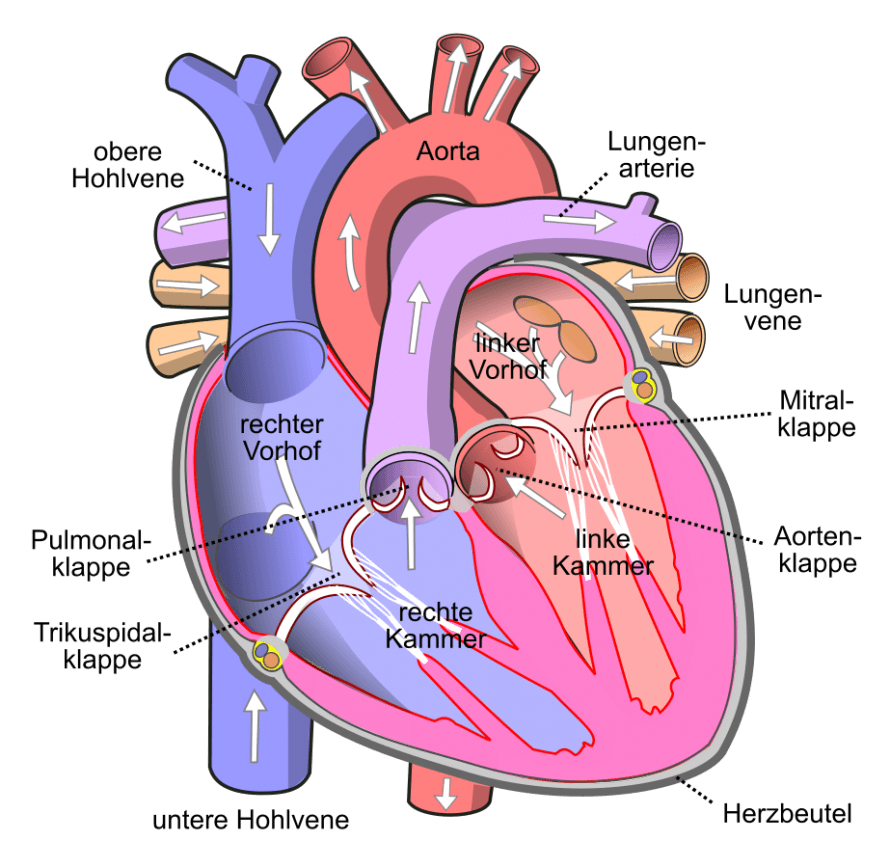
Mitral and tricuspid regurgitation are heart valve diseases in which the corresponding heart valves—the mitral and tricuspid valves, respectively—do not close properly. This causes blood to flow backward into the atria with each heartbeat, rather than flowing only into the ventricles. In mitral regurgitation, the valve between the left atrium and the left ventricle is affected, while tricuspid regurgitation affects the valve between the right atrium and the right ventricle. Over time, this backflow can strain the heart, lead to heart failure, and cause various symptoms.
“Ultimately, heart valve diseases—particularly mitral and tricuspid regurgitation—are closely linked to age. As people get older, the likelihood of developing such a condition increases, especially between the ages of 70 and 80. In the general population, the prevalence is about three percent, meaning that three out of every hundred people may have one of these valve leakages.
The main symptoms reported by patients are shortness of breath on exertion and increasing shortness of breath when climbing stairs. These symptoms are usually progressive, meaning they worsen over time. For this reason, it is often helpful to ask early on about the patient’s exercise tolerance—for example, how it was a year ago—to detect the progression of the disease. Another common symptom is cardiac arrhythmia, particularly atrial fibrillation.
Atrial fibrillation can both cause valvular insufficiency and be exacerbated by it. This often makes clinical assessment in everyday practice difficult, as it is necessary to distinguish which is the primary problem and which treatment should be administered first. Fluid retention is particularly common in tricuspid regurgitation: people suddenly gain weight, their lower legs and ankles become swollen, and their socks feel tight and constricting. “These symptoms are typical signs of functional insufficiency caused by changes in the heart chambers, such as enlargement of the atrium or ventricle,” explains PD Dr. Büllesfeld at the beginning of our conversation, and continues:
“A basic distinction is made between primary, structural valve problems—in which the valve itself is damaged, for example by tears or inflammation—and secondary, functional problems. In the latter case, the heart valves themselves are intact, but the heart chambers—especially the enlarged atrium or the weakened ventricle—alter the geometry so significantly that leaks occur. Particularly after heart attacks or in cases of cardiomyopathy, where the left ventricle becomes increasingly dilated, the valve eventually begins to leak due to the altered geometry of the valve apparatus and the heart chambers.
Distinguishing between these causes is crucial for treatment. Primary valve problems, which are caused by the valve itself, are usually acute events that can be treated surgically quickly, allowing for rapid and typically permanent improvement in symptoms and findings. In such cases, patients are often still relatively healthy, which facilitates surgical treatment. In contrast, patients with secondary regurgitation resulting from enlargement and changes in the heart chambers usually suffer from other severe heart diseases and are generally in poorer health. For them, catheter-based techniques are often an option, as they are less invasive and can specifically address the complex changes in the heart.”
Early diagnosis is essential for targeted treatment and to prevent disease progression, although modern therapeutic procedures often already allow for minimally invasive treatment.
“In the diagnosis of heart valve diseases, particularly mitral and tricuspid regurgitation, the process typically begins with a thorough clinical examination and a review of the patient’s medical history. For patients complaining of shortness of breath and fluid retention, an ultrasound of the heart—known as transthoracic echocardiography—is performed first. This technique is the primary tool for assessing the valves and their function, as well as the severity of the leaks. To gain even more detailed insights, a supplementary transesophageal echocardiogram is often performed, in which a flexible probe is briefly inserted into the esophagus.
This allows for more precise visualization of the valve and helps identify the exact causes and mechanism of the closure defect. If complex or diffuse changes are suspected, a cardiac catheterization is also performed. This allows for inspection of the coronary arteries to identify narrowings or previous heart attacks that may have affected valve function. This approach ensures that all relevant factors are taken into account in order to initiate targeted treatment. At the same time, drug therapy—particularly diuretics—is often initiated to alleviate symptoms.
These medications can reduce fluid retention, which can already lead to improved cardiac geometry and partially improve valve function. “It is an important first step toward stabilizing the heart’s condition and slowing the progression of the disease,” says PD Dr. Büllesfeld.
In contrast to another common heart valve disease, aortic stenosis—which represents a fixed mechanical problem that can only be treated by valve replacement—heart valve regurgitation, especially when it is functionally based, can often be improved with medication alone.
PD Dr. Büllesfeld comments: “Only when these conservative measures do not yield the desired results is interventional or surgical treatment indicated. It is important to note that treatment is only possible after careful diagnosis and optimization.
The transition from medical management to invasive therapy occurs only after a patient-specific assessment and when conservative measures have failed to produce sufficient improvement. In addition to medical factors, lifestyle factors also play a decisive role. Smoking, obesity, and alcohol and nicotine consumption are all factors that strain the heart and can contribute to the development and worsening of heart valve problems.
Being overweight contributes to increased strain on the heart through high blood pressure, while smoking increases the risk of coronary artery disease. Alcohol and nicotine consumption are also known triggers for atrial fibrillation, which in turn affects the heart’s geometry and thus valve function. Delays in making lifestyle changes can significantly impair treatment success, which is why both approaches—medication and lifestyle modifications—should always go hand in hand.”
In the interventional treatment of mitral and tricuspid regurgitation, various catheter-based procedures are now used that are designed to repair or stabilize leaky heart valves without open-heart surgery. In the field of heart valve therapy, catheter-based procedures represent a relatively recent innovation that has become increasingly established in recent years.
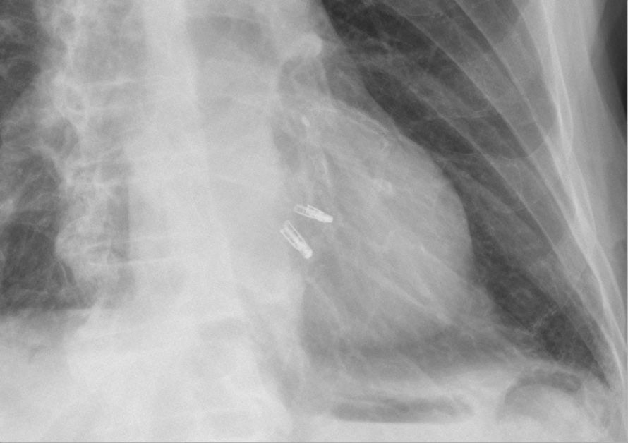
“While traditional surgical techniques have been in use since the 1950s and 1960s and were the primary alternative to drug therapy until about two decades ago, minimally invasive catheter-based procedures now offer a gentler option, particularly for high-risk patients. The best-known example is the mitral clip, a minimally invasive procedure in which a small clip is delivered via a catheter from the groin to the heart valve and placed at the site of the leak.
This procedure is now considered a first-line therapy when conservative measures fail. This technique was introduced in 2001 and has been scientifically validated in recent years by several studies, including the recently published European cardiology guidelines. In recent years in particular, the amount of available data on the mitral valve clipping technique—also known as “clipping”—has grown significantly compared to that for surgery. The so-called Matterhorn study, a randomized trial involving 200 patients, showed that clipping is not only non-inferior to surgery but is even superior in some respects, primarily due to a lower complication rate—with fewer bleeding episodes and strokes—as well as lower 30-day mortality.
In addition, patients can usually leave the hospital after just two to three days following a catheter procedure, whereas surgical treatment requires a hospital stay of several days followed by follow-up care. For the tricuspid valve, the technique is even newer, but the clip has also been used successfully in clinical practice for several years. Several studies are currently underway to scientifically investigate the effectiveness of this procedure for treating tricuspid valve regurgitation.
Despite all these positive results and experiences, catheter-based procedures are currently suitable only for certain patient groups. Younger patients and those with a low surgical risk are generally not candidates for the catheter-based technique at this time, as there is not yet sufficient data available. In general, catheter-based procedures are particularly suitable for high-risk patients who, due to other medical conditions or poor overall health, usually do not tolerate surgical treatment well. In particular, for younger and surgically viable patients with significant structural valve defects, traditional surgery remains the standard of care, as it still offers the best prospects for success in these cases.
“Overall, the development of these minimally invasive methods represents a significant advance in treatment, so that today we can offer a less invasive treatment option to many patients who previously could not be treated curatively or could only be treated at considerable surgical risk,” explains PD Dr. Büllesfeld.
Interventional heart valve therapy for mitral and tricuspid regurgitation is particularly suitable for patients for whom conventional open-heart surgery would entail significant risks due to various factors.
The procedure for interventional heart valve therapy is generally predictable, minimally invasive, and involves several phases. First, in close collaboration with an interdisciplinary team, a thorough diagnostic evaluation is conducted to assess the exact nature and severity of the valve regurgitation using echocardiography, and sometimes additional imaging techniques such as cardiac MRI or cardiac CT. Based on these results, a decision is made as to whether the patient is a suitable candidate for catheter-based treatment.
On the day of treatment, the procedure is prepared in a specialized cardiac catheterization lab. Patients are usually given sedation or local anesthesia and are continuously monitored. The procedure usually takes 1–2 hours. In MitraClip therapy, the catheter is inserted through the femoral vein to repair the valve. During the procedure, the team monitors heart function and blood flow to ensure success.
The procedure is performed under ultrasound guidance and general anesthesia to precisely control the degree to which regurgitation is reduced. Care is always taken not to close the valve too tightly to avoid complications. In some cases, multiple clips may be placed side by side to achieve a better sealing effect. After the treatment, patients are closely monitored for several hours to up to one day, during which vital signs and potential complications such as bleeding or cardiac arrhythmias are checked. Patients can usually resume mobility shortly thereafter. Finally, they receive instructions for follow-up care.
“There are no specific restrictions on the patient’s recovery following the procedure. Although there are risks, which are explained in detail beforehand, these are generally manageable. The most common risk is bleeding at the access site—that is, in the femoral vein—which can be effectively treated with a pressure bandage.
The risk is about five percent, and patients should take it easy physically for a week, although no strict recovery periods are required. There is also a possibility that, in rare cases, the clip may partially come loose. For the mitral valve, the probability of this occurring is 1 to 3 percent; for the tricuspid valve, it is 5 to 6 percent. The clip is attached so that it fastens the opposing leaflets of the valve together, similar to a paper clip that is securely fastened. “Loosening” means that one of the leaflets detaches from the clip, but the clip remains attached to the other leaflet. This loosening of the clip is often due to poor imaging quality, which can be detected in a timely manner through ultrasound monitoring.
Detachment usually occurs immediately after the clip is finally released or within the first few days. A later occurrence is virtually impossible, as tissue bridges form over the clip within weeks, further stabilizing it. If this complication occurs, the clip can usually be stabilized during a second catheter procedure, and the valve can be resealed by implanting an additional clip.
There are generally no long-term limitations. The first follow-up after a clip procedure takes place three months later; thereafter, patients are monitored annually on an outpatient basis. Experience with this procedure now spans over 17 years, dating back to the first CE certification in 2008. Initial long-term data are therefore now available, albeit only to a very limited extent. So far, however, no major signs of problems have been identified, which is why the therapy is also being used in younger patients, around the ages of 50–60,” explains PD Dr. Büllesfeld.
Currently, a range of innovative technologies and devices are being used in the treatment of mitral and tricuspid regurgitation, significantly improving the minimally invasive repair and treatment of these heart valve diseases. The introduction of catheter-based treatment methods for heart valve diseases is already at an advanced stage in Germany.
PD Dr. Büllesfeld comments: “Although these techniques have become increasingly important in recent years and are well-established in many centers, they are not yet widely available. This is primarily due to the strict guidelines regulated by the Federal Committee, as well as the high demands on infrastructure and interdisciplinary collaboration. Currently, these minimally invasive procedures are primarily offered at specialized centers that have the necessary experience and collaborative partners, such as cardiac surgeons.
The technique has long since achieved the status of a standard procedure and is classified as a clear recommendation (Class 1) for the treatment of functional atrial valve insufficiency in the current guidelines of the European Society of Cardiology. Catheter-based techniques are increasingly preferred, particularly for the mitral valve, a trend underscored by their comparatively low invasiveness, low mortality rate, and short hospital stay. Although the procedure is still relatively new for the tricuspid valve, experience is growing, and the technique is gaining importance. It is expected that in the future, an increasing number of patients will be treated interventionally, while traditional surgery will be used only for specific indications.
“Further advancements in catheter-based techniques, including options such as reducing the size of the valve annulus and implanting replacement valves, will continue to improve treatment options in the coming years.”

If it is too late to use a clip, this does not automatically mean that treatment must be switched to valve replacement. Rather, replacement is an alternative that becomes necessary in certain cases—especially when the valve structure is severely calcified or the leaflets are very short.
“In general, current practice—both in surgery and in interventional procedures—focuses on preserving the patient’s own valve as much as possible rather than replacing it with a prosthesis. In the past, valves were often completely replaced surgically, but this led to poorer outcomes. Today, the principle of saving the valve through minimally invasive procedures and replacing it only when structural damage makes repair impossible has become standard practice.
However, if it is too late for a clip or a replacement, this usually means that the disease has progressed and the prognosis is palliative—that is, symptom-oriented and associated with a poor outlook. In such cases, the primary goal is to alleviate symptoms, with no prospect of a permanent cure. We perform approximately 70 clip procedures each year. This is a substantial volume and ensures a high quality of care. We have been performing these procedures for over 10 years.
I myself have over 15 years of experience with the clip technique, which I introduced and have continuously refined during my time at the University Hospital of Bern in Switzerland. I also offer a consultation hour specifically aimed at private practice colleagues who treat patients with valve problems. During these consultations, I see the patients and provide detailed advice before a decision is made regarding further treatment. “This service has been very well received and helps prepare patients optimally for the next steps,” emphasizes PD Dr. Büllesfeld, and with that, we conclude our conversation.
Thank you very much, Dr. Büllesfeld, for this informative explanation!
- PD Dr. med. Lutz Büllesfeld, MHBA, FESC, FSCAI, is Chief of Internal Medicine with a focus on cardiology at the GFO Clinics in Bonn.
- He is an expert in interventional cardiology, offers state-of-the-art treatment options for cardiovascular diseases, and specializes in interventional heart valve therapy—particularly the treatment of mitral and tricuspid regurgitation—as well as other highly specialized procedures.
- He is known for his comprehensive expertise and for treating complex heart conditions with personalized care and modern technology.
- Director of the Cardiac Catheterization and Arrhythmia Center in Bonn, which performs over 3,000 procedures annually and ranks among the leading facilities in Germany.
- Recognized with professional certifications from the FESC (European Society of Cardiology) and the FSCAI (Society for Cardiovascular Angiography and Interventions).
- Areas of expertise: Interventional treatment of coronary heart disease, structural heart disease, and cardiac arrhythmias.
- Committed to innovation, high-quality medical care, and compassionate, personalized care.
- Sets standards in cardiac care through professional expertise, state-of-the-art technology, and a warm atmosphere.
Share this article
Expert Interviews
Read next
- Expert interview
Expert interview with Professor Dr Elmar Herbst, PhD
Oct 15, 2026
Patient-centred and personalised knee surgery
Read more - Expert interview
Cardiogeriatrics: Significance and benefits in the context of hospital reform
Sep 21, 2026
Cardiogeriatrics: Significance and benefits in the context of hospital reform
Read more - Expert interview
Expert Interview with Professor Dr. Amadeus Hornemann, MPH
Sep 18, 2026
Prof. Amadeus Hornemann on Precision Medicine in Gynecology: Modern Surgery, New Techniques, Better Quality of Life
Read more