Expert Interviews
Spinal Surgery

Alexandra Pfitzmann ·
In Germany, an estimated 8 to 10 million people suffer from spinal conditions requiring treatment; in Austria, the figure is about 1 to 1.5 million; and in Switzerland, there are approximately 600,000 to 700,000 people affected. Modern spinal surgery now offers both conservative and surgical procedures to relieve pain, correct misalignments, and improve mobility in the long term. The editorial team of the Leading Medicine Guide spoke with spinal specialist Dr. med. Christoph Weber about this topic.

Spinal surgery deals with the treatment of diseases, injuries, and malformations of the spine that can significantly impair mobility, posture, and quality of life.
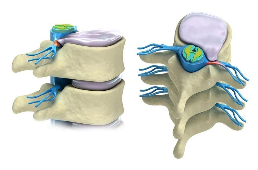
“When discussing surgically relevant spinal conditions, degenerative changes—that is, age-related and wear-and-tear changes in the spine—are by far the most common. While injuries are also seen in a clinical setting—such as osteoporotic vertebral fractures—severe trauma resulting from accidents, such as skiing or traffic accidents, is typically treated in an acute care hospital. Therefore, degenerative conditions clearly dominate daily practice. In older patients, spinal stenosis is the primary concern—a narrowing of the spinal canal caused by joint wear, osteoarthritis, herniated discs, and general wear and tear of the spine. The nerves that supply the legs run through this spinal canal. As the canal becomes increasingly narrow, these nerves eventually run out of space. Pressure builds up on the nerves, initially causing pain and sensory disturbances in the legs. In cases of more severe narrowing, this can eventually lead to muscle weakness and even symptoms of paralysis. This process is slow and gradual. There is no need to fear that such symptoms will develop overnight. However, it is precisely this slow progression that poses the danger: Many older people get used to the increasing limitations and simply attribute them to aging. Typically, the pain worsens when walking. Their range of activity becomes increasingly limited; those affected go out less often, eventually spending most of their time at home, and end up getting far too little exercise overall. This is a significant risk factor in old age, because a lack of physical activity leads to general physical decline—with effects on the cardiovascular system, lungs, digestion, and overall health,” explains Dr. Weber.
The goal of any treatment is to reduce or eliminate pain and, above all, to restore and improve mobility.
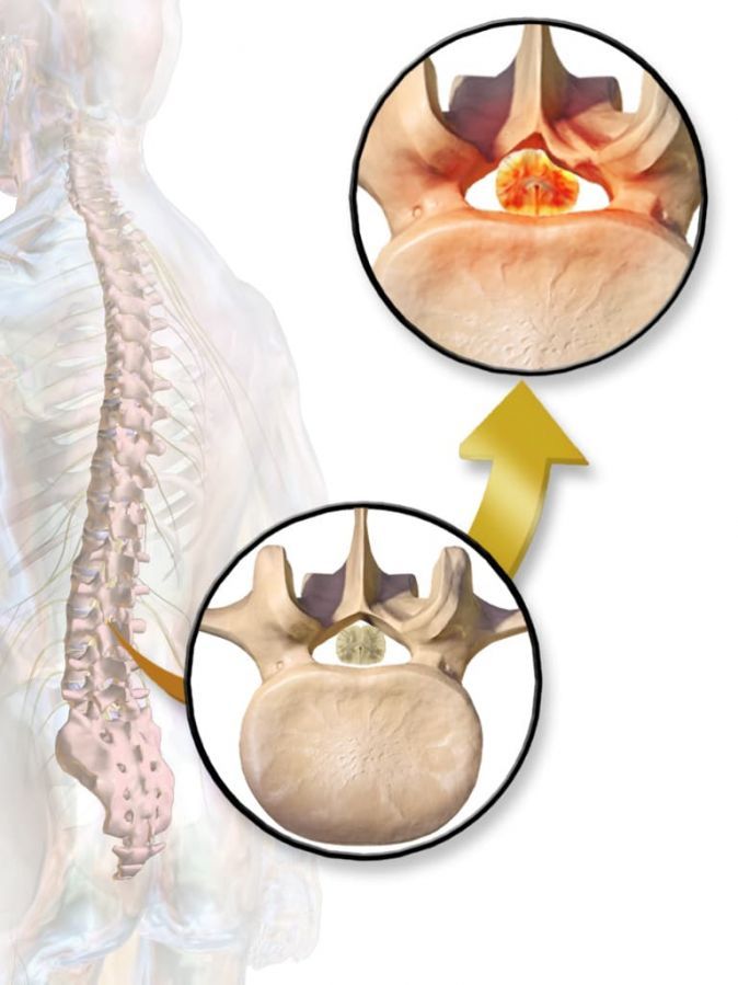
Dr. Weber describes some typical symptoms in patients with spinal canal stenosis: “A typical outward sign of this condition is a stooped gait. Many people with this condition walk with a slight forward lean because this slightly widens the spinal canal and alleviates their symptoms. They can only walk short distances and often have to stop, sit down, or bend their upper body forward. This temporarily relieves the leg pain, allowing them to walk a little further. This condition is called claudicatio spinalis—also known as ‘shop-window disease’—because those affected have to stop repeatedly while walking, as if they were looking at shop windows,” and he continues:
The term “window-shopping disease” is usually used in connection with peripheral arterial disease (PAD). This is a circulatory disorder in the legs caused by arterial calcification, which produces symptoms similar to those of spinal stenosis—namely, increasing leg pain the farther one walks. This makes it all the more important to talk with patients, conduct a thorough physical examination, and perform targeted diagnostic tests to identify the cause of the symptoms and initiate appropriate treatment.
“Of course, many people first wonder what conservative measures can be taken. As a general rule, exercise is always beneficial and generally recommended. In the past, bed rest was often prescribed for back pain, but today we know that this is precisely what is not advisable. The goal is rather to alleviate the pain to the extent that those affected are once again able to move and manage their daily lives. However, which conservative measures are appropriate depends heavily on the cause. If there is confirmed, severe spinal stenosis—that is, a significant narrowing of the spinal canal—studies now clearly show that surgery is the most effective treatment in these cases. The reason is simple: only surgery addresses the root cause directly. It restores space in the spinal canal and provides lasting relief to the nerves. Nevertheless, treatment always begins with conservative therapies. These include physical therapy, targeted exercises, and injections—such as cortisone injections into the spinal canal—to reduce swelling in the inflamed nerves. However, in cases of severe narrowing, this is not a sustainable solution because these measures only treat the symptoms—not the actual cause of the problem.”
Older people are more likely to have degenerative changes such as spinal canal stenosis, while younger patients tend to experience problems such as herniated discs. Furthermore, genetic predisposition and lifestyle factors play an important role.
“More and more people are working in a seated position, often for many hours at a time. This puts a great deal of strain on the spine because the human body is not actually designed to spend most of its time sitting. The lever forces acting on the spine create unphysiological stresses that cause pain. At the same time, the muscles atrophy, excess weight places additional strain on the spine, and chronic overload sets in. In the long term, this can also lead to herniated discs, even if no clear cause is initially visible on an MRI. Today, surgery for herniated discs is not performed as readily as it used to be—it always depends on the individual case. The decisive factor is how much pressure the herniation is exerting on a nerve. A herniated disc itself is not the problem; rather, it is the pressure on the nerve. If this pressure is severe enough to potentially damage the nerve and cause sensory disturbances or paralysis, the benefits of surgery clearly outweigh the risks. If, on the other hand, the patient only has localized back pain and nerve function is intact, conservative treatment is the first line of defense—for example, with physical therapy and, if necessary, nerve blocks. The intervertebral disc contains a lot of fluid, which the body gradually reabsorbs. In most cases—about 60 to 70%—the herniation resolves, the pressure on the nerve subsides, and symptoms improve without surgery. This process takes several weeks to a few months. The key factor is the progression of the condition: Even if symptoms have not completely resolved after six weeks, a noticeable trend toward improvement supports continuing conservative therapy. However, if significant nerve dysfunction is already present, surgery should be performed. “The surgery relieves pressure on the nerve by directly removing the herniated disc tissue, allowing the nerve to recover,” emphasizes Dr. Weber.
Preoperative diagnostics play a central role in spinal surgery, as they form the basis for precise, safe, and individually tailored surgical planning. Imaging techniques such as MRI (magnetic resonance imaging) or CT (computed tomography) provide detailed information about the anatomy of the spine, the condition of the intervertebral discs, vertebral bodies, nerves, and soft tissues, as well as any possible misalignments or narrowings.
Surgical treatment of the spine is always considered when natural healing processes, conservative therapies, or medication are insufficient to relieve pain, restore spinal function, or prevent neurological damage.
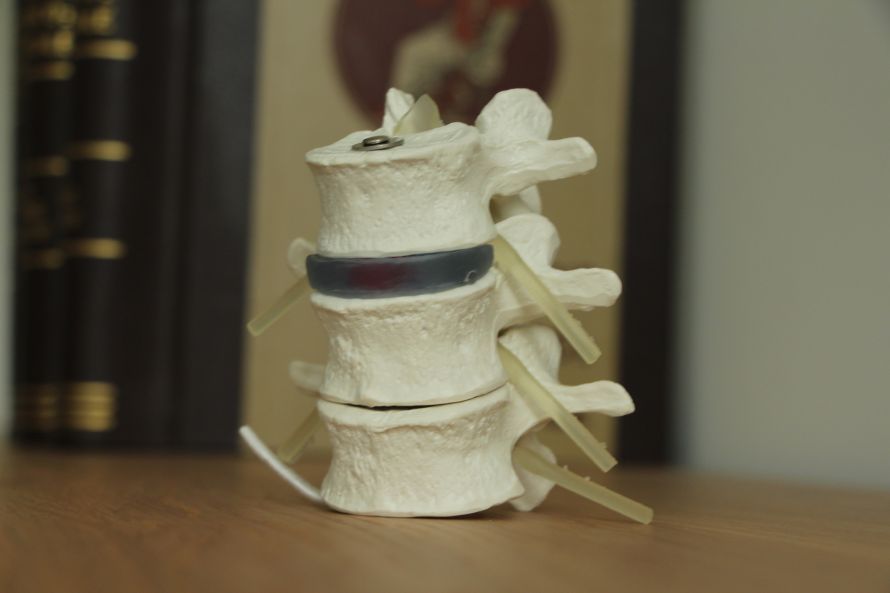
“The only truly causal treatment for spinal canal stenosis is decompression—more specifically, microsurgical decompression. It is considered the gold standard today. During the procedure, excess tissue is carefully removed or shaved away through a small incision using a surgical microscope and fine instruments, precisely at the point where the spinal canal is narrowed. The goal is to remove just enough material so that the nerves once again have the space they need. The narrowing is usually located in the area of the intervertebral discs, most commonly between the fourth and fifth lumbar vertebrae, followed by the segment between the third and fourth lumbar vertebrae. Often, two segments are affected at the same time, and sometimes more. Decompression of a single segment usually takes about half an hour to three-quarters of an hour. It is important to note that decompression does not automatically mean that stabilization or “fusion” is also necessary. These are two different procedures that must be evaluated individually for each patient. The current trend is toward decompression alone, because it has been shown that in many cases, widening the spinal canal is sufficient on its own, and additional stabilization is not absolutely necessary. This is good news for patients. Microsurgical decompression is a relatively minor procedure: shorter operating time, smaller incisions, less blood loss, less muscle damage, and a lower risk of infection. In summary, decompression surgery is significantly less invasive overall than stabilization surgery. However, there are situations in which decompression alone is not sufficient because it would otherwise be impossible to achieve long-term relief from symptoms. “In such cases, additional stabilization is necessary,” says Dr. Weber regarding decompression.
During stabilization, two screws are inserted per vertebra. The intervertebral disc between them is removed and replaced with a spacer, known as a cage. The screws are connected to each other via rods, so that this segment—that is, two vertebrae with the intervertebral disc between them—is no longer mobile.
Dr. Weber explains: “The procedure can be performed as open surgery or minimally invasively through small incisions using navigation technology. Which method is appropriate must always be decided on a case-by-case basis. Despite the term ‘fusion,’ no one needs to worry about being immobile afterward. In fact, only individual vertebrae are fused together. The body’s overall mobility is therefore preserved. Once healing is complete, there are normally no restrictions in daily life, work, sports, or hobbies—most people are just as mobile afterward as before, often even more so than before the surgery, because the pain has disappeared.”
Modern spinal surgery now offers a wide range of highly advanced surgical techniques aimed at relieving pain, alleviating pressure on neurological structures, and maintaining mobility in the long term.
A key trend is minimally invasive surgery: Procedures such as microsurgical discectomy or endoscopic spinal canal decompression make it possible to precisely remove diseased tissue without causing extensive tissue damage.
“If a procedure can be performed using a minimally invasive approach, this has a particularly positive effect on the immediate recovery phase. The surgery is shorter, the incisions are smaller, the extent of the surgery is less, and blood loss and the risk of infection are lower. All of this makes the initial period after the procedure easier. In the long term, however, it makes no significant difference whether open or minimally invasive surgery was performed. Once everything has healed, the results in terms of function and freedom from symptoms are essentially equivalent. The decision between minimally invasive and open surgery depends on many factors. These include the individual patient, their age, their general health, and any pre-existing conditions. The specific condition of the spine also plays a major role. For example, if vertebrae need to be repositioned or “repositioned,” an open procedure is typically chosen. If, on the other hand, the spinal alignment is stable and the procedure involves only screw fixation, this can often be performed using a minimally invasive approach. Each situation is assessed on a case-by-case basis. The question of cement often arises in this context as well. Generally, cement is not routinely used in spondylodesis, i.e., the fusion of vertebrae with screws. However, there are exceptions: In patients with osteoporosis—that is, reduced bone density—the screws can be additionally reinforced with bone cement. After the screws are placed in the vertebra, a viscous cement is injected through the screws; it envelops the screw like a cloud and anchors it more firmly in the bone. Within a few minutes, the cement hardens, thereby improving the screws’ stability. “This technique is used specifically for patients with osteoporosis, but it is not standard practice for everyone,” Dr. Weber clarifies.
Spinal Implants: The Function of the Screws, Formation of the Bone Bridge, and Risks of Adjacent Segment Degeneration.
Dr. Weber provides further important details regarding the implants to be used: “In spinal fusion, the implants generally remain in the body permanently. However, the actual goal of the surgery is for a bony connection to form between the fused vertebrae. The screws essentially serve as temporary fixation so that the body can reliably form the bony bridge. During the surgery, bone grafts or artificial materials are also inserted to support bone growth. This way, the body does not have to produce every bone cell on its own but has a substrate on which ossification can occur. As soon as this bony bridge forms, it takes over the stability, and the load on the screws decreases. One aspect that must be carefully discussed beforehand is what is known as adjacent segment degeneration. Fusion of a segment alters the distribution of stress in the spine: the adjacent segments must compensate for the loss of mobility and are therefore subjected to greater stress. This additional stress can cause these segments to degenerate more rapidly than under normal circumstances. Such adjacent segment degeneration occurs in up to 10% of cases. In such cases, it may become necessary to extend the existing spondylodesis—for example, if the adjacent segment begins to cause problems after a few years. The screws and implants themselves remain in place; the stabilization is simply extended to include the new segment.”
Recovery time is usually significantly shorter with minimally invasive procedures. Many patients can get back on their feet after just a few days, and rehabilitation can begin sooner. This allows for an earlier return to daily life and work, whereas open surgeries often require longer hospital stays and a more intensive postoperative phase.
“The length of the hospital stay depends primarily on the patient’s age, general health, and the extent of the surgery. Take, for example, a 70-year-old patient in good general health with no serious comorbidities who underwent microsurgical decompression at two segments; this patient can usually go home on the third day, or at the latest on the fourth day after surgery. In most cases, no special rehabilitation is necessary. The patient goes home, and the best therapy consists of taking relaxed walks. A follow-up examination takes place after about six weeks. By then, pain medication is usually significantly reduced or has already been discontinued. By this point, patients usually already notice a clear improvement. Although the final treatment outcome is often not yet fully achieved—since nerves need a lot of time to recover—a noticeable and significant improvement is generally evident within the first few days and weeks after surgery. Movement plays a central role from the very beginning. In fact, mobilization begins immediately: as soon as the patient wakes up from anesthesia, they are helped to their feet on the day of the surgery. Physical therapy and nursing care support this process several times a day. Initially, patients walk within their room, then around the ward, and later climb stairs. The walking distances are increased step by step until, after three to four days, patients are able to move around safely and can be discharged home,” explains Dr. Weber regarding the recovery process.
Patient anxiety always plays a major role in spinal surgery. Many have nightmarish fears: they think they might end up in a wheelchair, that their nerves might be damaged, or that they might never be able to walk again. This fear is often based on misinformation.
Dr. Weber clarifies: “Of course, it is theoretically possible for serious damage to occur during surgery, but the actual risk is extremely low. To draw a comparison with airplanes or ships: There is no such thing as an absolutely safe system, but the probability that something will actually happen is very low. Thanks to modern technology such as surgical microscopes, navigation systems, and the years of experience of spinal surgeons, such complications are extremely rare today. Spinal neuronavigation, which is now widely used, is a technology that enables even more precise screw placement and thus offers patients additional safety. Interestingly, the risk of suffering nerve damage is often even higher if surgery is not performed at all. As the disease progresses naturally, nerves can be continuously damaged, which can lead to paralysis. Such paralysis does not have to be complete—it can develop gradually, for example, when a patient can no longer fully shift their body weight onto the tips of their toes on one leg, while other movements continue to function normally. Often, only individual muscle groups are affected, so those affected may not even be aware of the limitations at first. They may only notice that their gait is unsteady or that their leg sometimes buckles. In such cases, surgery can help: By decompressing the nerves, they can often recover and regain strength. This means that the underlying condition poses a greater risk of nerve damage than the surgery itself.”
Movement is everything!

To conclude our conversation, spine specialist Dr. Christoph Weber recommends: “When it comes to preventive advice for the spine, the key phrase is clear: exercise is everything! It doesn’t have to be a specific sport—what’s important is that people do something they enjoy, because then they’ll do it regularly and willingly. Above all, physical monotony should be avoided. Sitting at a desk or in front of a computer for several hours at a time puts a lot of strain on the spine. A simple but effective measure is to schedule regular breaks for movement. For example, an alarm or a reminder on your cell phone can help you stand up briefly every hour, stretch, or take a few steps around your workspace. Even these small breaks relieve pressure on the spine and promote long-term back health. In addition, a balanced diet, avoiding excess weight and nicotine, and—especially for older patients—ensuring an adequate intake of calcium and vitamin D are highly recommended.”
Thank you very much, Dr. Weber, for this helpful information on spinal surgery!
- Specialist in neurosurgery, spinal surgery, and radiosurgery in Zurich.
- Use of Switzerland’s first ZAP-X system for high-precision, outpatient radiosurgery of brain tumors without surgery.
- Comprehensive microsurgical and minimally invasive spinal surgeries for degenerative, traumatic, and neoplastic conditions.
- Many years of clinical expertise, supported by an interdisciplinary network, including second opinions and modern patient care.
Share this article
Expert Interviews
Read next
- Expert interview
Expert interview with Professor Dr Elmar Herbst, PhD
Oct 15, 2026
Patient-centred and personalised knee surgery
Read more - Expert interview
Cardiogeriatrics: Significance and benefits in the context of hospital reform
Sep 21, 2026
Cardiogeriatrics: Significance and benefits in the context of hospital reform
Read more - Expert interview
Expert Interview with Professor Dr. Amadeus Hornemann, MPH
Sep 18, 2026
Prof. Amadeus Hornemann on Precision Medicine in Gynecology: Modern Surgery, New Techniques, Better Quality of Life
Read more