
Expert Interviews
Cystic Tumors of the Pancreas. When to Operate, and When Not to?

Alexandra Pfitzmann ·
Cystic tumors of the pancreas are increasingly being discovered incidentally today—for example, during an MRI or CT scan. The challenge lies in distinguishing benign cysts from those that carry a significant risk of malignancy. Therefore, precise diagnosis, accurate assessment of biological behavior, and careful consideration are crucial: When is surgery advisable, and when is close monitoring sufficient?

“The pancreas can be affected by both benign and malignant tumors, with the dreaded pancreatic cancer—known as ductal adenocarcinoma—accounting for the largest proportion of malignant tumors, at around 90 percent. However, such carcinomas do not arise suddenly but develop over a series of prolonged precursor stages. Recurrent inflammation of the pancreas, particularly chronic pancreatitis, can contribute to such changes through the constant cycle of inflammation and tissue repair.
Certain histopathological abnormalities are also considered potential starting points for tumor development. In addition to these classic pathways, there are other “routes” that can ultimately lead to pancreatic carcinoma. One of these is primarily benign, cystic changes in the pancreas. These non-solid, fluid-filled structures can develop into malignant tumors over time.
“It is therefore crucial to detect such precursor changes early, monitor them closely, and, if necessary, remove them surgically in a timely manner so that a carcinoma does not develop in the first place,” explains Prof. Dr. Bahra at the beginning of our conversation, adding:
“Pancreatitis is most commonly caused by chronic alcohol consumption, particularly in cases of alcohol dependence or chronic heavy drinking. Another major risk factor is heavy smoking, which—depending on an individual’s predisposition—can also trigger severe and recurrent inflammation of the pancreas. Such repeated episodes of inflammation cause permanent changes in the tissue, which must constantly regenerate.
It is precisely within this cycle of inflammation and repair that the risk of developing pancreatic cancer can increase significantly, especially in people with chronic pancreatitis who also smoke. These are solid tumors. When it comes to cystic changes in the pancreas, another question arises: Which of these cysts are harmless, which pose a risk, and which require surgery? Cysts do not contain air, but rather fluid—and it is precisely this fluid that is crucial.
If the fluid is clear and watery, they are called serous cysts. They are considered biologically harmless and have virtually no potential to become malignant. However, if the cyst contains mucous, viscous fluid, it is a mucinous lesion. With these mucus-filled cysts, the question always arises as to whether they could be a precursor to cancer, since they can indeed develop into an adenocarcinoma over time.
Therefore, distinguishing between serous and mucinous cysts is a key diagnostic step. If a mucinous lesion is detected, it can be further classified to more accurately assess the individual’s risk.”
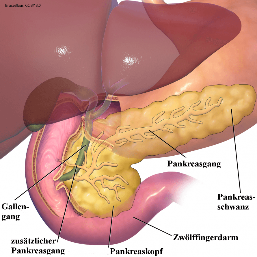
Serous cystadenomas are benign lesions of the pancreas filled with clear fluid and carry no risk of malignant transformation. In contrast, mucinous cystic neoplasms (MCN) and certain forms of so-called intraductal papillary mucinous neoplasms (IPMN) are clearly considered precancerous, that is, precursors to a potential cancer. Pseudocysts, on the other hand, usually develop following pancreatitis; they are not true cysts and do not carry a risk of malignant transformation. In addition, there are rare cystic tumors whose potential for malignant transformation can vary greatly depending on the type and is clinically relevant in some cases. The precise differentiation of all these lesions—from benign serous cystadenomas to precancerous MCN and IPMN, and ranging from harmless pseudocysts to rare high-risk scenarios—therefore constitutes the crucial step in any therapeutic decision.
Imaging plays a central role in assessing the risk of cystic pancreatic tumors because it allows us to determine whether a lesion is harmless, potentially precancerous, or already requires treatment.
“In most cases, cystic changes in the pancreas are discovered incidentally. Often, an MRI is initially performed for entirely different reasons—such as spinal symptoms or to investigate other conditions—and suddenly a cyst in the pancreas is detected. Interpreting such findings is often not straightforward, as it immediately raises alarm and the radiological description usually remains nonspecific. It is then crucial to outline a clear, reassuring diagnostic pathway for the patient, because not every cyst automatically indicates a high risk or even an impending cancer. Many cases can be clarified in a calm and structured manner. However, there are also patients who do develop symptoms.
This becomes particularly relevant in the case of a so-called IPMN—an intraductal papillary mucinous neoplasm. In this condition, the pancreatic duct system undergoes changes: The normally smooth lining forms small papillary protrusions, which are biologically similar to adenomas and produce mucus. This mucus can obstruct the flow of digestive secretions and thus trigger obstructive pancreatitis. Typical symptoms include elevated pancreatic enzymes (such as lipase or amylase) in the blood or upper abdominal discomfort. If such a change occurs in the head of the pancreas, it can—depending on its severity—even press on the bile duct.
This leads to jaundice because bile can no longer flow into the small intestine. This is rare, but it is a clear warning sign. It is precisely at this point that the question of when an IPMN is dangerous and when it is not becomes important. So-called “high-risk stigmata” play a central role in risk assessment: bile duct obstruction with jaundice, contrast-enhancing nodules in the cyst wall measuring approximately five millimeters or larger, or a main duct dilated to over one centimeter due to mucus accumulation. If such features are present, there is strong evidence of a precancerous lesion that can no longer be reliably monitored. “In that case, surgical removal must be seriously considered,” explains Prof. Dr. Bahra.
Surgical removal is medically indicated when imaging or the clinical situation suggests that a cystic pancreatic tumor carries a significant risk of malignant transformation or that high-grade dysplasia is already present.
Prof. Dr. Bahra elaborates: “If there are no high-risk stigmata, one can breathe a sigh of relief for the time being. Nevertheless, there are criteria that, while less threatening, still require attention—the so-called ‘worrisome features.’ These include cysts up to about three centimeters in size, a only slightly dilated main duct, a thickened cyst wall, detectable growth over the course of multiple examinations, a moderate increase in certain tumor markers, or mild pancreatitis resulting from mucus stasis.
Such findings are not immediately life-threatening, but they understandably cause anxiety because they indicate a potential risk profile. There are no conservative treatment options in the form of drug therapy. If there is no clear indication for surgery, structured monitoring remains the option. Established monitoring intervals exist for this: for cysts larger than one centimeter, an MRI is performed every two years; for cysts between one and two centimeters, annually; for cysts between two and three centimeters, every six to twelve months; and for larger cysts, every three to six months.
In practice, this close monitoring is stressful for many patients because their lives revolve around one examination after another. Therefore—despite the lack of a compelling indication for surgery—surgical removal may be considered during an individual consultation if the psychological strain is high and the patient desires clarity. “Ultimately, the only therapeutic option is surgery,” he says, adding regarding gender-specific considerations:
“Cystic lesions such as IPMNs can be further subdivided into main duct, side duct, and mixed forms, with involvement of the main duct in particular being considered critical. In addition, there are so-called mucinous cystic neoplasms (MCNs), which occur exclusively in female patients and are almost always located in the body or tail of the pancreas. We now know that these consist of ectopic ovarian tissue that remained in the upper abdomen during embryonic development and forms mucus-producing stroma there.
These lesions are also considered relevant precancerous conditions. In young female patients with MCN and a corresponding cystic marker profile—such as a markedly elevated CEA level—surgical removal is generally recommended. Both groups, IPMN and MCN, are classified as mucin-filled cysts with the potential for malignant transformation, whereas purely serous, water-filled cysts are considered benign.”
The type of surgery to be considered depends on the location of the cyst.
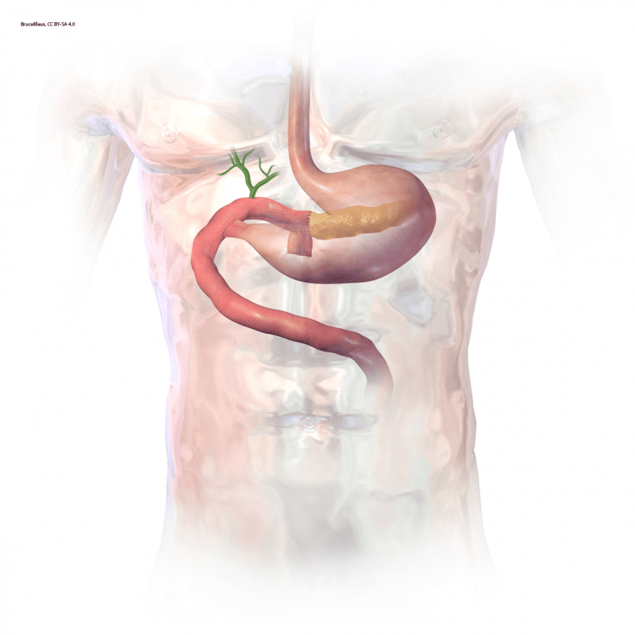
“If the cyst is located in the head of the pancreas, a major Kausch–Whipple procedure would be necessary—a surgery that is not recommended lightly (named after the surgeons Allen Oldfather Whipple and Theodor Kausch). However, if the lesion is located in the body or tail of the pancreas, a tail resection can often be performed today using minimally invasive techniques, such as with the aid of a surgical robot. The growth pattern of the cyst also plays a role.
Although growth of about 30 percent within a year is not yet a high-risk criterion, it does indicate that the condition is progressing. How this is managed depends heavily on the patient’s age and overall health: A 45-year-old family man will receive different advice than an elderly, seriously ill patient for whom major surgery would not be an option anyway. Since there are no conservative treatment options, monitoring in younger patients often means very long-term, sometimes lifelong follow-up. While European guidelines recommend ongoing monitoring, American guidelines discuss the possibility of discontinuing monitoring after five years of stability—an assessment that remains controversial because carcinomas can develop even later.
Cases in which patients discontinue follow-up and return years later with a carcinoma underscore this problem. Of course, the patient has the freedom to make their own decisions. If the burden of constant monitoring is significant or if additional “worrisome features” arise, the patient may wish to undergo surgical removal. Such decisions are made on an interdisciplinary basis—in collaboration with radiology, gastroenterology, and surgery. It is important that communication does not exacerbate fears or suggest premature surgery.
Not every cyst requires surgery. In cases of high-risk stigmata, there is a clear indication for surgery; in cases of “worrisome features,” close monitoring is performed, supplemented by procedures such as endosonography with fine-needle aspiration. This allows for the analysis of cyst fluid and cells to more accurately assess the risk. “If there are no relevant risk factors and the patient is primarily seeking peace of mind, imaging-based monitoring remains the approach—because any invasive diagnostic procedure or anesthesia also places a burden on the patient,” notes Prof. Dr. Bahra.
The surgical options depend heavily on the cyst’s location and the complexity of the procedure. For procedures on the body or tail of the pancreas, the current standard is that they should ideally be performed using minimally invasive, robot-assisted techniques. Open surgery should no longer be an option in this area, because robotic technology is more precise, more controlled, and significantly less invasive for patients.
“The situation is different for surgery on the head of the pancreas—the classic Whipple procedure. It is one of the most challenging procedures in visceral surgery. Robotic procedures are not yet widely established in German-speaking countries, but are already being offered at specialized centers. Due to its high complexity, this procedure must be performed by experienced surgeons. Anyone with a cyst in the head of the pancreas—whether due to high-risk stigmata or worrisome features—should therefore consult a center like ours, which specializes in robotic pancreatic surgery.
There, the procedure can be performed using a minimally invasive approach, which significantly reduces the burden on the patient. From the surgeon’s perspective, open surgery for cystic lesions in the head of the pancreas is no longer considered state-of-the-art. In general, cystic masses can almost always be treated with minimally invasive surgery. Only in exceptional cases—such as after major prior surgeries, in the presence of severe adhesions, or when a carcinoma involving blood vessels is already present—must the feasibility be critically assessed. In cases of true tumor surgery, robotic technology may reach its limits because the tumor grows differently than expected.
Cystic lesions, on the other hand, are ideally suited for robotic surgery, as they are generally clearly demarcated and technically accessible. For patients, this means: no large abdominal incision, less pain, faster recovery, and an overall much less invasive procedure. We use a state-of-the-art da Vinci system for this, specifically the XI console.
Our center also serves as a training and center of excellence for robotic pancreatic surgery and benefits from my many years of experience at Charité as well as in the U.S. (UPMC Pittsburgh), where I intensively studied robotic pancreatic surgery and subsequently refined the technique before establishing the current robotic surgery center,” emphasizes Prof. Dr. Bahra.
Monitoring is always sufficient when the biological risk is low, the imaging is stable, and the surgical risk outweighs the potential benefit. The intervals follow a clear risk-tiered approach and are adjusted individually over time.
At Waldfrieden Hospital in Berlin, approximately 60 pancreatic surgeries are performed each year. However, the development of cystic changes cannot be actively prevented. They usually arise due to a genetic predisposition and develop, so to speak, on their own, without lifestyle factors such as diet, exercise, or certain behaviors having any influence.

“While smoking, heavy alcohol consumption, or severe obesity increase the overall risk of pancreatic cancer, they do not influence the formation of these typical cysts. Rather, it is crucial to correctly classify the type of lesion and assess the individual’s risk. Studies show that about 15 percent of people in Central Europe develop cysts in the pancreas after a certain age, though this does not automatically indicate a need for surgery.
For many affected individuals, the psychological aspect plays a major role, as the term “pancreas” often triggers anxiety. The question of the prognosis for a possible pancreatic cancer frequently arises. Pancreatic cancer remains one of the most aggressive tumors in the abdominal cavity. Since it causes no symptoms for a long time, it is usually detected at a late stage; 80 to 85 percent of those affected are already at a stage where surgery is no longer possible at the time of diagnosis.
Only about 15 percent are eligible for surgery at all. Even with modern therapies, the five-year survival rate after successful surgery is around 30 percent—figures from studies that are often even lower in everyday clinical practice. Compared to other types of cancer, such as breast or colorectal cancer, the prognosis is significantly worse. This makes it all the more important to detect precancerous changes, such as cystic lesions, at an early stage and—if necessary—remove them in a timely manner so that carcinoma does not develop in the first place,” states Prof. Dr. Bahra.
Today, the care of patients with cystic pancreatic tumors is consistently interdisciplinary, because a sound treatment decision is only possible when radiology, gastroenterology, surgery, pathology, and, if necessary, oncology combine their respective perspectives. Each of these disciplines makes an indispensable contribution to risk assessment and treatment planning.
At Waldfriede Hospital, all types of cancer are treated in certified cancer centers. For pancreatic, colorectal, and anal cancers, there is a visceral oncology center, supplemented by specialized centers for gynecological tumors and a large breast center.
“All departments are certified according to the guidelines of the German Cancer Society. In addition, the hospital serves as a training center for robotic surgery in both the colorectal region and the upper gastrointestinal tract. In colorectal surgery, procedures are performed almost exclusively using robotic technology, and modern minimally invasive techniques are also used in the upper abdomen—for example, on the stomach, liver, or pancreas—provided that the anatomical and disease-specific conditions are met. Cystic lesions of the pancreas, particularly in the body and tail regions, are always treated using minimally invasive techniques and never via open surgery.
For the future, a more consistent centralization of pancreatic surgery would be desirable. Procedures on the pancreas should be performed exclusively in certified pancreatic centers so that care is provided by teams with high case volumes, well-established protocols, and specialized infrastructure. Studies from the Netherlands clearly show that such structures can significantly reduce complication rates.
The point is not that only a few large centers perform hundreds of procedures per year, but rather that hospitals with sufficient experience—for example, more than 50 procedures annually—take on the treatment. “However, many hospitals in Germany perform only single-digit or low double-digit numbers of procedures and thus meet neither the quality requirements nor the criteria for certification,” Prof. Dr. Bahra emphasizes at the conclusion of our conversation.
- Chief of Visceral Surgery at Waldfriede Hospital in Berlin; recognized expert in complex upper abdominal and tumor surgery
- Specialist in pancreatic and upper abdominal tumors, with a focus on cystic, benign, and malignant pancreatic tumors
- Robotics & minimally invasive surgery for precise, gentle procedures
- Hernia surgery at a certified center, including robot-assisted procedures
- Director of a DKG-certified tumor center
Share this article
Expert Interviews
Read next
- Expert interview
Expert interview with Professor Dr Elmar Herbst, PhD
Oct 15, 2026
Patient-centred and personalised knee surgery
Read more - Expert interview
Cardiogeriatrics: Significance and benefits in the context of hospital reform
Sep 21, 2026
Cardiogeriatrics: Significance and benefits in the context of hospital reform
Read more - Expert interview
Expert Interview with Professor Dr. Amadeus Hornemann, MPH
Sep 18, 2026
Prof. Amadeus Hornemann on Precision Medicine in Gynecology: Modern Surgery, New Techniques, Better Quality of Life
Read more



