Expert Interviews
Expert Interview with Prof. Bisdas on Aortic Aneurysms: Early Detection and Modern Treatment Methods Save Lives

Alexandra Pfitzmann ·
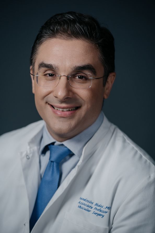
Professor Dr. Theodosios Bisdas is a renowned specialist in vascular surgery who heads the Department of Vascular Surgery at the Athens Medical Center. His extensive training began with medical studies at Aristotle University of Thessaloniki, followed by a fellowship in the Department of Cardiac, Thoracic, and Vascular Surgery at Hannover Medical School. There, he earned his doctorate with a groundbreaking dissertation on the safety of vascular grafts.
His career took him to renowned research institutions such as the Crossbit Research Center in Hanover and the Mount Sinai School of Medicine in New York, where he gained further expertise in vascular surgery. Prof. Dr. Bisdas has made significant contributions to vascular surgery, particularly through his work as a senior physician at the specialized clinic at St. Franziskus Hospital in Münster, where he performed over 2,000 endovascular procedures.
His academic achievements include over 150 publications in international journals and the supervision of numerous doctoral theses. He is a respected editor and reviewer for leading medical journals and serves on scientific committees for international conferences. His dedication has been recognized with numerous awards, including prizes from the German and European Societies for Vascular Surgery, as well as the Vascular Career Advancement Award. As an expert in the development of innovative endovascular techniques, he advises medical technology companies worldwide.
His leadership role in the multicenter CRITISCH Registry study reflects his commitment to excellence and innovation in the treatment of complex vascular diseases. Prof. Dr. Bisdas is also a co-founder of the Summer Academy for Vascular and Endovascular Surgery and the founder of the Vascupedia platform, which disseminates knowledge about vascular surgery worldwide. His humanitarian commitment is evident in initiatives such as the Mission Heart Help in Eritrea, which provides cardiac surgical care.
Prof. Dr. Bisdas embodies professional brilliance and a deep commitment to advancing vascular surgery for the benefit of his patients. The editorial team of the Leading Medicine Guide spoke with Prof. Dr. Bisdas about aortic aneurysms—a condition that is often life-threatening—and learned more about modern approaches and treatment options.
An aortic aneurysm is a serious vascular disease characterized by a dangerous dilation of the aorta. This condition can remain asymptomatic for a long time and is often not detected until it reaches life-threatening proportions. Thanks to advances in medical diagnostics and innovative treatment methods, such as minimally invasive endovascular procedures, aortic aneurysms can be treated more effectively today. Early detection is crucial to minimizing risk and enabling affected patients to enjoy a better quality of life.
The risk factors for developing an aortic aneurysm are diverse and often interrelated. Among the most important is arterial hypertension—or high blood pressure—which places additional strain on the wall of the aorta and increases the risk of dilation. Smoking is a major risk factor, as it damages the vessel walls, promotes inflammation, and accelerates atherosclerosis, all of which increase the risk of an aneurysm developing. High cholesterol levels also contribute to plaque buildup in the blood vessels and promote thinning of the vessel walls.
“An aortic aneurysm is a specific type of dilated section of the aorta that usually develops gradually and goes unnoticed for a long time. The most important risk factors for the development of such an aneurysm are high blood pressure—that is, arterial hypertension—as well as smoking and lipid metabolism disorders. A family history of the condition also plays a significant role. In addition to these factors, advancing age—particularly being over 65—significantly increases the risk.
Furthermore, being male is an important risk factor, especially among men who smoke and are older than 65. A targeted ultrasound examination is particularly recommended for this group, as it offers great benefits for early detection. “This examination has now become standard practice in many countries and is strongly recommended as part of preventive care for high-risk patients to detect potential aneurysms early and treat them appropriately,” explains Prof. Dr. Bisdas at the beginning of our conversation.
An aortic aneurysm is so dangerous because it usually goes unnoticed and is discovered only by chance, often during cardiological examinations recommended due to other risk factors.
“Since the aneurysm causes no symptoms in most cases, patients often don’t notice anything until it’s already too late. Without a timely diagnosis, the aneurysm can continue to grow and, in the worst case, reach the critical size of 5.5 centimeters, which significantly increases the risk of rupture. This rupture, which can occur in the abdomen or chest, is fatal in most cases, which is why it is considered extremely dangerous and is referred to in medical literature as a ‘life-threatening condition.’
If the aneurysm is discovered by chance, a precise diagnosis is made using advanced imaging techniques. The first step is usually an ultrasound examination, known as an abdominal sonography. If an enlarged aneurysm is suspected, a CT angiography or an MRI is performed, which provides precise information about the aneurysm’s size, shape, and extent. Based on these results, the doctor decides whether treatment is necessary. Treatment is indicated if the aneurysm has a diameter greater than 5.5 centimeters or if it is growing rapidly by more than one centimeter within a year. In such cases, surgical procedures are an option.
The decision depends on the patient’s individual condition, genetic predisposition, comorbidities, and life expectancy. “Thanks to advances in minimally invasive techniques, nearly 90 percent of aneurysms are now treated using catheter-based procedures with stent grafts, making gentle, low-risk therapy possible,” explains Prof. Dr. Bisdas.
For high-risk groups, such as individuals with a family history or known risk factors, regular ultrasound examinations (sonography) of the abdominal and thoracic aorta should be performed. These non-invasive, painless procedures make it possible to detect aortic dilation at an early stage, even before symptoms appear. Consistent monitoring and modification of risky lifestyle habits, such as smoking and high blood pressure, are the most important measures for significantly reducing the risk of developing an aortic aneurysm.
Ultrasound, particularly transabdominal sonography, is the primary screening tool when an aneurysm is suspected. It can detect even small aneurysms as small as approximately 3 cm. Computed tomography (CT), specifically computed tomography angiography (CTA), provides a high-resolution view of the aorta and its wall structure. It is particularly effective when a detailed anatomical assessment is necessary, such as in cases of unclear findings or planned surgical procedures.
CTA enables precise measurement of aneurysm dimensions, assessment of wall thickness, calcification, and possible associated conditions such as blood clots or narrowings. Magnetic resonance imaging (MRI) is primarily used for patients in whom radiation exposure should be avoided, such as those requiring repeated follow-up examinations or pregnant women. It also provides high-resolution images and, with the use of special contrast agents, can reveal even more details about the wall structure and associated conditions.
The decision to perform surgical or minimally invasive treatment of an aortic aneurysm is based primarily on the size of the aneurysm, its rate of growth, and the patient’s individual risk factors.
For an abdominal aortic aneurysm (AAA), treatment is recommended when the aneurysm reaches or exceeds 5.5 centimeters in diameter, as the risk of rupture increases significantly at this size. For thoracic aortic aneurysms, the standard threshold is usually 5 centimeters, although earlier treatment may be considered in cases of increasing risk, a family history of the condition, or certain associated factors.
Even for smaller aneurysms that are still below the threshold values, an individualized risk assessment is performed: Rapid growth (more than 0.5 cm per year), accompanying symptoms, wall instability, or the presence of connective tissue disorders may necessitate early intervention. In addition, risk factors such as high blood pressure, smoking, or cardiovascular disease are taken into account in treatment planning.

“When deciding on traditional open surgery, we primarily use this method for certain patient groups. In particular, young patients suffering from syndromes such as MAFAN (mitral and aortic malformations, anomalies, and urticaria) or LDS (Loeys-Dietz syndrome)—in which the body’s connective tissue is genetically compromised—typically require open surgery. These genetic disorders are associated with connective tissue weakness, which significantly increases the risk of large aneurysms forming and growing rapidly.
In addition, we opt for open surgery in patients who do not meet the anatomical criteria for minimally invasive, endovascular therapy. This applies, for example, to very small vessels that make it difficult or impossible to use catheter-based techniques, as well as to very large aneurysms where endovascular treatment carries the risk of being insufficiently effective or causing complications. “These are the three main indications for which traditional open surgery is the preferred option,” said Prof. Dr. Bisdas.
MAFAN (Mitral and Aortic Malformations, Anomalies, and Urticaria) is an acronym used in medicine to describe a syndrome characterized by certain genetic malformations and disorders. However, in the context of an aortic aneurysm, MAFAN refers to a specific spectrum of genetic disorders that leads to an increased susceptibility to the development of aneurysms, particularly in the aorta.
Loeys-Dietz syndrome (LDS) is a rare genetic disorder that affects connective tissue and is characterized by dysfunction of collagen and structural genes. The syndrome is named after the researchers Loeys, Dietz, Debakey, and Vogt and leads to generalized connective tissue weakness, which is particularly evident in the formation and risk of aneurysms (including those in the aorta). It increases the risk of developing large and usually very rapidly growing aneurysms, which, if not detected and treated in time, can lead to life-threatening ruptures or dissections.
Several modern, innovative procedures are now available for the treatment of aortic aneurysms, which differ in their techniques, risk profiles, and prognosis. The two main approaches are traditional open surgery and the minimally invasive endovascular aneurysm repair method.
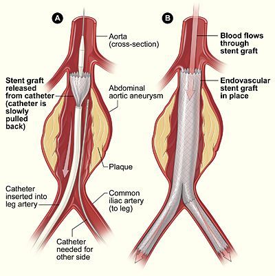
“Endovascular treatment of an aneurysm is now the preferred method for many patients, as it is less invasive, allows for shorter hospital stays, and carries a lower perioperative risk. The procedure is performed through two small incisions in the groin, through which wires are inserted to serve as a pathway for stents, balloons, and other devices. The aorta is visualized using an iodine-based contrast agent. One challenge with this method, however, is that the contrast agent must be carefully dosed in patients with impaired kidney function—such as those with renal insufficiency—to prevent kidney damage.
During the surgical portion of the procedure, the stent is precisely placed in the aorta so that blood flows through it, relieving the aneurysm—that is, the weakened section of the vessel wall—from blood pressure. This significantly reduces the pressure on the aneurysm wall, effectively eliminating the risk of rupture. The duration of the surgery depends on the size of the aneurysm.
For an isolated abdominal aortic aneurysm, the procedure takes about an hour and can even be performed without general anesthesia. However, if important side branches of the aorta—such as the renal or intestinal arteries—are involved, the surgery becomes more complex. In such cases, custom-made prostheses with built-in collateral branches are required, which extends the surgery time to three to four hours. “Despite the longer duration, the safety and effectiveness of these procedures remain at the same high level as with standard surgeries,” states Prof. Dr. Bisdas, adding:
“Particular challenges arise with very complex aneurysms in which the entire aorta is affected. In such cases, there is a risk that blood supply to vital organs—such as the spine, kidneys, or intestines—will be compromised, which could potentially lead to paraparesis or paraplegia. Endovascular therapy is generally considered a safe and effective option.
In contrast, there is open surgery, which involves making an incision in the abdomen. This procedure is significantly more invasive, requires longer recovery times, and involves a longer hospital stay. However, it allows the entire aorta to be replaced with an artificial prosthesis, which effectively eliminates the risk posed by the aneurysm once and for all. However, this method carries risks, such as complications involving surrounding organs, injuries to the intestines or ureters, and the risk of prosthesis infection, which can occur in about two percent of cases over the long term.
Such open surgeries typically last three to four hours and require admission to an intensive care unit. For younger patients, open surgery is often well tolerated because they generally do not have significant pre-existing conditions. For older patients or those with cardiovascular disease, however, endovascular treatment is the preferred option, as it is less invasive and allows for a quicker return to daily life.”
If an aneurysm is detected at an early stage, before it enlarges significantly or symptoms appear, there is a chance to significantly minimize the risk of a tear or a life-threatening rupture.
At this early stage, patients can often be placed under watch with regular follow-up visits to monitor growth and intervene only if the aneurysm exceeds a certain size or shows accelerated growth. This allows procedures to be performed under stable, well-predictable conditions, which significantly increases the chances of success and reduces complications.
Prof. Dr. Bisdas comments: “Many patients come in with a diagnosis of an abdominal aortic aneurysm and ask why surgery isn’t performed immediately when there is uncertainty. Especially in the case of aneurysms around four centimeters in size, which do not yet pose an acute risk, the decision is often made to wait and see. The answer to this question is based on extensive studies, particularly a large European, multicenter study.
The study found that, in the case of small aneurysms, the benefits of early endovascular treatment are not clear-cut because the vessels are still very small and narrow. This leads to a high rate of early occlusion of the stents, which offers no survival benefit. It is entirely possible that a patient with a four-centimeter aneurysm will not experience any increase in diameter through consistent conservative treatment—that is, through medications such as statins, blood pressure-lowering drugs, and aspirin.
As long as the aneurysm does not grow, the patient can go up to ten years without surgery. An exception is made for saccular aneurysms, in which only one side of the aorta is affected and the integrity of the wall may be compromised. Such aneurysms pose a higher risk of rupture, and treatment is recommended in all cases. Most small aneurysms cause no symptoms. If symptoms do occur, they are usually associated with aneurysms measuring around 3.5 to 4.5 centimeters, and the causes are often related to inflammation of the wall, which irritates the nerves in the intestines. In such cases, reducing the inflammation is the focus of treatment.”
For an aneurysm measuring three centimeters, an annual ultrasound examination is sufficient to monitor its progression.
“Factors that promote growth vary from person to person, but there are clear recommendations that every patient should follow. Blood pressure should be well controlled and, if necessary, managed with appropriate medication. Smoking must be stopped immediately, as it is directly linked to the growth of the aneurysm.
Likewise, treating lipid metabolism disorders and taking statins are important because these stabilize the aneurysm and strengthen the wall. It is also important to avoid physical overexertion and to maintain a healthy body weight,” notes Prof. Dr. Bisdas.
If an aortic prosthesis has been implanted in a patient following open surgery, the general rule is that treatment is complete. Nevertheless, there is a possibility that additional aneurysms may develop later in the area of the aorta where the prosthesis did not replace the native aorta.
“Although this risk is relatively low, we occasionally observe aneurysms in the area of the anastomosis—that is, where the prosthesis was sutured to the aorta. Therefore, we do not recommend routine follow-up within the first five years, but rather individually tailored follow-up care. For patients treated with a stent, the follow-up protocol is different. After treatment, an immediate follow-up is usually performed, followed by ultrasound examinations at three, six, and twelve months, and annually for at least five years.
If a decrease in the aneurysm’s diameter is observed during these checkups, further intervention is usually not necessary. An exception is the so-called endoleak, in which blood continues to be detectable outside the prosthesis within the aneurysm sac. This means that the physician must thoroughly investigate the leak to determine its cause. Various types of endoleaks are distinguished, with the most dangerous being the so-called Type 1 variant, in which the stent graft was not optimally adapted to the vessel. This can result in blood continuing to flow into the aneurysm sac, which is usually due to an inappropriate choice of prosthesis or further aneurysm progression. Type 2 endoleaks are common and describe blood flow into the aneurysm sac, which is usually sealed within the first twelve months by thrombosis of the lateral sutures and often requires no further treatment.
However, if blood continues to flow through large lateral sutures or the aneurysm grows significantly, a second-level intervention, such as embolization, may be necessary. Other types, such as so-called “disconnection” (where the prosthesis is no longer fully connected to the natural vessel) or perforation of the prosthesis, occur extremely rarely—usually with very early generations of prostheses—and, in extreme cases, may require removal of the prosthesis. Overall, it can be said that modern stents generally remain in the body for a lifetime. Replacement is necessary only in the event of complications, such as an aneurysm that continues to grow despite the stent, or in cases of severe stent malfunction—which is a very risky surgery,” emphasizes Prof. Dr. Bisbas.
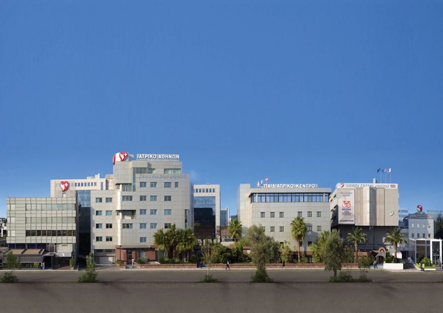
At the Athens Medical Center, approximately 100–150 aneurysm surgeries are performed annually. Endovascular treatment is considered the gold standard in this field.
“In this field in particular, patients benefit significantly from modern technologies, especially in the so-called hybrid suites we have—a combination of high-quality imaging and a sterile environment. In these specially equipped rooms, we can visualize the structures very precisely using X-rays, which is essential for performing the surgeries safely. These hybrid suites are designed to remain sterile while also allowing for open surgery at any time in the event of complications, without compromising sterility.
Thanks to the software and technical advancements in these rooms, it is now possible to perform surgeries with lower radiation exposure and fewer nephrotoxic contrast agents. We also have such hybrid rooms at the Athens Medical Center. Our experience encompasses complex procedures ranging from treatment of the aortic arch to the specialized vessels in the abdominal region. At our center, we perform a wide range of procedures, and the technological capabilities significantly enhance the safety and efficiency of these surgeries,” emphasizes Prof. Dr. Bisdas.
To prevent an aortic aneurysm or slow its progression, lifestyle changes and careful medical care are particularly crucial.
At the conclusion of our conversation, Prof. Dr. Bisbas offers some recommendations for prevention: “The most important preventive measures for avoiding an aneurysm are a healthy lifestyle. This includes consistently monitoring blood pressure, avoiding smoking, maintaining a balanced and nutrient-rich diet, and engaging in regular physical activity. It is also crucial to treat lipid metabolism disorders early on and to avoid physical overexertion if an aneurysm has been diagnosed. Patients with a family history of aneurysms, in particular, should seek medical advice. Even after diagnosis, targeted lifestyle changes can help slow or even stop the growth of an aneurysm, which significantly improves the prognosis!”
Share this article
Expert Interviews
Read next
- Expert interview
Expert interview with Professor Dr Elmar Herbst, PhD
Oct 15, 2026
Patient-centred and personalised knee surgery
Read more - Expert interview
Cardiogeriatrics: Significance and benefits in the context of hospital reform
Sep 21, 2026
Cardiogeriatrics: Significance and benefits in the context of hospital reform
Read more - Expert interview
Expert Interview with Professor Dr. Amadeus Hornemann, MPH
Sep 18, 2026
Prof. Amadeus Hornemann on Precision Medicine in Gynecology: Modern Surgery, New Techniques, Better Quality of Life
Read more