Expert Interviews
Bursitis and Impingement

Alexandra Pfitzmann ·
Millions of people develop bursitis at some point in their lives, often in the shoulder, elbow, hip, or knees. To learn more about this—and to what extent bursitis and impingement are related—the editorial team at Leading Medicine Guide spoke with Prof. Dr. Sepp Braun, a specialist in sports and joint surgery.
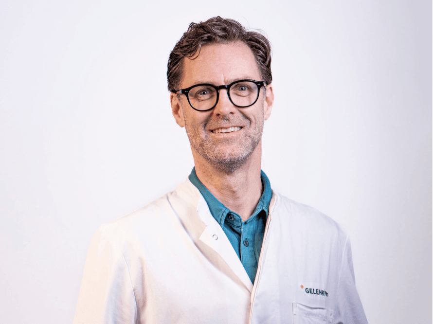
Bursitis and impingement are common causes of discomfort in the musculoskeletal system, particularly in the shoulder area. The condition can limit mobility and cause pain, though a precise diagnosis and modern treatment methods enable a quick recovery.
The causes of bursitis are varied and can generally be divided into acute and chronic triggers.
In cases of acute inflammation, the main cause is usually a sudden trauma or injury. This can result from a careless fall, a violent impact, sudden overstretching, or a sports-related injury—for example, during overhead throws in handball, tennis, or weightlifting.
Direct trauma resulting from an accident, such as a blow to the joint, can also irritate or injure the bursa so severely that it swells acutely and becomes painful. In addition, local infections can trigger acute bursitis, for example, through a skin injury that allows bacteria to enter the bursa and cause an infection.
“Bursitis and impingement are ultimately caused by mechanical factors. The bursae of the shoulder—which, in an anatomical sense, are not actually true sacs but rather a thin, slippery layer of tissue—lie over the rotator cuff, beneath the acromion.
When inflamed, this layer of tissue can cause pain. The inflammation is primarily triggered by mechanical stress. As a rule, impingement—that is, the pinching of a muscle or tendon between bony structures—is the main cause of this inflammation. Impingement itself is not a distinct clinical condition, but rather a consequence of misalignment of the shoulder joint, in which the head of the humerus is not properly centered in the glenoid cavity.
This can be caused by misalignment or instability—either functional, such as due to poor muscle coordination of the rotator cuff, or structural, such as tendon tears. When tendons are damaged or torn, the joint can no longer be stabilized, leading to misalignment. This results in impingement, in which structures such as the bursa become irritated and inflamed, which then triggers pain, for example, when lifting or moving,” explains Prof. Dr. Braun, adding regarding the causes:
“Another cause of impingement is what’s known as external impingement, in which bony structures narrow the space and thus irritate the tendons and bursa. A common example is the so-called acromial spur, which results from ossification of the acromion.Anatomical variations, such as a wide, outwardly overhanging portion of the acromion or a change in scapular position in which the shoulder blade tilts forward, can also restrict mobility and lead to impingement symptoms. The so-called “lateral acromion downslope,” in which the acromion tilts downward laterally, is also a common change that can develop over the course of a person’s life due to muscle pull from the deltoid muscle. All of these bony and anatomical factors can be the cause of impingement symptoms; however, impingement itself is never the actual diagnosis, but rather always a consequence of an underlying cause that must first be identified and then treated.”
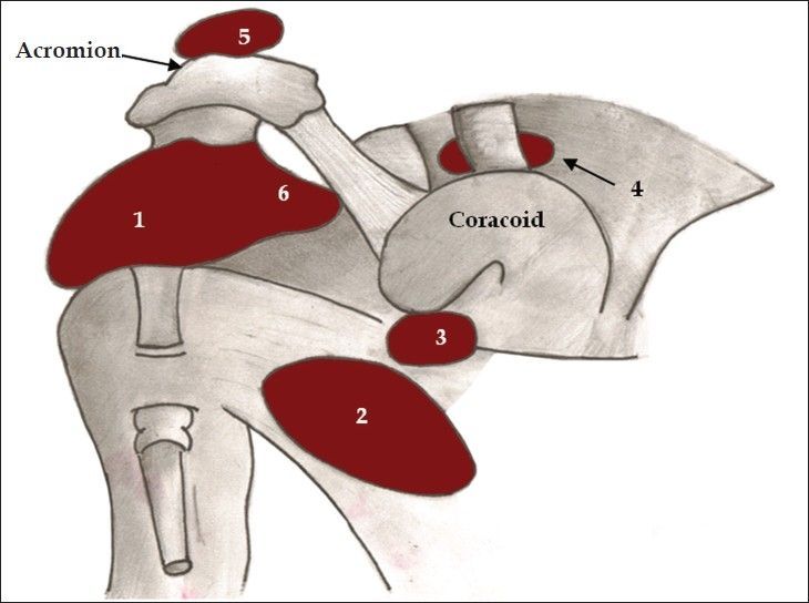
The subacromial bursa is a bursa located between the acromioclavicular joint and the tendon of the supraspinatus muscle._ Zameer Hirji, CC BY 3.0
Prolonged strain caused by repetitive movements—such as heavy lifting, overhead work, poor posture, or prolonged sitting—can lead to chronic irritation of the bursa. Chronic bursitis can develop particularly in occupational activities that require repetitive movements.
Congenital as well as degenerative changes in bones or tendons—such as osteoarthritis in the joint or tendon changes—can irritate the bursa over an extended period and cause chronic inflammation. It is also important to note that, in chronic cases, the formation of the bursa is repeatedly stimulated by ongoing microtrauma or inappropriate movement patterns, leading to persistent inflammation.
Impingement itself is not the actual diagnosis, but rather always merely a consequence of an underlying problem.
“A good example can be found in the elbow area. If you spend a lot of time sitting at a desk, your elbows may rest on the edge of the table. With this posture, a small sac of tissue sometimes forms; while it doesn’t necessarily cause pain, it can result in visible swelling.
There is also a so-called bursa in the elbow—a lubricating sac over the tip of the ulna (the elbow bone)—that allows the skin to move with every bend of the elbow. The pressure and constant rubbing against the table cause mechanical irritation there, which can trigger an inflammatory reaction. As a result, the tissue swells and fluid accumulates, causing a doughy swelling.
This reaction is usually not inflammation in the sense of an infection, but rather purely mechanical irritation. In rare cases, this bursa can become infected, leading to true bacterial bursitis, which must be treated with antibiotics. “If the strain persists and the irritation recurs repeatedly, the tissue can become chronically inflamed and, in severe cases, may even need to be surgically removed to allow for new tissue growth, since the bursae keep reforming,” says Prof. Dr. Braun, adding:
“Especially in overhead sports like tennis, the risk of shoulder problems should not be underestimated. Repeated overhead movements, such as during a serve, often cause problems attributable to capsular shortening, joint decentration due to certain changes in the shoulder capsule, and differences in the mobility of the joint structures.
These stresses can lead to internal impingement, in which tendons and bursae become irritated and inflamed. It is important here to diagnose the exact cause in order to take targeted countermeasures, since treating the inflammation alone provides only short-term relief, while the actual cause persists in the long term.
A lasting solution always requires a clear diagnosis, as this is the only way to effectively treat a sports injury. Therefore, I am firmly convinced that it is not enough to treat only the acute symptoms, for example through anti-inflammatory measures or symptom relief. The actual cause—the biomechanical stress or anatomical change—must be identified and addressed in order to achieve lasting improvement.”

The development of impingement, particularly in the shoulder joint, involves complex biomechanical and pathophysiological processes. Impingement syndrome arises primarily from repeated mechanical stress on the tendons and structures in the so-called subacromial space, which causes the synovial membrane in the subacromial space to be constantly irritated and damaged. The common consequence is recurrent bursitis, which keeps recurring due to the persistent irritation. The resulting inflammation further increases friction, which exacerbates wear and tear and limits shoulder mobility.
Various diagnostic procedures are used to differentiate between bursitis and impingement syndrome in the shoulder region.
“The diagnostic evaluation always begins with a comprehensive, thorough clinical examination. This is the central element of the diagnostic process, because only through a precise analysis of range of motion, limitations, and pain points can a clear picture be obtained. It is essential to observe movement patterns dynamically—including the movement of the scapula—in order to detect functional disorders at an early stage.
It is also important for the patient to undress during the examination, as an examination through a T-shirt is not effective; only in this way can one truly assess the movement and positioning patterns of the shoulder or shoulder joint. In addition, imaging techniques are used to ensure a complete and accurate diagnosis: First, an X-ray in three planes is very helpful for precisely assessing bony changes and bone structures.
In addition, magnetic resonance imaging (MRI) has become virtually indispensable in orthopedic diagnostics today. While MRI images used to be considered secondary, their importance has increased significantly in recent years because they provide very detailed insights into the soft tissues, tendons, ligaments, and structures in the shoulder region, thereby enabling targeted treatment planning.
However, it is important to note that MRI images should only be considered after a clinical examination in order to approach the matter with an open and objective mind. “Ultimately, imaging is just an additional tool to confirm or rule out the clinical diagnosis,” explains Prof. Dr. Braun, adding:
“In addition to X-rays and MRI scans, ultrasound is also a valuable option for assessing the condition of the shoulder. It should be noted, however, that ultrasound results are highly examiner-dependent, and the quality depends heavily on the device used and the examiner’s experience. Modern high-resolution devices make the assessment much easier, but the physician’s expertise plays a crucial role in making the best use of the findings.
I personally use ultrasound frequently, but I consider it much more sensible to make surgery-related decisions based on the clinical examination and MRI images, as these offer a more comprehensive means of assessment and ensure greater certainty in treatment planning. A combination of careful clinical examination and targeted imaging, particularly MRI and ultrasound, forms the basis for a precise diagnosis and sets the course for the correct treatment.”
Conservative treatment is generally sufficient when symptoms are mild, inflammation is still limited to local irritation, or symptoms occur only with certain movements.
These measures include rest, avoiding strenuous activity, physical therapy, anti-inflammatory medications, local injections, or specific exercises for muscle relaxation and postural correction. This often leads to a significant improvement in symptoms, making surgery unnecessary at first.
“If we’re talking solely about functional impingement, without significant structural damage, it’s clear from the outset that conservative therapy is needed first. To me, that’s actually a given. However, if I detect major or multiple rotator cuff tendon injuries in a patient—which are also evident during the examination as functional impairment, significant loss of strength, and more severe pain—especially in active, demanding patients—it often makes sense to proceed with surgery right away.
This is because, in such cases, conservative treatment is highly unlikely to be successful in the long term, and surgery can significantly shorten the patient’s recovery time. As for conservative measures, we primarily rely on physical therapy, supplemented by medication or, in some cases, injections. I am generally cautious about using injections. There are situations in which they make sense, such as to alleviate acute symptoms and thus facilitate the actual therapy.
Injections can be particularly helpful when the pain is so severe that physical therapists initially need a lot of time to reduce the pain enough to allow for active exercise—though the primary focus here is on muscle strengthening, not massage. However, I’m cautious because the effect is usually only short-term. Local anesthetics and cortisone are usually used. I’m very sparing in my use of them because potential side effects can occur both in the joint and in the tendon area, and the long-term effectiveness is questionable,” Prof. Dr. Braun clarifies, and goes on to explain therapy with autologous plasma:
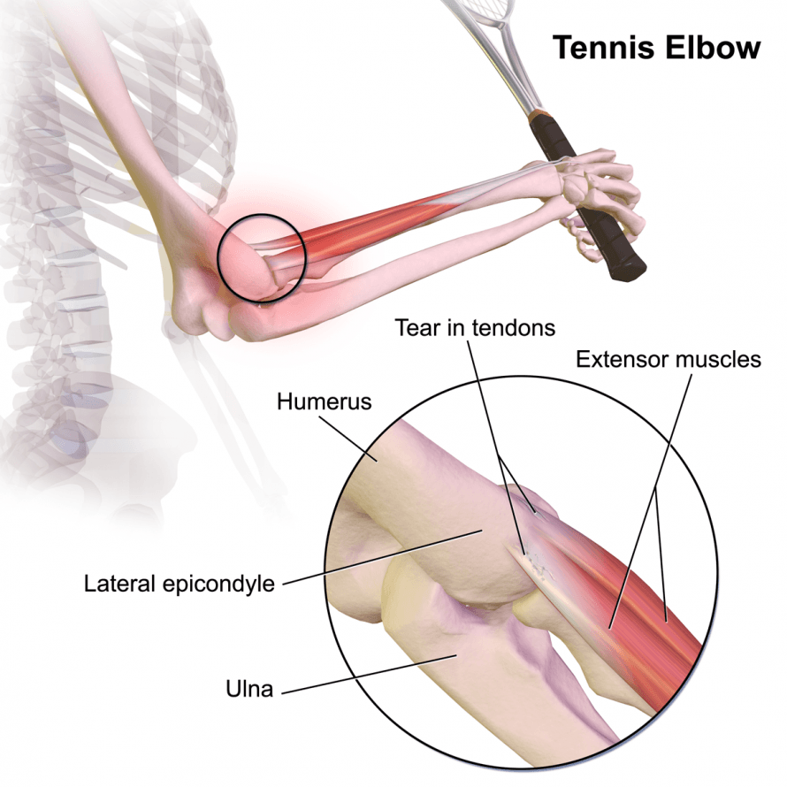
“There’s also the option of using autologous plasma, or PRP (platelet-rich plasma). I like to use it, for example, in cases of so-called epicondylitis such as tennis or golfer’s elbow, and I’ve had good results with it. PRP contains white blood cells that can have an anti-inflammatory effect. In the area of the bursa, which is often treated with cortisone, I tend not to use PRP. I also use it occasionally for partial tendon ruptures, but only in selected patients—this is not a standard treatment, but an option that promises success in individual cases.”
For chronic problems where conservative measures do not yield the desired results, minimally invasive surgical treatment—such as shoulder arthroscopy—may be indicated to specifically address the underlying causes, for example by removing bone spurs, pain points, or the repair of damaged tendons.
“In cases of isolated impingement, I do not perform surgery in my practice. The patient receives effective physical therapy and a conservative treatment regimen and, as a rule, does not undergo surgery because it is usually not necessary. In the last ten years, I haven’t operated on anyone just to remove a bone spur on the acromion; I probably haven’t done that at all.
If impingement leads to structural damage—for example, to the rotator cuff—the rotator cuff is the primary focus of the surgery, and the bone spur is removed during the same procedure. I have never performed surgery for isolated subacromial decompression alone. Of course, this may also be because I rarely see such patients in my practice. However, it cannot be ruled out that there are situations in which such a procedure might be appropriate—for example, in cases of very pronounced bone spur formation—to prevent incipient tendon damage.
Most often, I don’t see patients until structural damage to the tendon has already occurred. For the postoperative rehabilitation phase following rotator cuff surgery, the rule of thumb of “two times six weeks” has proven effective. The first phase involves six weeks of rest with a very cautious rehabilitation protocol so that the tendon can heal undisturbed. This is followed by a second, more active phase of another six weeks, during which the tendon is slowly subjected to structural stress and range of motion is gradually increased to promote complete healing.
Without stress, the tendon cannot heal in a permanently stable manner. After these two periods of six weeks, the worst is over. “During the first six weeks, the affected side is not yet ready for everyday activities; in the second six weeks, patients can usually resume small household tasks that do not require a large range of motion or significant strength,” says Prof. Dr. Braun regarding the treatment and recovery process.
There are no clear differences between athletes and non-athletes when it comes to the development of shoulder tendon injuries—no one is consciously doing anything wrong. However, factors such as posture and regular exercise are key to preventing symptoms. Those who maintain good posture and strong muscles can prevent problems.
“Posture is a key factor. Posture is important in general, but especially for shoulder health. Anyone who sits in front of a computer all day or—even worse—slumps with a laptop on their lap, letting their shoulders hunch forward and developing a rounded back, puts undue strain on the shoulder and the rotator cuff.
An upright upper body posture with the shoulder blades pulled back, on the other hand, can have a preventive effect and help avoid shoulder problems. Another important aspect is physical activity. The muscles should be kept as fit as possible, because a certain level of core strength is essential for a joint to function properly. “As with all joints, regular exercise within a safe range is very beneficial for long-term health,” advises Prof. Dr. Braun as a preventive measure.
In addition, it is advisable to avoid repetitive movements that push the body to its limits or to modify them through targeted adjustments to technique and load. Taking regular breaks during strenuous activities and consciously practicing proper posture can prevent long-term damage. Learning and adhering to ergonomic movement patterns while working and exercising also help to optimally relieve strain on the shoulder and avoid peak stress levels.
These preventive strategies, combined with targeted training for mobility and stability, help maintain muscular balance in the shoulder joint and thus sustainably reduce the risk of impingement and bursitis. Overall, the earlier and more consistently these measures are implemented, the more effectively the development of chronic symptoms can be prevented.
In practice, very similar basic concepts are applied to all joints—shoulder, knee, elbow, hip, and ankle. This approach allows for focused and specialized diagnosis and treatment of the joints. Although the basic principles are comparable, the individual characteristics of each joint differ, in some cases significantly.
Many thanks, Prof. Dr. Braun, for these important insights into the treatment of bursitis and impingement!
- Dr. Sepp Braun: a leading expert in sports and joint surgery; practices at the “Gelenkpunkt” clinic in Innsbruck. Extensive training at, among others, the Klinikum rechts der Isar (Technical University of Munich), the BG Clinic in Murnau, and the University Medical Center Freiburg. One-year fellowship at the Steadman Clinic in Vail, USA—a world-renowned center for sports orthopedics.
- Specializations: arthroscopic and open shoulder surgery, shoulder arthroplasty, complex sports injuries.
- President of the AGA—Europe’s largest professional society for arthroscopy and joint surgery (over 6,000 members).
- Co-founder of the German Arthroscopy Registry (DART) for quality assurance and the further development of innovative techniques.
- At the “Gelenkpunkt” practice: high level of expertise in minimally invasive surgery, reconstructive joint surgery, and complex joint injuries. Focus on minimally invasive procedures for rapid healing and a quick return to sports and daily life. Collaboration within an international network of experts for state-of-the-art, scientifically sound treatment.
- Active research, regular publications, and the development of new technologies to optimize outcomes.
- A globally sought-after specialist, treating elite and recreational athletes as well as patients of all fitness levels.
Share this article
Expert Interviews
Read next
- Expert interview
Expert interview with Professor Dr Elmar Herbst, PhD
Oct 15, 2026
Patient-centred and personalised knee surgery
Read more - Expert interview
Cardiogeriatrics: Significance and benefits in the context of hospital reform
Sep 21, 2026
Cardiogeriatrics: Significance and benefits in the context of hospital reform
Read more - Expert interview
Expert Interview with Professor Dr. Amadeus Hornemann, MPH
Sep 18, 2026
Prof. Amadeus Hornemann on Precision Medicine in Gynecology: Modern Surgery, New Techniques, Better Quality of Life
Read more