Expert Interviews
Structured Treatment Protocol in Visceral Surgery: From Tumor Board Diagnosis to Minimally Invasive Surgery

Alexandra Pfitzmann ·
Visceral surgery involves the surgical treatment of internal organs, particularly the digestive tract, the liver, the bile ducts, and the thyroid gland. It combines modern surgical techniques with precise diagnostics, which are often performed using minimally invasive methods today, thereby enabling a less invasive evaluation. In cases of oncological diseases, the interdisciplinary tumor board also plays a central role: Each patient’s case is discussed there with experts from various specialties to arrive at an individually tailored, medically sound treatment decision. The editorial team of the Leading Medicine Guide spoke with Sven Henne, a specialist in general and visceral surgery, about the specific process and how treatment strategies are developed.

The path from initial diagnosis to surgery for gastrointestinal tumors follows a clearly structured, interdisciplinary process designed to accurately assess the disease and plan the best possible treatment.
“In visceral surgery, there are two typical ways patients come to us: through the emergency room with acute symptoms such as sudden abdominal pain, vomiting, or signs of intestinal obstruction—or on an elective basis through the outpatient clinic, after they have already been preliminarily examined by a primary care physician, gastroenterologist, or private practice surgeon and have been recommended for surgery. In elective cases, the process works as follows: the patient first schedules an appointment through the administrative office and is then seen by a specialist during the outpatient clinic. There, a detailed medical history is taken, including any comorbidities and medication regimen. We always appreciate it when patients bring previous test results or medical reports with them. The specific clinical presentation is then examined and medically evaluated—for example, an inguinal hernia, reflux symptoms, or proctological conditions such as hemorrhoids or fistulas. Depending on the findings, a treatment recommendation is made: Some conditions can be monitored or treated conservatively, while many—such as a symptomatic inguinal hernia—require surgery, provided there are no serious pre-existing conditions that would preclude it. During the same appointment, the surgeon provides information about the surgical procedure and the risk profile, ensuring the patient has sufficient time to consider the decision and that the explanation is provided by the surgeon who will actually perform the procedure. One to two weeks before the surgery, patients come in for outpatient preparation; they bring lab results from their primary care physician or have blood drawn, have a consultation with the anesthesiologist, and are prepared for inpatient admission. After three to four hours, they return home and arrive on an empty stomach at the agreed-upon time on the day of surgery. They are greeted at the reception area, the surgical site is marked, and, upon request, we notify family members by phone after the surgery, as it often takes a while for patients to regain consciousness and become oriented. Depending on the procedure, patients are then transferred to the intensive care unit, the general ward, or—in the case of minor procedures—discharged home the same day,” explains Sven Henne, who then describes the procedure for emergencies:
“The situation is different in the emergency room. Patients usually arrive there via ambulance or on their own with acute symptoms. Our colleagues in the emergency room examine them first and consult us—the visceral and general surgeons—if necessary. Blood is drawn, the test results are evaluated, and the patient undergoes a clinical examination, often supplemented by an ultrasound. If further diagnostic tests are needed—such as a CT scan—they are ordered. Once a clear diagnosis is established, appropriate action can be taken—not every case requires surgery. In unclear cases—those that are “neither fish nor fowl”—we admit patients for one night of clinical monitoring and reassess them the next day. This is how the flow of patients through the emergency department works.”
In the tumor board, the treatment strategy for patients with oncological diseases is developed collaboratively by experts from various specialties. Surgery, oncology, gastroenterology, radiology, pathology, and, if necessary, radiation therapy each contribute their respective perspectives.
The discussion is based on all available diagnostic information—imaging, endoscopic findings, histological results, laboratory values, and the patient’s general state of health. It is crucial to precisely determine the tumor stage and to clarify whether primary surgery is appropriate or whether preliminary treatment—such as chemotherapy or radiation therapy—promises better therapeutic outcomes.
“When a patient is diagnosed with a tumor, most arrive at the surgical department with a confirmed diagnosis—often following a screening colonoscopy or because blood was noticed in their stool. Screening is recommended starting at age 50, or earlier if there is a family history of the disease. If a biopsy confirms colorectal cancer, the so-called staging—that is, diagnostic testing to assess the extent of the disease—usually takes place even before the preoperative consultation. This process determines whether lymph nodes are affected, how deeply the tumor has invaded the intestinal wall, and whether there are metastases in the liver, lungs, or other organs. This information is crucial for planning further treatment. For colon cancer, a CT scan is usually sufficient for this purpose, possibly supplemented by an ultrasound and blood tests for tumor markers. In most cases, this tumor is treated primarily with surgery, without prior chemotherapy or radiation therapy. With rectal cancer, the situation is more complex. Here, the different tumor stages may necessitate preoperative treatment with radiation and chemotherapy. For this reason, an MRI is also performed, which provides a better view of the soft tissues, as well as a transrectal ultrasound to assess the extent of the tumor even more precisely. A rigid rectoscopy is also necessary to determine the exact lower margin of the tumor, as this influences the treatment. “High-seated tumors are treated like colon cancer, whereas tumors in the middle and lower thirds are treated according to specific protocols,” explains Sven Henne.
Once all findings have been collected, the case is discussed at a tumor board meeting. This meeting brings together all the medical disciplines involved in diagnosis and treatment.
Sven Henne elaborates: “The gastroenterologist reports on the endoscopy findings and points out any special features, such as large polyps or very early-stage tumors that would otherwise not be detectable from the outside during minimally invasive surgery. In such cases, he recommends marking the tumor in advance with medical ink—a ‘tattoo’ that facilitates navigation in the operating room. The radiologist presents the imaging findings, explains the extent of the tumor, and describes its relationship to surrounding structures such as the ureter or organs. The oncologist assesses the situation from the perspective of chemotherapy, while the radiation oncologist determines whether radiation therapy is possible and at what dosage—which is particularly important for patients who have already undergone prior radiation therapy in the pelvis. The surgeon contributes his or her assessment; ideally, the surgeon already knows the patient and determines whether primary surgery is possible or whether the patient’s general condition should be improved first—for example, by treating anemia or through physical activity. The result is a joint decision based on current guidelines but always tailored to the individual patient. The recommendation may be primary surgery, prior chemotherapy or radiation therapy, or—in cases of advanced metastasis—a palliative approach focused on quality of life and symptom control. For rectal cancer, this tumor board meeting always takes place both preoperatively and postoperatively. For colon cancer, a postoperative discussion is usually sufficient. However, if a patient receives a multi-stage treatment plan, they are presented to the board again after each stage—after diagnosis, after preoperative treatment, and after surgery. The same applies to follow-up care: Any new relevant findings—such as the occurrence of a liver metastasis two years after surgery—are discussed again by the tumor board. This ensures that every patient receives the best possible, interdisciplinary treatment at all times.”
If surgery is being considered, the next step is to determine whether a minimally invasive or open procedure offers the greater benefit. Minimally invasive techniques are preferred because they deliver oncologically equivalent results while also offering advantages such as less pain, fewer wound complications, and a faster recovery. In some cases, “open” surgery may be more appropriate, e.g., in cases of significantly impaired heart function, following extensive previous surgeries, or due to anatomical peculiarities.
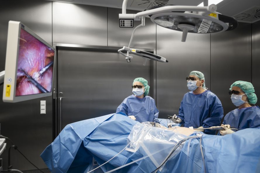
Sven Henne in the OR._Uwe Niklas
“During the tumor board meeting, we not only decide on the treatment plan but also on whether the procedure should be performed using open or minimally invasive techniques. Patients are then seen during the clinic visit, informed about their options, and undergo a clinical examination. This often makes it clear which surgical method is appropriate. If a patient has undergone multiple previous abdominal surgeries, that may be a reason to opt for open surgery initially. Very large stomach tumors or severe liver cirrhosis with collateral circulation—comparable to varicose veins in the abdomen—also increase the risk of bleeding and limit the options for laparoscopy. Nevertheless, most patients can undergo minimally invasive surgery. A previous surgery is not a contraindication in this context. Often, the surgeon first uses a laparoscope to examine the interior. If the adhesions are manageable, the procedure can continue using a minimally invasive approach. Only when it becomes apparent that the situation inside the abdomen is too complex or that there is excessive adhesions is the procedure switched to open surgery. There are, therefore, reasons for performing open surgery from the outset, but they are rare overall. The advantages of minimally invasive surgery are clear. The 3D camera provides an excellent view, allowing surgeons to work very closely and precisely on the tissue. For patients, this method means, above all, significantly less disruption to the abdominal wall, since only small incisions are necessary. This has a direct impact on pain management: recovery is faster, fewer pain medications are needed, and the risk of postoperative bowel paralysis is reduced as a result. Morphine derivatives, in particular, slow bowel motility, and this effect is exacerbated after bowel surgery. Minimally invasive procedures result in significantly fewer and shorter-lasting problems in this regard. Thanks to the faster recovery, many patients can be discharged from the hospital sooner. On average, the hospital stay is about seven days. Some go home after just five days, while others stay for ten—depending on how quickly the bowel function resumes and on the individual’s pain tolerance. On average, however, minimally invasive surgery noticeably accelerates the recovery process,” emphasizes Sven Henne.
For benign and malignant conditions, surgical techniques differ primarily in their objectives and extent. These differences significantly determine how the surgery is performed—and whether robotic systems offer additional benefits.
“For malignant tumors, the surgery always follows the principle of oncological resection. This means that not only is the tumor itself removed, but also its potential metastatic pathways: the inflowing and outflowing blood vessels as well as the entire lymphatic tissue, including the associated lymph node stations. This is familiar, for example, in breast cancer, where lymph nodes in the armpit are often removed as well. In the intestinal region, the fatty tissue containing the blood and lymph vessels that supply the affected section of the intestine is also resected. This ultimately determines the extent of the surgery, as the intestine can only be re-sutured where it has a good blood supply on both sides. If the vessel supplying the tumor is ligated centrally, it is necessary to carefully assess exactly where the intestine can be safely reconnected. In cases of benign conditions, this procedure is not necessary. Here, neither the lymphatic drainage areas need to be removed nor the blood vessels centrally ligated. As a result, the resection is usually smaller, and potential side effects—such as postoperative intestinal paralysis—may also be less severe,” states Sven Henne.
Oncological or minimally invasive procedures are followed by structured, closely coordinated postoperative care designed to prevent complications, accelerate recovery, and safely guide patients through the initial postoperative period.
A central component of follow-up care is the so-called fast-track or enhanced recovery concept, which combines early mobilization, a rapid resumption of nutrition, and targeted pain management. These measures help stabilize circulation, reduce the risk of thrombosis and infection, and restore bowel function more quickly. Another important component is the close monitoring of vital signs and wound healing. Although modern minimally invasive techniques result in smaller incisions and less pain, blood values, inflammatory markers, and the function of the affected organs are still monitored regularly to detect complications early. For oncology patients, interdisciplinary follow-up care also plays a major role: surgery, oncology, radiology, and, if necessary, radiation therapy work together to determine whether further treatments such as chemotherapy or immunotherapy are necessary and when they can begin. Patient-centered support is also crucial. This includes structured educational consultations, personalized nutritional counseling, physical therapy guidance, and clear instructions for the period following discharge. Many hospitals now use digital tools or telephone-based follow-up care to detect symptoms early and quickly address any concerns.
Sven Henne explains: “Follow-up care differs significantly between benign and malignant conditions. This can be clearly illustrated using colorectal cancer as an example, as clear, guideline-based protocols apply in this case. If colorectal cancer is surgically treated with the intent to cure—that is, with the realistic goal of restoring the patient to health—and the pathologist subsequently confirms that the tumor has been completely removed and no metastases remain, structured follow-up care begins. Patients receive a tumor passport and a doctor’s letter specifying exactly which tests are necessary and at what intervals. For the first two years, follow-up visits take place every six months. During these visits, the patient’s well-being is assessed, any changes in symptoms are noted, and blood tests and ultrasound examinations are performed. The first colonoscopy after surgery is also crucial: If the tumor had previously narrowed the bowel and the section beyond it could not be visualized before surgery, the first colonoscopy is recommended as early as six months later. In all other cases, it is performed one year after surgery and then again after five years—unless there are abnormalities that necessitate an earlier follow-up. Follow-up appointments take place annually until the fifth year after surgery. In addition, a CT scan after one year may be advisable, as it is known to enable earlier detection of liver metastases. This recommendation has been incorporated into the current guidelines. The well-known “five-year threshold” still applies: If no relapses occur during this period, the risk of recurrence is very low. That is why structured follow-up is conducted over precisely this period.”
Visceral surgery was introduced at Rummelsburg Hospital because the facility wanted to evolve from a purely orthopedic specialty clinic into an acute care hospital. A gastroenterology department was already planned, and it was therefore clear that a visceral surgeon would also be needed to complement it. This is how the idea to establish the new specialty came about.
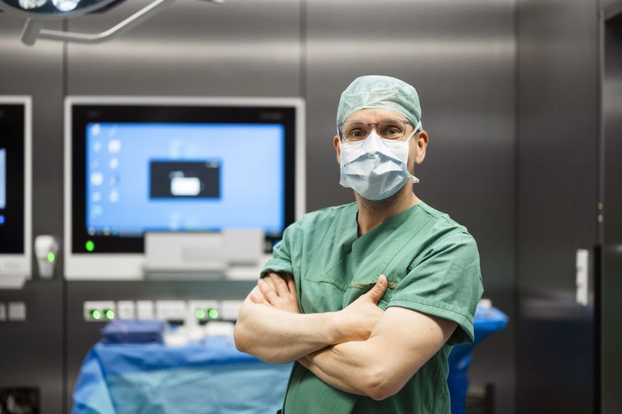
Sven Henne in the OR_Uwe Niklas
“I accepted this challenge and, together with the hospital administration, charted the course to get there. First came the search for a team. A colleague from my previous hospital joined a month after I did; another senior physician will start on March 1; and two resident physicians are also part of the team. The on-call system is integrated with that of the orthopedic residents, ensuring that a surgical on-call physician is on site around the clock—supplemented by a senior physician on call who can be called in at any time. Before work could even begin, a great deal of structure had to be put in place. This involved defining the requirements for the ward, the operating room, and the entire technical infrastructure. For minimally invasive surgery, we needed a new laparoscopic tower, including CO₂ insufflation, scopes, and camera units. Various systems were tested, and quotes were obtained, until a 4K-3D system was finally purchased—with 10-mm optics and 5-mm optics for smaller patients. The surgical department closely oversaw this process and restructured the instrument trays accordingly. Consumables also had to be selected: drapes, meshes for inguinal and abdominal wall hernias, and specialized suture material. New rectoscopes, complete with light sources, were needed for the diagnosis of rectal tumors. Many procedures had to be mentally run through in advance to ensure nothing was overlooked—and yet, in day-to-day practice, there is always something that needs to be fine-tuned. “The entire setup process went very smoothly and opens up broad prospects for the clinic’s future development,” summarizes Sven Henne, adding at the end of our conversation:
“In addition to minimally invasive colorectal surgery, we cover the entire spectrum of general and visceral surgery—with a few exceptions, such as pancreatic surgery or highly complex liver operations. This includes thyroid surgery as well as the treatment of benign conditions, such as abdominal wall hernias or symptomatic gallstones, which we perform on a regular basis. We also treat benign and malignant tumors of the stomach here, as well as anti-reflux surgeries for chronic heartburn or when the stomach has migrated into the chest cavity. The entire oncological spectrum of colorectal surgery is also part of our portfolio, as are acute conditions: appendicitis, intestinal obstructions caused by adhesions, and other emergencies are treated here. Our services are complemented by proctology—ranging from fistula surgery to pilonidal cysts, which we treat with or without plastic closure. Rummelsberg Hospital thus offers a wide range of procedures that cover the entire spectrum of visceral surgery.”
Thank you very much, Mr. Henne, for this discussion on visceral surgery!
- Chief of General and Visceral Surgery at Rummelsberg Hospital, specializing in minimally invasive and oncological surgery
- Surgical treatment of tumors of the digestive tract; presentation of all oncological cases to the tumor board
- Surgery on the liver, bile ducts, spleen, and thyroid
- Treatment of hernias as well as surgeries for reflux disease and appendicitis
- Consistent use of minimally invasive keyhole techniques
- Close interdisciplinary collaboration with gastroenterology and radiology to ensure optimal diagnosis, treatment, and follow-up care
Share this article
Expert Interviews
Read next
- Expert interview
Expert interview with Professor Dr Elmar Herbst, PhD
Oct 15, 2026
Patient-centred and personalised knee surgery
Read more - Expert interview
Cardiogeriatrics: Significance and benefits in the context of hospital reform
Sep 21, 2026
Cardiogeriatrics: Significance and benefits in the context of hospital reform
Read more - Expert interview
Expert Interview with Professor Dr. Amadeus Hornemann, MPH
Sep 18, 2026
Prof. Amadeus Hornemann on Precision Medicine in Gynecology: Modern Surgery, New Techniques, Better Quality of Life
Read more