Cholecystectomy is the medical term for the removal of the gallbladder. The procedure is a safe surgery with a very low complication rate. Both minimally invasive and open gallbladder removal are routine procedures. Cholecystectomy is the only satisfactory method for treating symptomatic gallstone disease.
The Gallbladder—Structure, Function, and Possible Diseases
The gallbladder is a thin-walled, pear-shaped organ with a capacity of about 30–50 ml. It is connected to the liver by connective tissue and is located on the underside of the liver in the right upper abdomen.
The gallbladder serves as a storage organ for the bile produced in the liver.
The Bile Ducts and Their Connections
Liver cells produce bile, which flows through tiny bile ducts into progressively larger ones. Outside the liver, two large bile ducts eventually merge to form the common hepatic duct (ductus hepaticus communis).
Bile is released from the liver (bile produced on demand) and the gallbladder when food is ingested.
In this process, it flows through the cystic duct (Ductus cysticus), which originates from the gallbladder, into the common hepatic duct. After the cystic duct and the common hepatic duct join, the main bile duct is called the hepatocholedochal duct (DHC).
This, in turn, joins the pancreatic duct (ductus pancreaticus). It ultimately empties into the duodenum via a papilla that functions as a valve (Papilla of Vater).
Immediately before entering the duodenum, the bile duct has a sphincter muscle that contracts during resting digestion.
The resulting reflux causes the gallbladder to fill with bile via the cystic duct. Shortly after a meal, the opening of the bile duct (Papilla of Vater) opens, and the bile flows out.
Functions of the Bile Acids Produced in the Gallbladder
Bile acids serve a number of important metabolic functions, primarily:
- Digestion of fats and
- Transport of fat-soluble substances
The small intestine absorbs the fat-soluble substances almost completely and transports them back to the liver via the bloodstream. They are then ready to be excreted again via bile.
If the gallbladder has been removed, the liver continues to produce bile, which is drained into the small intestine via the common bile duct.
The only thing missing is a reservoir for the bile. The patient does not notice this as long as they do not consume excessive amounts of fat at one time.
Otherwise, the undigested fats could cause diarrhea. Given our modern dietary habits and the constant availability of food, the gallbladder’s reservoir function is no longer necessary.
Gallstones as an Indication for Cholecystectomy
Gallstones—a common condition
Every day, our body produces about 250–1,000 ml of bile. The gallbladder thickens and concentrates the bile.
Due to the composition of bile (water, salts, cholesterol, bilirubin), an excess of one of these components may occur.
When oversaturated, the secretion tends to crystallize and form various types of gallstones, such as:
- cholesterol stones
- Bilirubin stones, etc.
At least 15% of the general population has gallstones. Women are more likely to develop gallstones than men.
Risk factors for the development of gallstones include:
- female gender
- high cholesterol levels
- being overweight
- extreme fasting
- diabetes mellitus
- certain medications
- genetic factors
- Pregnancy, and many more
The number of people affected increases with age. However, not everyone with gallstones experiences symptoms. Most people with gallstones (about 75%) remain symptom-free throughout their lives.
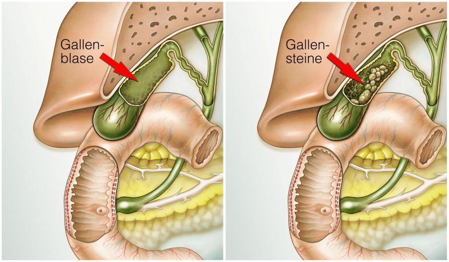
However, if the following symptoms occur, treatment is necessary to prevent serious complications from gallstones:
- episodic pain (colic) after large meals
- Constant pain in the upper abdomen that radiates to the back and right shoulder, accompanied by nausea or vomiting
In such cases, the gallbladder must be removed, as it is the primary site where gallstones form.
Complications of Gallstones
Gallstones can vary greatly in shape and size, ranging from a few millimeters to several centimeters.
Smaller stones pose a greater risk, as they can pass through the bile ducts or become lodged, causing colic.
This can obstruct the flow of bile into the intestines, leading to a backup of bile. This may result in pale stools and dark-colored urine.
This can also lead to the passage of bile pigment (bilirubin) into the blood, causing jaundice (obstructive jaundice).
It can also lead to inflammation of the bile ducts (cholangitis), which is often accompanied by fever and chills.
At the same time, gallstones can also obstruct the common duct of the gallbladder and pancreas. A backup of digestive juices can lead to inflammation of the pancreas (biliary pancreatitis).
A long-lasting, chronic obstruction of bile flow can lead to damage to the liver cells. As a result, the liver cells die off and are replaced by scar tissue (biliary cirrhosis).
Gallstones themselves can also damage the gallbladder wall and cause inflammation of the gallbladder (cholecystitis).
A severely inflamed gallbladder can rupture (perforate), which can lead to the leakage of bile into the abdominal cavity. This, in turn, can cause life-threatening peritonitis (biliary peritonitis).
Chronic inflammation of the gallbladder can lead to scarring and shrinkage of the gallbladder (atrophic gallbladder).
The same applies to a diagnosis of a porcelain gallbladder, which gets its name from chronic inflammation and calcium deposits. Both conditions are prone to degeneration, which can lead to the development of gallbladder cancer.
Particularly large gallstones with a diameter of more than 3 cm are also a reason to remove the gallbladder (cholecystectomy), even if the patient has no symptoms. Here, too, there is a higher risk of developing gallbladder cancer.
The same applies if gallbladder polyps are detected. In such cases, the gallbladder should also be removed, as it cannot be ruled out that this growth is a malignant tumor.
In rare cases, gallbladder inflammation can develop following severe trauma or major surgery (stress-induced cholecystitis).
What is the treatment for gallbladder diseases?
Upon diagnosis of gallbladder inflammation, treatment initially involves antibiotics and an early cholecystectomy. An early cholecystectomy is the surgical removal of the gallbladder within three days.
If symptoms subside, the surgery can be performed during a symptom-free interval after the inflammation has resolved. This is usually the case after four to eight weeks.
Early removal of the gallbladder is associated with fewer complications. This is because inflammation of the gallbladder leads to changes in the gallbladder wall, as well as adhesions and scarring. These complications can make the surgery more difficult.
A gallbladder empyema (accumulation of pus in the gallbladder) or gangrenous cholecystitis (tissue death due to inflammation) requires immediate surgery. In these cases, there is a risk of perforation leading to peritonitis.
If a malignant tumor of the gallbladder is suspected, doctors remove the gallbladder via open surgery, depending on the extent of the tumor.
In cases of advanced tumors, the procedure must be extended to include removal of adjacent liver tissue. In such cases, doctors should always remove the adjacent lymph nodes (ligamentous lymph nodes) as well.
Simply removing or breaking up the gallstones (extracorporeal shock wave lithotripsy, ESWL) while preserving the organ is not medically satisfactory.
Furthermore, the stones would soon reform, necessitating another procedure. The use of medication to dissolve gallstones (litholysis) also does not provide lasting success.
Satisfactory results and a cure for gallstone disease are not possible without removing the gallbladder.
The Procedure for Cholecystectomy (Gallbladder Removal)
More than 190,000 cholecystectomies are performed annually in Germany. In over 90% of cases today, the gallbladder can be surgically removed via laparoscopy. Medical professionals also refer to this method as keyhole or minimally invasive surgery.
Minimally Invasive Cholecystectomy (Gallbladder Removal)
During minimally invasive gallbladder removal, doctors first introduce carbon dioxide into the abdominal cavity through a blunt cannula while the patient is under general anesthesia.
The gas causes the abdominal wall to lift slightly, providing sufficient visibility and space for the operation inside the abdominal cavity.
Using port sleeves with diameters of 3–10 mm, doctors insert a camera and surgical instruments into the abdominal cavity. The images captured by the camera are displayed magnified on a monitor in the operating room for the surgeon to view.
Consequently, the skin incisions that doctors must make to insert the trocars into the abdominal cavity are very small.
Typically, four skin incisions are made (above the navel, as well as in the center and on the side of the upper right abdomen). Doctors usually have to enlarge the incision in the navel area to about 2 cm after detaching the gallbladder from the liver bed. This allows them to remove the gallbladder, especially if it contains stones.
Under certain conditions, and to minimize scarring, doctors can also remove the gallbladder through three incisions.
The incisions are then located:
- above the navel, as well as
- in the right and left lower abdomen
A single, larger incision above the navel (single-port technique) is also possible.
Before detaching the gallbladder from the liver bed, doctors must sever the cystic duct along with the cystic artery (A. cystica).
This is done after clamping the vessels with metal clips or absorbable plastic clips. These dissolve on their own after a few weeks. Only then can doctors peel the gallbladder out of the hepatic bed and remove it along with the stones.
Before closing the incision, doctors release the gas that was previously introduced into the abdominal cavity. The body can metabolize any small amounts of carbon dioxide that may remain.
A cholecystectomy typically takes about 30–60 minutes. The patient is allowed to get out of bed and drink fluids on the same day.
This is followed by a rapid return to a normal diet. The patient can be discharged from the hospital after two to four days.
Open Gallbladder Removal (Cholecystectomy)
In open (conventional) cholecystectomy, surgeons access the abdominal cavity through an incision in the upper right abdomen or in the midline. The midline is located halfway between the sternum and the navel.
Surgeons typically make an incision at least 8–10 cm long along the right costal margin. The length of the incision usually depends on the patient’s build and the conditions within the abdominal cavity.
The surgeon then ligates and clamps the cystic duct and the vessels supplying the gallbladder. Next, under direct visualization, the surgeon dissects the gallbladder, along with its contents, from the liver bed. Finally, the surgeon closes the abdominal wall.
If there are no complications, the patient typically stays in the hospital for about four to seven days.
Complications and Risks of Gallbladder Removal (Cholecystectomy)
Gallbladder removal is a safe procedure with a very low complication rate. Both minimally invasive and open cholecystectomies are routine procedures.
Complications from an elective cholecystectomy depend largely on factors such as the patient’s comorbidities and complications related to gallstone disease. Emergency cholecystectomies carry a higher risk and a higher complication rate.
General surgical risks and injuries to the bile ducts or adjacent organ structures are extremely rare.
General surgical risks include:
- Bleeding
- Postoperative bleeding
- Infections
- Impaired wound healing or
- thromboembolism
Repeat surgery is necessary only in some cases and for more severe injuries, such as those involving the common bile duct.
Bile stasis can result from stones remaining in the bile ducts.
If stones are present in the bile ducts before or after surgery, doctors can remove them during an endoscopic procedure. This procedure is also known as endoscopic retrograde cholangiopancreatography (ERCP).
ERCP allows for:
- Visualization of the bile ducts and the pancreatic duct using X-ray contrast dye
- Performing therapeutic procedures such as the removal of impacted gallstones in the bile ducts (but not stones from the gallbladder)
Sometimes, during laparoscopic removal, the doctor must switch to an open procedure. The doctor must inform the patient of this before the procedure.
For example, if unforeseen circumstances arise, the gallbladder can be removed through a larger abdominal incision, which carries less risk for the patient.
Possible circumstances include:
- severe adhesions or scar tissue,
- unclear anatomical conditions,
- significant bleeding, or
- the presence of a gallbladder tumor or other conditions
Sore muscle-like discomfort in the upper abdomen and shoulders may occur a few days after the procedure. This is caused by nerve irritation resulting from the gas in the abdominal cavity during surgery.
Findings on Minimally Invasive Cholecystectomy
Laparoscopic gallbladder removal offers significant advantages over open cholecystectomy, such as:
- faster recovery
- the patient experiences less pain
- there is less scarring and, as a result, a lower risk of incisional hernias, and
- a lower risk of wound infection due to the smaller incisions
In addition to avoiding a large incision, the trauma is also significantly reduced because fine, miniaturized instruments are used. The surgical trauma is thus limited to the affected area of the body. A wide opening of the abdominal cavity is not necessary.
For the patient, this results in a significant reduction in overall stress and a much faster recovery and return to normal activities.
Patients who undergo laparoscopic surgery typically leave the hospital sooner and are able to return to work more quickly.
Follow-up Care After Cholecystectomy
Follow-up care is only necessary if symptoms recur.
There is no need to avoid certain foods or follow a special diet afterward. Normal, unrestricted physical activity is usually possible again after about 14 days.
Conclusion on Cholecystectomy
This surgery provides a cure for symptomatic gallstone disease. There are no satisfactory alternative methods available. Surgery is the treatment of choice.
Cholecystectomy is a safe procedure with a very low complication rate.
It is possible to live a normal life without a gallbladder. Quality of life is also higher than it would be with a diseased gallbladder and recurring symptoms.