Interventional radiology is a branch of radiology that combines therapeutic procedures with real-time imaging using CT, ultrasound (sonography) and other techniques. This enables surgeons to perform minimally invasive procedures with even greater precision and the greatest possible preservation of healthy tissue.
Here you will find further information as well as a selection of specialists in interventional radiology.
What is interventional radiology?
Interventional radiology is officially classified as a branch of diagnostic radiology. Radiology encompasses all imaging examination procedures, such as
- ultrasound,
- magnetic resonance imaging (MRI),
- computed tomography (CT) or
- angiography (X-ray contrast imaging of blood vessels).
Interventional radiology refers to minimally invasive procedures carried out under radiological guidance – these are therefore also referred to as interventional radiology procedures.
These minimally invasive surgical procedures are carried out without a direct view of the interior of the body. This
- spares the tissue,
- minimises the risk of infection and
- enables faster healing.
To aid orientation and control, the surgeon monitors their progress using radiological imaging.
These imaging techniques are used to precisely locate the area to be treated. With the aid of interventional radiology, the surgeon can also perform precision surgical procedures that would otherwise only be possible via ‘open’ surgery. Open surgery involves making longer incisions to access the surgical site.
Areas of application for interventional radiology
Common areas of application include:
- Angioplasty (widening of narrowed blood vessels)
- Diagnosis and treatment of the bile ducts or the biliary tract system (PTC, percutaneous transhepatic cholangiography, possibly as PTCD)
- Tumour ablation procedures (tumour treatment)
- Haemostasis
- Vertebroplasty (stabilisation of damaged vertebral bodies)
- Periradicular therapy (treatment of nerve pain and infiltration for back pain)
Vascular procedures, i.e. procedures on blood vessels, are the main area of application in interventional radiology. In particular, peripheral arterial stenoses and occlusions, such as
- carotid stenosis,
- renal artery stenosis or
- narrowed coronary arteries (coronary heart disease)
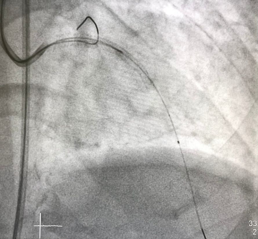
are treated quickly and easily using this imaging modality. In the case of coronary heart disease, for example, PTCA (percutaneous transluminal coronary angioplasty) may be used.
Placement of a drug-eluting stent using intravascular ultrasound (IVUS) © Belezapoy | AdobeStock
Angioplasty
The procedure is carried out under local anaesthetic.
During angioplasty, the doctor administers a contrast agent to the patient and then takes X-ray images. The resulting angiogram shows the blood vessels and provides information about pathological changes (such as blood clots or calcifications). Their exact location can also be seen on this angiogram.
The doctor then makes a small incision through which they insert a catheter into a suitable blood vessel. With the help of the angiogram, the surgeon can precisely guide the catheter into the affected blood vessel.
In the event of a narrowed blood vessel, they use what is known as a balloon catheter or a stent (a vessel-stabilising implant): both lead to the recanalisation of blocked vessels by widening the narrowed area and keeping it open in future.
Tumour treatment
Computed tomography is used to treat certain tumours locally. Under CT guidance, the doctor can administer an injection of medication with pinpoint accuracy. Alternatively, radioactive particles can be delivered into the relevant blood vessels as part of selective internal radiotherapy (SIRT).
Furthermore, it is possible to cut off a tumour’s blood supply using a catheter. Transarterial chemoembolisation (TACE), for example, specifically induces a blood clot in the vessels supplying the tumour.
Chemosaturation, on the other hand, temporarily isolates the liver from the body’s blood circulation. This allows the liver to be treated with chemotherapeutic agents whilst sparing the rest of the body. Each chemotherapeutic agent used acts locally on the liver without placing a strain on the entire body.
Another treatment method is radiofrequency ablation (RFA). This precisely destroys tumourous tissue using alternating current.
Haemostasis
Trauma surgeons use interventional radiology to stop major bleeding using minimally invasive techniques. Using a catheter, the doctor obliterates the damaged vessel with vessel-occluding materials or closes it with a membrane-covered stent.
The doctor can also treat damage to the aorta (main artery) in this minimally invasive way.
Vertebroplasty
Fractured or collapsed vertebrae can cause pain and sensory disturbances. The injection of bone cement stabilises the injured vertebrae.
For the procedure, the patient lies face down on a treatment table. They are given a general anaesthetic to ensure they do not move even slightly.
The doctors then use an X-ray machine or a CT scanner. This enables them to pinpoint the exact position of the vertebrae. Under image guidance, the surgeon then inserts a hollow needle through which he injects the stabilising bone cement into the vertebra.
Treatment of nerve pain
Nerves that send constant or excessive pain signals severely impair quality of life. This is precisely where interventional pain management comes in.
The doctor can treat this with
- anti-inflammatory,
- desensitising and
- pain-relieving or
- anaesthetic
medicines.
To do this, they visualise the affected part of the spine using fluoroscopy or computed tomography. They then insert the needle at the correct angle and to the appropriate depth, before injecting the medication.
If necessary, the patient is given a local anaesthetic. Where required, the doctor also uses a contrast agent.
Other areas of application in interventional radiology
In dialysis patients, shunts (or dialysis shunt systems) are created for the purpose of blood filtration. Shunts are artificial connections between an artery and a vein.
Other areas of application include, for example, tissue sampling (biopsy) and the insertion of drains and feeding tubes. Where findings are unclear, targeted biopsies are also taken.
After treatment
Patient care
Patient care varies greatly depending on the clinical presentation and the procedure performed. For example, following vertebroplasty, one day of bed rest is recommended so as not to interfere with the cement setting. If a drain has been inserted, it is usually left in place for only a few days.
A day of rest is also advised following the administration of anaesthetics. You must not drive a vehicle on this day.
Prognosis
The prognosis also depends on the type of procedure the doctor has performed. Catheterisation of the bile ducts or blood vessels, for example, has a very high success rate even in the long term. Patients suffering from nerve pain also generally benefit enormously from the treatment.
How promising a tumour treatment is depends on
- the treatment method as well as
- the type, size, location and stage of the tumour
. Some tumours are not (or are no longer) curable, whilst for others the prospects are very good. The treating doctor can provide a more detailed assessment.
Possible complications of the procedures, such as bleeding
Interventional radiology is a purely minimally invasive procedure. As a result, the complication rate and the extent of side effects are extremely low.
The following complications are rarely possible with injections:
- infections
- accidental misinjections
- Nerve damage
- Bleeding
In vertebroplasty, secondary fractures or symptoms of paralysis due to pressure on the spinal cord may occur. The latter can result, for example, if bone cement leaks out of the vertebra.
The treatment of nerve pain may temporarily cause
- sensory disturbances,
- paralysis or
- a worsening of symptoms
.
Some people are intolerant to the injected medicines or contrast agents.
Conclusion
Interventional radiology is a therapeutic method with a wide range of applications.
Thanks to real-time imaging, it enables gentle, highly precise treatment. As a result, the complication rate is much lower than with an invasive procedure.
FAQ
Does it replace conventional surgery?
In many cases, yes: instead of open surgery, image-guided, minimally invasive procedures are used – for example, via a catheter or needle.
Who performs the procedures?
Specially trained interventional radiologists, usually in close collaboration with other specialists.
Which conditions are treated?
Commonly vascular diseases, peripheral circulatory disorders, and benign and malignant tumours such as liver tumours.
Is it also used in emergencies?
Yes – in cases of acute bleeding or for thrombosis prevention, for example by inserting a Cavafilter, it plays an important role in emergency care.

About the medical author
Sabine Schneider
Editor-in-Chief
Sabine Schneider – medical author: Explore expert articles and medical expertise in the Leading Medicine Guide.
More about the medical author →Sources
- DocCheck Medizinlexikon: https://flexikon.doccheck.com/de/Interventionelle_Radiologie
- Radiologie-Netz: https://www.radiologie.de/radiologische-therapie/