A disc prosthesis is a modern surgical procedure in which a damaged intervertebral disc is replaced with an artificial disc. The goal of the surgery is to reduce pain and preserve spinal mobility. Unlike spinal fusion, a disc prosthesis allows for continued movement in the affected segment, both in the lumbar spine (LS) and the cervical spine (CS).
Particularly for selected patients with a herniated disc or degenerative changes, the implantation of an intervertebral disc prosthesis represents a sensible alternative to traditional spinal fusion surgery.
A Herniated Disc as an Indication for an Artificial Disc
Signs of wear and tear in the spine
The mobility of all vertebrates—including humans—depends on the intact structure and function of the spine.
As we age, an unhealthy lifestyle—such as a lack of exercise or smoking—can lead to signs of wear and tear in the spine.
These degenerative changes lead to pain and impaired mobility. A genetic predisposition can also contribute to these symptoms.
These changes usually begin in the intervertebral discs. These are elastic shock absorbers located between the bony vertebral bodies; their flexibility enables the spine to move.
Structure and Function of the Intervertebral Disc
A healthy intervertebral disc consists of a tough, firm ring of connective tissue fibers (collagen), the “annulus,” which surrounds the disc nucleus (nucleus).
The nucleus has a high capacity to bind water and consists of 80 percent water. Since water cannot be compressed, the nucleus is not actually soft. However, it is deformable enough to shift within the fibrous ring and stretch it (Fig. 1).
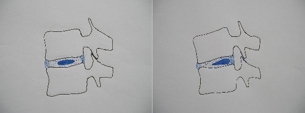
Displacement of the intervertebral disc nucleus during forward bending @ R. Schönmayr
High compressive pressure prevails inside the intervertebral disc, which is why it has no blood vessels or nerves.
Nevertheless, metabolic processes take place here that serve to supply the cartilage cells with nutrients and remove metabolic waste products.
This occurs via interstitial fluid. When the intervertebral disc is under load, the fluid passes through small, sieve-like openings into the bone of the vertebral bodies.
When the disc is unloaded—especially at night while lying down—the body draws the interstitial fluid back out of the vertebral bodies.
The more frequently the alternation between loading and unloading occurs, the better the metabolism of the intervertebral discs. Or, to put it another way: The less we load and unload our intervertebral discs, the poorer the exchange of substances and the faster the aging and wear of the discs.
Causes of a Herniated Disc
Much like a car tire that loses air, the intervertebral disc loses its ability to retain water and, with it, its water content. It collapses and becomes brittle and cracked.
Because the intervertebral discs are no longer fully hydrated, the vertebrae can now make small sliding movements. These movements also place unnatural stress on and damage the annulus fibrosus.
Cracks can form in the annulus fibrosus, allowing parts of the nucleus pulposus to protrude. This results in a herniated disc.
Since the annulus fibrosus is thicker at the front and sides than at the back, herniated discs most commonly occur at the back of the vertebral bodies. This is where the spinal canal and the nerve canals are located.
This means that, in addition to the back or neck pain caused by a herniated disc, nerve pain may also occur. This pain can manifest as severe, sometimes electric-like pain.
Symptoms of a Herniated Disc
Patients perceive the pain in the area of the body where the affected nerve is located. Therefore, herniated discs in the lumbar spine cause leg pain, while herniated discs in the cervical spine cause arm pain.
This pain is very severe and usually depends on the position and movement of the spine. As a result, people suffering from this pain adopt a cautious, protective posture and avoid movements and strain on the spine.
In doing so, the back muscles reflexively tense up strongly to prevent movement of the affected section of the spine.
If the herniated disc exerts significant pressure on a nerve or does so for an extended period, it can cause the following sensory disturbances:
- Tingling sensations (pins and needles)
- Numbness
- Loss of sensation
Paralysis may also occur in the nerves supplying the muscles.
Large herniations in the lumbar spine can impair bladder function and the anal sphincter. Patients are then unable to empty a full bladder or control bowel movements.
Herniation in the cervical spine can put pressure on the spinal cord. This does not usually result in paraplegia, but it can lead to the following problems:
- significant unsteadiness when walking (ataxia)
- increased muscle stiffness in the legs (spasticity) and
- sensory disturbances in various parts of the body

If severe pain radiates down the leg, this may indicate a herniated disc @ Microgen /AdobeStock
Conservative Treatment for a Herniated Disc
As long as herniated discs do not cause nerve damage, doctors treat them with non-surgical “conservative” measures.
These include:
- Medications
- Physical therapy
- Manual therapy or
- Physical therapies (heat, water, stretching)
Surgery should only be considered if these treatment options fail or if nerve damage is present.
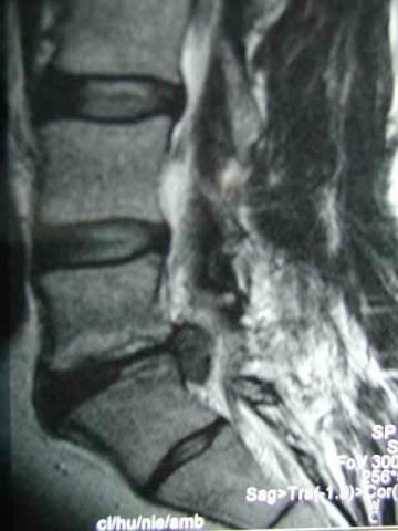
Lumbar disc herniation on an MRI scan @ R. Schönmayr
Surgery for a Herniated Disc
Before surgery, imaging studies are required following a physical and neurological examination. These include X-rays, magnetic resonance imaging (MRI), and computed tomography (CT). Sometimes a contrast study (myelography) followed by a CT scan is also necessary.
These images allow the herniation to be visualized. Its size and exact location can be identified (Fig. 2). This information is essential for planning the subsequent surgical procedure.
During surgery on the lumbar spine, a very small incision is made in the center or slightly to the side of the back. The surgeon locates the herniation using an endoscope or microscope and removes it. Sometimes, the surgeon must remove crumbly material from inside the intervertebral disc.
Goals and Prognosis of Intervertebral Disc Surgery
After surgery, leg pain usually subsides quickly, since the pressure on the nerves has been relieved. Nerve damage, on the other hand, takes longer to recover. In cases of very severe damage, it is also possible that the condition will not improve at all or will improve only partially.
As for back pain, the surgery is not always as successful: the damage to the intervertebral disc has not been repaired, and the abnormal slippage has not been eliminated.
If consistent strengthening of the back muscles does not stabilize the damaged intervertebral disc, persistent back pain may result.
This can lead to secondary damage to the vertebral joints (osteoarthritis) and the bony endplates of the vertebral bodies (osteochondrosis). These, in turn, cause pain. It stands to reason that insufficiently trained back muscles also cause pain, especially in cases of poor posture or spinal curvature.
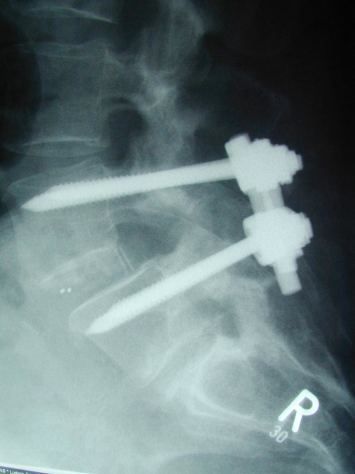
Fusion of two vertebral bodies by screwing the vertebrae together and inserting a plastic and bone implant between the vertebral bodies via a surgical approach from the back @ R. Schönmayr
If chronic back pain cannot be managed by other means, there are various surgical procedures that can be used.
Spondylodesis for a herniated disc
Spondylodesis is a surgical procedure that has been known for decades and has been further refined with modern technology. In this procedure, surgeons firmly fuse two adjacent vertebrae together (Fig. 3).
This fusion results in the two vertebrae growing together as a single bone unit. The remaining vertebrae, however, remain mobile, so that the overall mobility of the spine is only minimally impaired.
Nevertheless, this method has disadvantages: Not all patients who undergo the surgery become symptom-free. It places increased stress on the adjacent intervertebral discs. If these discs already show signs of wear and tear, this can worsen over time. Corresponding symptoms may occur.
Artificial Intervertebral Disc as an Alternative to Spinal Fusion
To prevent an unfavorable redistribution of stress following spinal fusion (spondylodesis), physicians attempt to replace a damaged intervertebral disc. In this way, doctors can preserve or restore mobility.
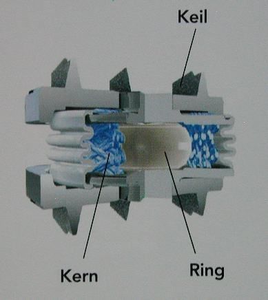
M6 intervertebral disc prosthesis with an elastic core, surrounded by a polyethylene mesh similar to a natural fibrous ring. The wedges on the end plates serve to anchor the prosthesis to the vertebral bodies @ SpinalKinetics Inc., manufacturer of the M6
However, early attempts with an artificial intervertebral disc (disc prosthesis) failed. In the 1950s, doctors failed in their attempts to implant a steel ball in place of the intervertebral disc.
They began implanting artificial intervertebral discs in the lumbar spine on a larger scale in 1984. Dr. Büttner-Janz and Prof. Schellnack developed a design consisting of a plastic sliding core and two prosthetic plates made of a metal alloy. They placed these between two lumbar vertebral bodies to replace the defective intervertebral disc there.
Over the years, they were able to overcome initial problems with material durability. As a result, the third-generation “Charité-Disc” available today is a mature implant used worldwide. There are now also a large number of artificial intervertebral discs from other manufacturers and based on different design principles.
The most interesting design currently available is a prosthesis that features an elastic core sandwiched between two titanium plates. This core is surrounded by a fibrous mesh that closely resembles a natural annulus (Fig. 4).
The movement pattern of the artificial intervertebral disc is similar to that of an intact intervertebral disc.
Disadvantages of an Artificial Intervertebral Disc
However, an artificial intervertebral disc is not recommended for every patient. The surgery to implant an artificial intervertebral disc requires a complex abdominal approach. During the procedure, surgeons must mobilize the large blood vessels in front of the spine (the abdominal aorta and the inferior vena cava) and move them to the side.
They may also injure or damage an important nerve plexus (responsible for sexual function) that runs in front of the vertebrae.
In cases of a normal herniated disc, therefore, the standard, significantly less invasive disc surgery is sufficient. This procedure is successful in about 90 percent of cases.
People with advanced disc damage and bony changes in the vertebrae and joints do not benefit from an artificial disc.
The reason is clear when we look at the vertebral joints. These joints connect two vertebrae to one another, providing guidance and limiting movement. Like all joints, they are subject to wear and tear and can cause very characteristic pain, particularly pain related to movement (Fig. 5).
If the joints are already damaged, increased movement and stress lead to intensified pain. In these cases, it is better to limit mobility through a fusion procedure.
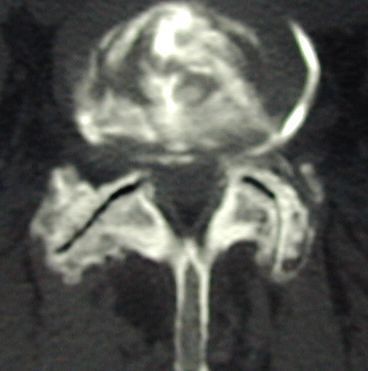
Advanced damage (osteoarthritis) of the facet joints @ R. Schönmayr
Who is a good candidate for an artificial disc replacement?
Who, then, is the right candidate for an artificial intervertebral disc? Patients with chronic back pain that cannot be adequately treated with conservative methods and that stems primarily from a damaged intervertebral disc.
The bony structures of the adjacent vertebrae should not show any advanced damage. In particular, the facet joints should still be functional and able to bear weight without pain. Doctors can assess whether this is the case before a planned surgery.
Under these conditions, patients who have already undergone surgery on the affected intervertebral disc may also be eligible.
The same applies to patients with intervertebral disc damage at multiple levels. However, various studies indicate that outcomes are not as favorable in such cases.
Sometimes it may also be advisable to combine surgical procedures: A disc with severe concomitant damage to the bony structures undergoes spinal fusion (spondylodesis). The adjacent, less severely damaged level (segment) receives an artificial intervertebral disc.
The advantage is that the patient does not have to place additional strain on the next intervertebral disc above it. This prevents a domino effect leading to “secondary instability.”
When is an artificial disc in the cervical spine appropriate?
In the cervical spine, the static load on the spine is lower and mobility is greater. For this reason, an artificial disc is used more frequently here. Nevertheless, it is important that bony degeneration has not yet progressed too far.
Especially in younger and middle-aged individuals, cervical disc herniations can occur even without significant joint damage (Fig. 6); therefore, it may be appropriate to use an artificial disc in this group.
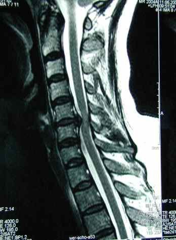
Herniated disc in the cervical spine on an MRI scan @ R. Schönmayr
The implantation of an artificial disc in the cervical spine is also significantly less complex and less invasive than in the lumbar spine.
Surgeons remove the diseased disc and the herniated portion through a simple incision at the front of the neck. In doing so, they carefully push the highly mobile soft tissues of the neck apart.
They then realign the intervertebral space slightly until it returns to its proper position. They insert the prosthesis under fluoroscopic guidance and subsequently check for mobility (Fig. 7).
Postoperative stabilization of the cervical spine with a neck brace (orthosis) is not necessary. Patients can be discharged from the hospital after a few days.
After about 3 months, the artificial disc is firmly anchored, allowing patients to resume normal daily activities. The surgeon must decide on a case-by-case basis when specific sports can be resumed.
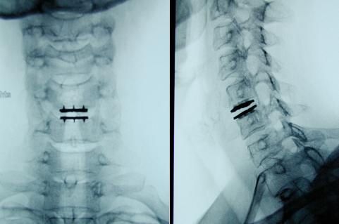
Implanted intervertebral disc prosthesis (M6) between the 5th and 6th cervical vertebrae in a postoperative X-ray view from the front and from the side @ R. Schönmayr
Complications and Risks of Artificial Disc Implantation
The potential complications associated with surgery involving the implantation of an artificial intervertebral disc should not be overlooked.
Medical professionals distinguish between:
- Complications arising from the surgical approach, which can occur in any surgery
- Complications arising from the implanted prosthesis
Fortunately, such complications are very rare. Experience to date shows that the durability of the implant materials does not pose any problems.
It is still unclear what happens to the prosthesis when the bone quality of the vertebrae deteriorates with age (osteoporosis). The relatively hard metal components could then sink into the bone. However, similar cases with other implants show that, in the worst-case scenario, ossification occurs around the implant. Spontaneous, slowly developing fusion is then possible. Medical professionals have observed such spontaneous bony bridging with varying frequency across the different available models.
However, this has no adverse effect on:
- surgical outcome
- Pain relief and
- neurological symptoms
Mobility in the operated segment, however, is lost.
Conclusion on artificial intervertebral discs
The success rate in terms of pain and neurological symptoms is just as good with spinal fusion surgery as it is with artificial disc replacement.
Whether mobility is maintained in the long term and whether wear and tear on the adjacent intervertebral discs can be prevented remains unclear to this day. In summary, it can be said that the artificial intervertebral disc has a firm place in the surgical treatment of intervertebral disc damage.
However, only patients who are suitable candidates should receive an artificial disc. This requires thorough preoperative diagnostic evaluations and extensive experience on the part of the operating surgeon. When used correctly and implanted with precision, the disc prosthesis represents a major advance and a valuable addition to treatment.
The implantation of a disc prosthesis aims to replace the diseased intervertebral disc and, unlike spondylodesis, to preserve the natural mobility of the spine. In this procedure, the surgeon places great emphasis on securely anchoring the intervertebral disc prosthesis to the adjacent vertebral bodies.
During the insertion of a disc prosthesis, the natural disc is removed, and an artificial ring with a polyethylene core is then placed between titanium end plates; the rough surface and coating of the metal plates ensure secure integration with the surrounding tissue.
Particularly in cases of herniated discs in the lumbar spine or cervical spine, the use of a disc prosthesis—such as a lumbar disc prosthesis or a cervical disc prosthesis—can offer benefits, provided that the disc is not severely degenerated.
Postoperative care plays a crucial role in ensuring long-term durability, as lifting and carrying heavy loads should be avoided during the first six weeks and in the weeks following surgery to preserve the spine’s natural mobility over the long term.
FAQ
What is an intervertebral disc prosthesis?
A disc prosthesis is an implant that replaces a damaged intervertebral disc and preserves the mobility of the spine. It is primarily used to treat degenerative changes or a herniated disc in the lumbar or cervical spine.
Who is a good candidate for an artificial intervertebral disc?
An artificial disc is suitable for patients with isolated disc degeneration without significant spinal instability. A clear medical indication is required, and other causes—such as severe osteoarthritis or instability of adjacent vertebral bodies—must be ruled out.
How is an artificial disc implant placed?
The implantation of an artificial disc is performed surgically under general anesthesia after the diseased disc has been removed. The prosthesis is then inserted between the vertebral bodies and anchored in such a way that natural mobility is preserved.
What are the advantages of an intervertebral disc prosthesis over spinal fusion?
Unlike spinal fusion, spinal mobility is preserved in the operated segment. This helps reduce the risk of overloading adjacent intervertebral discs and vertebral bodies.
What does postoperative care involve?
After surgery, a structured postoperative care plan is important to support the integration of the intervertebral disc prosthesis. During the first few weeks, heavy lifting and carrying should be avoided, supplemented by targeted physical therapy.
About the medical author
Prof. Dr. med. Robert Schönmayr
Medical writer