A keratoprosthesis is a specialized procedure used to restore vision in cases of severe corneal disease. A keratoprosthesis is implanted in the eye, particularly when a conventional corneal transplant is not possible. The osteo-odonto-keratoprosthesis is a specific type of this procedure in which the patient’s own tissue, such as a tooth, is used.
This procedure is rarely performed clinically but is used for selected patients. Implantation usually takes place in specialized centers, including in Germany, where extensive experience with this complex procedure has already been gained.
The prosthesis is designed to restore vision in cases of severe damage to the eye. This procedure may become necessary due to serious injuries such as burns or chemical burns.
Anatomical Background
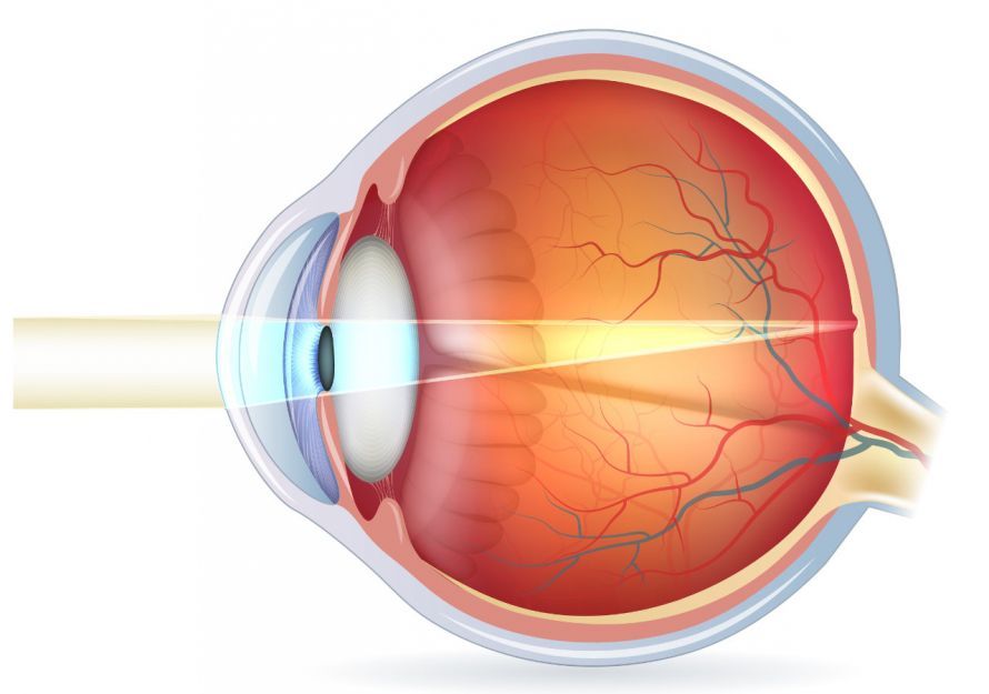
The cornea is the “windshield” of the eye. It protects the sensitive interior of the eye from external influences. It is transparent, allowing light to pass through it into the eye. Behind the cornea, the lens focuses the light and projects it onto the retina at the back of the eye.
There, the received information is converted into impulses that are transmitted to the brain via the optic nerves. A regularly structured, clear, and transparent cornea is therefore essential for a sharp image.
There are many diseases and disorders that can impair the eye’s ability to see. One of these is corneal opacity. This causes less light to pass through the cornea, and the light is also scattered. The result is reduced or blurred vision. Corneal opacity can be caused by
- scarring,
- infections,
- acid burns, or
- other diseases
.
A corneal transplant can help treat corneal opacity. In this procedure, the cloudy cornea is removed and replaced with a clear one. Unfortunately, the availability of human donor corneas is limited. In most cases, a corneal transplant does not involve replacing the entire cornea (thickness: approx. 500 micrometers). A thin layer (called a lamella) only 10–20 micrometers thick is sufficient in the majority of cases.

However, there are also diseases or injuries in which a corneal transplant fails. The recipient must regenerate the outermost layer of the cornea themselves; it cannot be transplanted. In rare cases, however, the eye is unable to do so—for example, in cases of severe chemical burns. In such cases, an artificial cornea (keratoprosthesis) can help restore the patient’s vision.
The Keratoprosthesis as a Last Resort
Researchers have developed various types of artificial corneas. Many patients experience a significant improvement in their vision after the implantation of an artificial cornea.
Implanting such a keratoprosthesis is generally difficult due to the unique nature of the cornea, but it is possible. Body tissue generally cannot bond firmly with artificial material.
Therefore, the use of artificial materials in the body is problematic. This leads to immune reactions and often to the rejection of the foreign material. Therefore, a material must be implanted that allows the body’s own tissue to fuse with it. Only then will it be firmly anchored, and the patient can benefit from improved vision.
A thorough ophthalmological examination is necessary before keratoprosthesis surgery.
To ensure a successful outcome with a keratoprosthesis, several conditions must be met:
- The affected eye should still be able to perceive light
- Intraocular pressure must be controlled (no excessive pressure!)
- The eye must not be inflamed, as this can interfere with the integration of the artificial cornea
In this article, we present several methods of keratoprosthesis.
Boston KPro (Boston Keratoprosthesis)
By far the most common procedure is the Boston keratoprosthesis, also known as the Boston KPro. It has been used in the U.S. since 1992 and has been continuously refined since then. In Germany, the Boston KPro has only carried a CE mark since 2015. However, it now represents a reliable alternative for affected patients.
The healthy iris controls the amount of light entering the eye. It dilates and constricts the pupil. With a Boston KPro, the patient’s iris is also removed. This function is then taken over by a part of the prosthesis.
A Boston keratoprosthesis is used when
- the patient is blind in both eyes (visual acuity of less than 5%), and
- a corneal transplant has failed or has too low a chance of success.
The Boston KPro requires careful and intensive monitoring after surgery. Only then can long-term success be achieved. In recent years, over 11,000 Boston KPro procedures have been performed worldwide. Nevertheless, the procedure is associated with a higher complication rate than a standard corneal transplant.

Boston KProthesis: Visible are the posterior metal plate, the sutured donor cornea, and the transparent anterior plastic rod. Transparent plastic is now used instead of metal © Mariagessa | Wikimedia CC4.0
Underlying Principle
Although the Boston KPro is referred to as a keratoprosthesis—that is, an artificial cornea—this is only part of the treatment. The procedure still relies on a donor cornea.
The donor cornea is implanted in a manner similar to a corneal transplant. However, it serves only as an anchor for the actual prosthesis. The donor cornea has no optical function.
This prosthesis consists of two parts:
- Front part made of transparent plastic: a sort of pin with a front plate attached
- Rear part: A larger, circular plate (now made of transparent plastic, formerly of metal) with holes and a metal fixation ring
The front part takes over the light-transmitting function of the cornea. The back part provides stability to the entire structure. The back plate also takes over the function of the iris that was previously removed.
The donor cornea and the posterior part of the prosthesis have a circular hole in the center that is the size of the pupil. The shaft of the anterior prosthesis is inserted through this hole. A ring secures it behind the ring plate. The plastic head of the anterior prosthesis is held in place by the sutured donor cornea and the ring plate.
Goals and Results of the Boston KPro
The target group consists of patients who are classified as blind due to corneal disease.
In most cases, patients tolerate the prosthesis well. Complications may include, among other things,
- the development of elevated intraocular pressure (glaucoma),
- mucosal overgrowth on the optical element,
- macular edema, or
- a retro-prosthetic membrane
. In many cases, however, these problems can be resolved.
The Boston KPro cannot provide perfect vision. After the procedure, patients may regain functional vision.
57–83% of patients with a Boston prosthesis can recognize letters on an eye chart from a distance of 6 meters that healthy individuals can identify from 60 meters. 19–23% of patients can even identify letters at 6 meters that healthy individuals can recognize at 12 meters. The cost of a Boston KPro is approximately 3,000 €.
Osteo-Odonto-Keratoprosthesis
In this treatment, physicians draw on findings from dentistry. Metal crowns and fillings can be permanently bonded to the mineral substance of the tooth. The Italian ophthalmologist Benedetto Strampelli made use of this observation in 1963. He used a tooth root to attach an artificial cornea to the surface of the eye.
This osteo-odonto-keratoprosthesis is used today at some clinics. For this procedure, the patient’s optic nerve and nerve cell layer must be intact. It is necessary to remove the lens and the iris. A tooth from the patient serves as the transplant material. This tooth is surgically removed along with its root and the surrounding jawbone.
The tooth root is then prepared so that an optical cylinder made of Plexiglas can be bonded to it. The resulting artificial cornea is then placed over the patient’s diseased cornea. As a rule, this keratoprosthesis enables orientation vision. After 10 years, 20–40% of patients have visual acuity greater than 5%. From a cosmetic standpoint, however, the result of such a keratoprosthesis surgery is not very appealing. Skin-colored buccal mucosa grows over the eye, covering the white areas and the colored iris.
If a patient does not have teeth suitable for this purpose, bone material can also be harvested from the tibia. This form of the procedure is also known as a “tibia corticalis” keratoprosthesis. A piece of bone can then serve to secure the optical cylinder in place.
Artificial cornea made of water-absorbent material
The procedure described below is still in the experimental stage.
Here, a material with a structure capable of absorbing water is used for the artificial cornea. Thanks to a special surface coating and a chemical modification of the edge, it can integrate more easily with the surrounding tissue.
The newly developed forms of the artificial cornea were tested in the laboratory for compatibility. To this end, they were implanted into several laboratory animals. The implanted variants integrated into the surrounding tissue with largely successful results. They proved to be sufficiently light-transmissive and firmly anchored.
Over a period of six months, no rejection of the keratoprosthesis occurred. Overall, the animals tolerated the keratoprosthesis well.
Artificial Cornea Made of Collagen Fibers
A keratoprosthesis made from aporous matrix of the protein collagen shows great promise. Collagen is also the main component of the natural cornea. Therefore, it is particularly well-suited for a keratoprosthesis.
The artificial cornea made from the collagen matrix has so far been used only in a pilot study. It is still in the experimental stage. Advantages of the artificial cornea made from collagen fibers would include:
- Improved vision similar to that achieved with a donor cornea transplant
- Better tolerance of this artificial cornea: Patients would need to take little or no medication to suppress a rejection reaction
- Pathogens cannot be transmitted with this type of artificial cornea
- In nine out of ten patients, the nerves severed during surgery grew into the new tissue. This ensured that the implant became sensitive to touch
Only two out of ten participants experienced a decline in vision following implantation. However, this impairment could be corrected with contact lenses.
Even better results could be achieved if sutures were not used during implantation. The sutures used to date delay the artificial cornea’s integration into the eye. This ultimately impairs healing and increases the risk of complications. Instead, a biological adhesive could be used to anchor the implant.
Complications and Postoperative Care
The procedure to implant the keratoprosthesis is usually performed on an inpatient basis under general anesthesia. Depending on how the surgery goes, the patient must remain in the hospital for between four and six days after the operation. The length of stay depends on whether complications arise.
Infections generally remain the biggest problem following the procedure. For this reason, follow-up treatment with antibiotics is very important. An untreated or undetected infection can, under certain circumstances, lead to blindness in the affected eye.
Furthermore, intensive follow-up examinations of patients are required after keratoprosthesis surgery. During these examinations,
- the fit of the artificial cornea,
- intraocular pressure, and
- visual acuity
are checked.
Outlook
The total number of artificial corneas transplanted is still low. Due to the complexity of keratoprosthesis surgery, the surgical center must have a certain level of experience.
In general, further improvements in the field of artificial corneas are expected. In particular, the material—and thus its biocompatibility—is the focus of research projects. If the costs of this complex procedure can be reduced, this method will see wider application and adoption.
Existing problems will also be resolved through future developments. These include, for example, the relatively low durability of the artificial cornea. Last but not least, this will also help address the shortage of available donor corneas.
FAQ
What is a keratoprosthesis?
A keratoprosthesis is an artificial prosthesis that replaces the cornea when it is severely damaged. It is implanted in the eye to restore vision.
When is an osteo-odonto-keratoprosthesis used?
The osteo-odonto-keratoprosthesis is used when a corneal transplant is not possible, for example due to chemical burns or thermal burns.
How is the implant inserted?
During the implantation procedure, a piece of jawbone with a tooth is first prepared, and then the implant is inserted into the eye. This procedure is complex and is performed in a clinical setting in several steps.
Who is this procedure suitable for?
The procedure is suitable for patients with severe eye damage in whom other treatments have been unsuccessful. Often, these patients also have glaucoma or a history of serious eye conditions.
How often is the keratoprosthesis used?
The keratoprosthesis is rarely used and is performed only at specialized centers in Germany that have sufficient experience with the procedure.

About the medical author
Univ.-Prof. Dr, med. Siegfried Priglinger
Medical Author · Head of the Department of Ophthalmology
Prof. Siegfried G. Priglinger FEBO is a specialist in ophthalmology in Linz. Here, he is also on the Board of the Department of Ophthalmology at the Aufnahmekrankenhaus in Linz.