Osteosynthesis is a proven surgical procedure for treating bone breaks and complex fractures. In this procedure, broken bones are stabilized using screws, pins, wires, or other implants. The goal of osteosynthesis is to precisely fix the bone fragments in place and facilitate optimal fracture healing. Different osteosynthesis techniques are used depending on the type and location of the fracture.
The procedure is frequently performed in orthopedics and trauma surgery for unstable or displaced bone fractures. Surgical stabilization allows the function of the affected bone to be restored more quickly. Modern techniques often allow for small skin incisions and precise repositioning of the bone fragments. Osteosynthesis plays a key role in the successful treatment of bone fractures.
Osteosynthesis—What Is It and How Is It Performed?
The term “osteosynthesis” can most simply be translated as “bone fixation.” Various surgical procedures are used for this purpose. They serve to restore overall stability to the bone following a fracture.
A bone fracture causes the bone to lose stability. During everyday movements, the fracture sites can shift repeatedly, preventing proper healing. Osteosynthesis is designed to address this issue. The goal of an osteosynthetic procedure is to realign the bone fragments into their original shape. At the same time, it is intended to stabilize the fracture site.
The following procedures are used to perform osteosynthesis:
- Plate osteosynthesis
- Screw osteosynthesis
- Intramedullary osteosynthesis
- Kirschner wire fixation—this is often used in children
- External fixation
- Tension band osteosynthesis
- Dynamic hip screw—this procedure is used when the femur near the hip is fractured.
The chosen procedure depends on the individual case, particularly the location of the fracture and its exact nature.
When is osteosynthesis performed?
Surgical treatment is not necessary for all bone fractures. However, osteosynthesis is recommended for the following conditions:
- open fractures or those that also involve a joint
- Fractures accompanied by injuries to nerves or blood vessels
- Fractures of the leg
- Fractures with multiple fragments (multifragmentary fractures)
- Fractures in patients who have sustained multiple life-threatening injuries
- in patients who have difficulty healing fractures, such as older adults and patients with osteoporosis
- if the patient needs to be mobilized more quickly than is usually the case, such as in the case of a competitive athlete
How are bones structured?
Basically, human bone consists of
- a relatively hard outer layer and
- a somewhat softer core, known as the cancellous bone.
Large bones also have a medullary cavity inside them, which contains bone marrow. However, as we age, the bone marrow is increasingly replaced by fat. Finally, the bone is surrounded by the periosteum.
What happens during osteosynthesis?
Before the actual procedure, the broken bone fragments must first be realigned into their correct position. This step is known as reduction.
In many cases, surgery isn’t even necessary for this. The doctor can return the bone fragments to their original position simply by manipulating and applying traction. If this isn’t possible, or if the fracture is complicated, reduction is performed during surgery.
Screw Osteosynthesis
The doctor drills a hole in the cortical bone large enough to accommodate a screw. Directly opposite this bone fragment, a slightly smaller hole is drilled, into which the doctor cuts the thread for the screw.
As the screw is screwed into the holes, the two bone fragments are pressed firmly together.
The cancellous screw has a long shaft with a short thread at the lower end. Here, too, the surgeon drills a hole in the bone through which the screw shaft can slide. The screw is then screwed into the hole so that the thread is located behind the fracture line. This, too, ultimately creates tension that presses the bone fragments firmly together.
Plate Osteosynthesis
The doctor first exposes the bone at the fracture site. He then selects a plate that matches the shape and size of the bone’s surface. The plate is placed across the fracture line and secured to all bone fragments with screws. In this procedure, the plate ensures a firm connection.
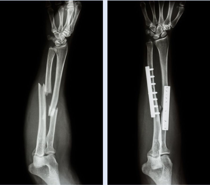
Intramedullary nail osteosynthesis
In intramedullary osteosynthesis, the doctor opens the bone’s medullary canal using an awl or a wire. The medullary canal is then enlarged, and a nail is driven into it, which should effectively bridge the fracture site. In a sense, it acts as an internal splint.
To prevent the nail from shifting, a transverse pin may also be driven into the bone (locking nail).
Kirschner Wire Technique
In this procedure, the fracture site is bridged with elastic steel wires. The doctor drives them through the cortical bone and anchors them in the cancellous bone.
However, the upper part of the wire should remain outside the bone. This allows it to be easily removed once the fracture has healed.
This method is particularly suitable for fractures of smaller bones or fractures in the growth plates. It is also commonly used for clavicle fractures.
Tension-band osteosynthesis
In this procedure, the tensile forces pulling the fracture fragments apart are converted into compressive forces. The surgeon first inserts two wires into the bone that run parallel to each other and perpendicular to the fracture line. He then places a soft wire loop around both wires. This loop is pushed through a canal into the bone so that it is tightly tensioned.
External fixator osteosynthesis
This procedure uses an external frame to hold the bones in place. To do this, small incisions are first made in the patient’s skin. Through these incisions, the doctor drills small holes into the fractured bone. He then inserts metal rods and connects them with metal struts. This stabilizes the fracture site.
Dynamic hip screw
This procedure is particularly well-suited for fractures of the femoral neck.
A guide wire is inserted into the part of the femoral neck located near the hip joint. Next, a screw with a short, thick thread is screwed into the femoral head. Finally, a metal plate is screwed onto the outer side of the femur. The fracture gap is compressed in the following weeks solely by the patient’s body weight.
Is there anything to keep in mind after the surgery?
Once the osteosynthesis is in place, the doctor sutures the muscles and the connective tissue of the skin. He then applies a wound dressing. Patients do not need to take any special precautions on their own after the procedure.
FAQ
What is osteosynthesis?
Osteosynthesis is a surgical procedure used to stabilize bone fractures. In this procedure, bone fragments are fixed in place using screws, pins, or wires to restore the bone to its natural position. Osteosynthesis means “bone joining” and ensures the safe healing of a fracture.
When is osteosynthesis necessary?
Osteosynthesis is necessary when a bone fracture cannot be adequately treated by immobilization with a splint or cast. Surgical treatment is often recommended, particularly for displaced fractures, unstable fractures, or malalignments. The goal is to achieve stable fixation and restore normal function to the affected bone.
What types of osteosynthesis are there?
The various osteosynthesis procedures include screw osteosynthesis, plate osteosynthesis, intramedullary nail osteosynthesis, tension band osteosynthesis, and external fixators. In screw osteosynthesis, the fracture fragments are fixed directly. In intramedullary nail osteosynthesis, an intramedullary nail is inserted through a canal in the medullary cavity and serves as an internal splint for the bone.
How is the surgery performed?
Before the surgery, the fracture is reduced, during which the bone fragments are anatomically realigned. Screws, plates, nails, or wires are then inserted to permanently stabilize the bone fragments. Depending on the type and location of the fracture, different surgical techniques are used to treat bone fractures.
What complications can occur after osteosynthesis?
Possible complications include infections, delayed fracture healing, loosening of the implants, or malalignment. In rare cases, screws in the bone or other implants may cause discomfort. Once the fracture has healed completely, the implants are either left in place or removed, depending on the situation. The individual healing process depends on the patient’s age, the type of fracture, osteoporosis, and body weight.

About the medical author
Sabine Schneider
Editor-in-Chief
Sabine Schneider – medical author: Explore expert articles and medical expertise in the Leading Medicine Guide.
More about the medical author →