CT angiography is a specialized imaging technique. It is a procedure used to visualize blood vessels within the human body.
Below you will find more information as well as a selection of specialists in CT angiography.
What is a CT scan?
Computed tomography is an important imaging technique in medicine. Doctors often refer to this imaging technique simply as “CT” or a “CT scan.” It is an examination that uses X-rays to produce cross-sectional images of tissues (bones, organs, blood vessels).
A CT scan is used, for example, in cases of severe traffic accidents (multiple trauma). This allows doctors to quickly and reliably detect or rule out injuries to bones, internal organs, or even bleeding. CT is also the imaging method of choice for the more detailed examination of tumors.
What is CT angiography (CTA)?
CT angiography is a CT scan that focuses specifically on the blood vessel system. However, since blood does not differ from normal tissue fluid, the administration of a contrast agent is necessary.
CT angiography makes it possible to visualize differences in density between various tissues. For example, water has a different density than bone or air.
To visualize the blood vessels even more clearly, the patient is given a contrast agent, just as in conventional angiography. These are iodine-containing, radiopaque liquids that do not allow X-rays to pass through them.
Doctors administer the contrast agent shortly before the CT scan through a vein in the arm. The contrast agent then circulates through the blood vessels with the bloodstream. This allows the vessels to be visualized and examined in detail—for example, to identify narrowings, blockages, or bleeding.
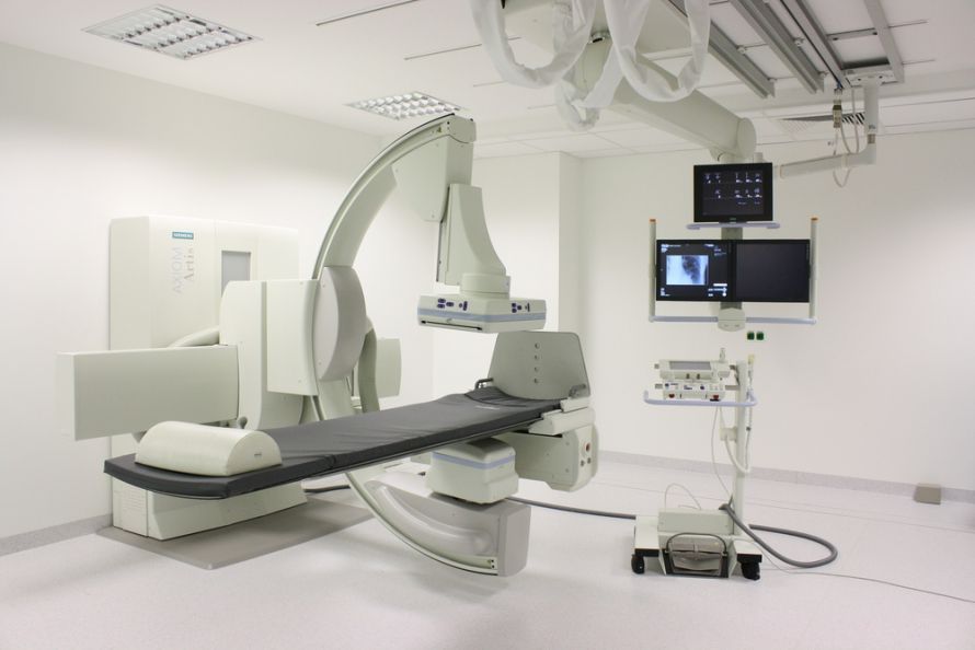 During CT angiography (CTA), the patient lies on their back on the examination table while the scanner moves through the circular opening @ Dirk Rothe /AdobeStock
Another variant is MR angiography, which uses magnetic resonance imaging (MRI).
For which conditions is CT angiography used?
What are the applications of CT angiography?
Typical diseases and conditions for which CT angiography is used include, for example:
- Aortic aneurysms
- medically known as aortic aneurysm
- The size and extent of the aneurysm are visualized, and
- in an emergency, whether it has ruptured
- Narrowing of the carotid arteries
Here, doctors examine narrowings of the carotid arteries more closely to determine whether surgery is necessary to prevent a stroke. Surgery is usually recommended if the degree of narrowing is > 70%
- Stroke
If a stroke has already occurred, CT angiography can show whether and where clots are present in the brain
This involves imaging the leg arteries, particularly to identify narrowings and blockages. Depending on the findings, further treatment may be recommended (e.g., balloon angioplasty or bypass surgery).
- Changes or abnormalities in the blood vessels, such as vascular tumors (hemangiomas)
- Renal artery stenosis (narrowing of the renal arteries)
- Thrombosis in the veins of the arms and legs, the abdominal vessels, and the vessels of the brain
- Suspected angina pectoris (chest pain) or heart pain caused by coronary artery disease (CAD)
- A pulmonary embolism
What makes CT angiography special?
The examination utilizes modern multislice computed tomography. Blood vessels such as arteries and veins can be segmented using high-speed computers and displayed in 3D images that can be freely rotated. While the raw images are always black and white, modern images can be color-enhanced.
One of the advantages of CT angiography is that it does not require any penetration into the patient’s body. There is no direct puncture, as is the case with conventional angiography. The examination techniques are the same for all vascular regions, so the patient does not notice any differences.
In MR angiography (magnetic resonance imaging), the patient lies inside a long tube. A CT scanner, on the other hand, is a large, ring-shaped device that surrounds the patient. The CT scan is therefore much faster and is also suitable for patients with a known fear of enclosed spaces.
CT angiography is particularly suitable for patients who have a pacemaker, as MRI is not an option for them. It also benefits very restless patients for whom a long MRI scan would be equally unsuitable.
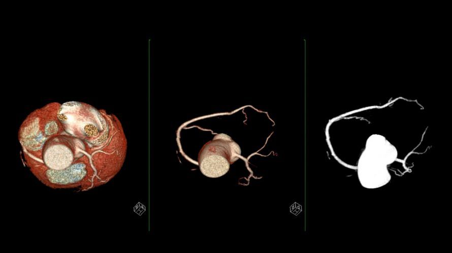 3D visualization of the coronary arteries using CTA @ samunella /AdobeStock
How does CT angiography work?
CT angiography is typically performed on an outpatient basis. The same guidelines apply as for other examinations using iodine-containing contrast agents. It is important to disclose any known allergies to contrast agents.
In such cases, it is usually necessary to administer certain medications to prevent an allergic reaction (shortness of breath, nausea, skin redness, shock). These include cortisone and antihistamines, which the patient receives shortly before the procedure. Prior to the CT angiography, doctors assess the patient’s kidney function through blood tests and laboratory analysis.
In total, CT angiography takes no more than ten minutes. For the examination, the patient lies on their back on an examination table.
Modern CT scanners use the multislice technique. This means they capture multiple cross-sectional images simultaneously. Newer machines also utilize what is known as the low-dose technique. This technique requires only 50 percent of the radiation to produce accurate images.
During the examination, the patient must hold their breath at specific intervals as instructed by the medical staff.
After the exam, the patient should drink plenty of fluids to quickly flush out the contrast agent. Additionally, in cases of diabetes mellitus or renal insufficiency, a deterioration in kidney function is a possibility.
Summary and Conclusion on CT Angiography
In summary, CT angiography is an imaging technique now available in nearly all hospitals for the diagnosis of all vascular diseases—from narrowings and blockages to hemorrhages.
The administration of an iodine-based contrast agent allows for the visualization of blood vessels in any region of the body. However, if the patient has a known contrast agent allergy, they should take medication to prevent allergic reactions.
The exam takes only a few minutes and can be performed even if the patient suffers from claustrophobia. Afterward, the patient should drink plenty of fluids to flush out the contrast agent and protect the kidneys.

About the medical author
Sabine Schneider
Editor-in-Chief
Sabine Schneider – medical author: Explore expert articles and medical expertise in the Leading Medicine Guide.
More about the medical author →