About one percent of all newborns are born with a heart defect. These heart defects vary in severity. Some require surgery as soon as possible after birth, while others are not detected until later. Children whose heart defects are successfully treated with surgery can generally lead completely normal lives. Here you will find further information as well as a selection of heart defect specialists and centers.
Prevalence of Individual Heart Defects
The prevalence of congenital heart defects is relatively high, affecting about one percent of newborns. However, the prevalence of individual heart defects is much higher. The following examples illustrate this:
- 31% ventricular septal defect
- 5–8% Aortic isthmus stenosis
- 7% Atrial septal defect
- 7% Persistent ductus arteriosus
- 7% Pulmonary valve stenosis
- 3–6% Aortic valve stenosis
- 5.5% Tetralogy of Fallot
A heart defect is not necessarily detected at birth. Symptoms often do not appear until later in life. Only in some cases are the symptoms severe enough that the heart defect is detected before birth or within the first few weeks of life. In such cases, the pulmonary artery and the pulmonary valve are usually affected.
Some Heart Defects in Detail
The spectrum of known heart defects ranges from relatively minor to life-threatening. In general, the more severe a heart defect is, the more likely it is that heart surgery will be the only remaining treatment option.
If more severe heart defects are treated during infancy, the children can go on to develop normally.
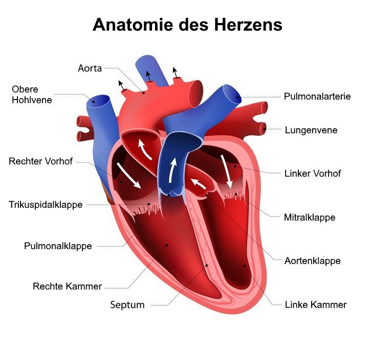
Pulmonary atresia
In this type of heart defect, the three leaflets of the semilunar valve do not open or are underdeveloped.
As a result, blood cannot flow from the right ventricle into the pulmonary artery. The blood therefore does not flow through the lungs and cannot be oxygenated.
Pulmonary valve stenosis
Pulmonary valve stenosis is also a defect of the valve leaflets of the pulmonary valve. During the heartbeat, this valve opens only partially, thereby narrowing the blood’s outflow tract.
Because of this narrowing, the heart must generate higher pressure to pump blood into the lungs.
Defects of the Septum
Children are often born with a heart defect involving the heart’s septum. This septum separates the left ventricle and left atrium from the right ventricle and right atrium of the heart.
The right ventricle and right atrium contain oxygen-poor blood returning to the heart from the systemic circulation. The left ventricle and left atrium, on the other hand, contain oxygen-rich blood from the pulmonary circulation.
When there is a defect in the heart septum, oxygen-poor blood can mix with oxygen-rich blood. This results in mixed blood with a lower oxygen content than necessary.
If the heart’s septum is permeable in the area of the atria, this is referred to as an atrial septal defect. Since there is increased pressure in the left atrium, oxygen-rich blood now flows into the right atrium.
A natural atrial septal defect is the so-called ductus arteriosus. This is a natural opening in the atrial septum that usually closes after birth. It primarily serves as a shunt to bypass the pulmonary circulation, which is not yet fully functional.
In a ventricular septal defect, the wall between the two ventricles is permeable.
Symptoms vary in severity depending on the size of the septal defect. If the hole is very large, it results in mixed blood that is very low in oxygen, and the body’s oxygen supply is compromised.
This can be recognized by a change in skin color—a bluish tint—and the child’s progressively decreasing ability to tolerate physical exertion. In these cases, only pediatric cardiac surgery can help by closing the defect through surgery. Smaller defects, however, usually go undetected for years due to their milder symptoms.
Heart defects are very often detected via
- an ECG,
- cardiac catheterization, or
- other imaging techniques
. The doctor will discuss the best course of action with you, the parents of the affected child. It’s also important to note that not every heart defect requires surgery.
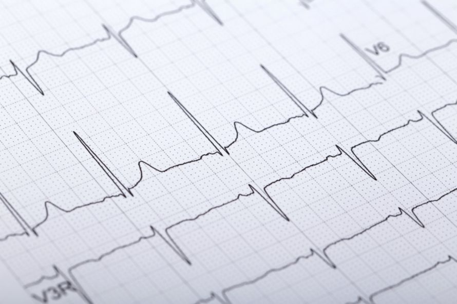
Often, it is sufficient to monitor minor septal defects regularly with an ECG at first. In infancy or childhood, many holes in the heart septum between the right and left sides of the heart close on their own. Fortunately, surgery is therefore usually unnecessary.
However, if a hole persists later on and such a heart defect is left untreated, serious complications can sometimes arise. These include, for example,
- inflammation,
- arrhythmias,
- heart valve disease, or
- permanent lung damage.
Transposition of the Great Arteries
In 5% of all cases, a very severe congenital heart defect occurs—known as transposition of the great arteries. This refers to a reversal of the connections between the aorta and the pulmonary artery and the heart’s ventricles.
This means that the heart pumps deoxygenated blood—rather than oxygen-rich blood—into the systemic circulation. This heart defect is fatal. To save the newborn, immediate postnatal surgery is necessary.
Tetralogy of Fallot
In what is known as Tetralogy of Fallot, four types of heart defects occur simultaneously:
- a ventricular septal defect,
- pulmonary valve stenosis,
- enlargement of the right side of the heart, and
- an aortic anomaly.
This clinical picture of a congenital heart defect is very complex.
Due to the increased pressure in the right ventricle resulting from pulmonary valve stenosis, blood is constantly forced through the ventricular septal defect. The resulting oxygen-poor mixed blood leads to the development of symptoms of oxygen deprivation in the systemic circulation.
In addition, Tetralogy of Fallot involves an aortic anomaly that can impair blood flow from the heart.
Congenital Heart Defects in Adolescents
During the growth phase, combinations of a previously corrected heart defect and newly acquired heart defects may occur. It is therefore possible that patients who have already undergone surgery may need to undergo heart surgery again later.
Minimally invasive procedures reduce the risk of scarring. Atrial septal defect surgeries can generally be performed using minimally invasive techniques today. The reduced scarring lowers the likelihood of later complications. The physical and mental strain is also less.
Symptoms of Congenital Heart Defects
A wide range of symptoms can indicate a congenital heart defect. The pediatrician is often the first point of contact when these symptoms appear.
The main cause of these symptoms is increasing oxygen deprivation. This is visible as cyanosis (a bluish discoloration) of
- skin,
- lips, and
- nail beds.
In addition, other symptoms may also occur, such as
- rapid or labored breathing,
- lack of energy,
- pale, cold, clammy skin,
- shortness of breath,
- fatigue,
- rapid heartbeat, and
- swelling in the feet, ankles, or abdomen
may occur.
Diagnosis of Congenital Heart Defects
Thanks to improved modern diagnostic methods, many heart defects can now be detected within the first year of life.
Prenatal diagnostics now also make it possible to identify severe heart defects prenatally, i.e., before birth. However, prenatal diagnostics are not intended to facilitate the planning of an early termination of pregnancy in cases of severe congenital heart defects. Rather, the goal is to ensure optimal care for the newborn after birth.
In cases of narrowing or defective heart valves, blood flow becomes turbulent or short-circuiting occurs. This causes loud heart murmurs that can be detected very easily with a stethoscope. Depending on the type of murmur, its origin can be determined.
The electrocardiogram (ECG) is also very important for diagnosing congenital heart defects. By recording the heart’s electrical activity, the doctor can determine the size and position of the heart and, above all, identify any arrhythmias.
However, the most important diagnostic method today is echocardiography (ultrasound of the heart). It provides a very precise image of the heart and all its structures. This makes nearly all heart defects visible. In addition, it allows for an assessment of heart function and the condition of the individual parts of the heart.
This examination method is used whenever a congenital heart defect is suspected. It is completely painless and risk-free and is therefore also used as a very gentle procedure for children.
Further, usually much more specialized examinations are performed depending on the nature of the suspected condition. These include the use of a cardiac catheter. Other imaging techniques include magnetic resonance imaging (MRI) and computed tomography (CT).
Severe Heart Defects in the Operating Room
In cases of very severe heart defects, a simple corrective surgery is often impossible. In such cases, the procedure must be carried out in several surgical stages. The top priority is to ensure blood flow to the body and lungs.
In most cases, doctors create artificial connections to produce mixed blood. This ensures at least a minimal supply of oxygen.
In some cases, the heart can be bypassed. This allows oxygen-poor blood to be routed directly from the large veins of the body into the pulmonary artery, where it is then oxygenated. By relieving the strain on the heart, this may improve blood flow and reduce arrhythmias.
One of the most challenging congenital heart defects is transposition of the great arteries. In these children, the artery leading to the lungs is located where the aorta should be, and the aorta, in turn, branches off toward the lungs. This makes it virtually impossible for oxygen-rich blood to reach the body.
Without life-saving surgery, these newborns die very shortly after birth. In the first few days of life, oxygen exchange occurs solely through so-called postnatal shunt openings. Therefore, surgery must be performed within a few days of birth.
During this surgery, the aorta and the pulmonary artery are detached from the heart, swapped with each other, and reconnected to the heart in their correct positions.
Is there a way to prevent congenital heart defects?
There are, in fact, a number of known risk factors that can have a harmful effect on the developing heart. The primary goal, therefore, is to avoid these risk factors.
- Girls should get vaccinated against rubella so they do not contract the disease during a future pregnancy.
- You should consult a doctor before taking any medications during pregnancy. Over-the-counter medications and vitamin supplements are among the products that pose the greatest risk.
- Alcohol and nicotine are strictly off-limits during and even after pregnancy (while breastfeeding).
It is also particularly important for expectant mothers to attend all prenatal checkups. During these regular checkups, a congenital heart defect can be detected early. To do this, the baby’s heart is examined in detail using ultrasound.
FAQ
1. What types of congenital heart defects are there? Heart defects
are classified as either congenital or acquired. Congenital forms include septal defects, valve malformations, and malformations of the major blood vessels, such as transposition of the great arteries or hypoplastic left heart syndrome (HLHS).
2. Are heart defects always detected in childhood?
Some heart defects are not detected until adulthood, such as when a patent foramen ovale remains open. In young adults, long-term complications such as heart failure may occur, which often affects the right side of the heart.
3. How are patients with congenital heart defects treated?
Treatment for adults and children is provided in pediatric cardiology or cardiology. In cases of complex malformations, shunts may be implanted to ensure that blood from the lungs reaches the systemic circulation—often, multiple procedures are necessary.
4. Can congenital heart defects be genetic?
Congenital heart defects can be genetic—for example, a heart malformation often occurs in cases of trisomy 21. Infections such as rubella during pregnancy are also among the known risk factors for this condition.
Share this article

About the medical author
Sabine Schneider
Editor-in-Chief
Sabine Schneider – medical author: Explore expert articles and medical expertise in the Leading Medicine Guide.
View full expert profile →