Cardiomyopathy is a disease of the heart muscle in which the heart muscle tissue undergoes changes. The causes and symptoms of such conditions can vary widely. Shortness of breath, chest pain, and rapid heartbeat are just a few of the symptoms that may occur. Cardiac output decreases, and the first symptoms of heart failure begin to appear. In medicine, cardiomyopathy is classified as a heart disease that involves structural and functional changes in the heart muscle.
Here you will find further information as well as a selection of cardiomyopathy specialists and centers.
Types of Cardiomyopathy —Primary and Secondary Cardiomyopathy
Generally, a distinction is made between primary cardiomyopathy, which directly affects the heart muscle, and forms caused by other diseases. These are referred to as primary and secondary cardiomyopathies. Primary cardiomyopathies develop directly in the heart muscle. Secondary cardiomyopathy occurs when other conditions damage the heart muscle tissue.
Based on changes in the structure and function of the heart muscle, cardiomyopathy can be classified into five types:
- Dilated cardiomyopathy (DCM): The most common form of cardiomyopathy, characterized by an enlarged ventricle and a significantly reduced pumping function of the heart.
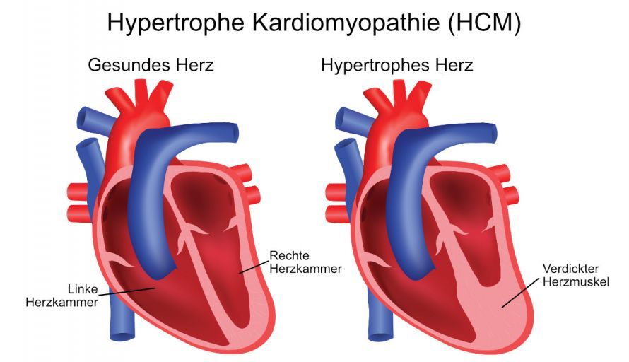
- Hypertrophic cardiomyopathy (HCM): The second most common form of cardiomyopathy. It is genetically determined and tends to run in families. The heart muscle is too thick and no longer sufficiently elastic. The diameter of the ventricle is normal or reduced, and pumping function is normal. This type of cardiomyopathy can also be obstructive (HOCM), in which case blood flow through the thickened heart muscle is impaired. In severe cases, the condition is referred to as hypertrophic obstructive cardiomyopathy, in which blood flow is additionally impeded.
- Restrictive cardiomyopathy: A rare form; the exact causes are unknown. The body incorporates increased amounts of connective tissue into the muscle; the ventricular walls harden and can no longer expand sufficiently. Less blood flows from the atria into the ventricles. This leads to blood congestion, and the atria enlarge. The ventricles themselves retain their normal size and are generally able to continue pumping normally.
- Arrhythmogenic right ventricular cardiomyopathy: Affects the right ventricle and can trigger arrhythmias during physical exertion. The cause is unknown. Some of the cells in the right ventricle die off and are replaced by fatty and connective tissue. The muscle tissue becomes thinner, and the heart’s conduction system is impaired. Young male athletes are frequently affected.
- Hypertensive cardiomyopathy: This form is triggered by chronic high blood pressure. The heart pumps more forcefully due to increased resistance in the arteries. As a result, the left ventricle gradually thickens and loses its ability to pump effectively.
Other heart muscle diseases include non-compaction cardiomyopathy. In this congenital form, only the left ventricle is affected. On an ultrasound image, the muscle tissue appears swollen, like a sponge. Remodeling processes lead to scarring of the heart muscle; the ventricles enlarge and can no longer function adequately.
Stress cardiomyopathy is caused by severe emotional or physical stress. It is also known as broken heart syndrome or Tako-Tsubo cardiomyopathy.
Causes of Cardiomyopathy
Primary cardiomyopathies, which develop directly in the heart muscle, are often genetically based. Genetic factors frequently play a central role, leading to structural changes in the heart muscle. Inherited heart muscle diseases are often present from birth, though symptoms usually do not appear until later in life.
Numerous diseases can damage the heart muscle. Medications, such as cancer treatments, can also trigger cardiomyopathy.
Possible causes include:
- Inflammation of the heart muscle caused by infections
- Autoimmune diseases such as rheumatoid arthritis or scleroderma (hardening of the connective tissue),
- Diseases that cause certain substances to accumulate in the heart muscle,
- Metabolic disorders, such as severe thyroid dysfunction or diabetes mellitus,
- Medications and treatments for cancer, such as chemotherapy and radiation therapy,
- Severe vitamin deficiencies, such as vitamin B deficiency in beriberi or vitamin C deficiency in scurvy
- Drugs and poisoning,
- Diseases that primarily affect the skeletal muscles (muscular dystrophy) or extend to the nervous system.
If the doctor identifies the cause of the cardiomyopathy, he or she can initiate treatment. This can prevent the disease from progressing rapidly. However, the exact cause of cardiomyopathy cannot always be clearly determined.
Symptoms of cardiomyopathy
Symptoms vary in severity depending on the type of cardiomyopathy. The timing of when symptoms first appear also depends on the type of heart muscle disease. In some cases, cardiomyopathy can go undetected for a long time. Early signs may be nonspecific, which is why any relevant symptoms should prompt an early evaluation for suspected cardiomyopathy.
In every type of cardiomyopathy, the heart muscle tissue (myocardium) changes. The heart’s function and performance decline. Patients experience various symptoms, depending on the specific form and severity of the condition.
In all forms of cardiomyopathy, certain parts of the heart muscle—or the entire muscle—are no longer able to function properly. This leads to characteristic symptoms.
Heart Rhythm Disturbances
The body requires more oxygen. This increases the heart rate; the heart beats faster and often irregularly. This results in heart palpitations. In addition, episodes of dizziness or even brief fainting spells often occur.
Fatigue
The heart cannot pump enough oxygen-rich blood into the arteries and to the brain. As a result, patients feel exhausted, tired, and confused. Due to the slowed blood flow, the tissues extract more oxygen from the blood.
Cold and bluish patches of skin, especially on the hands and feet, are typical signs of this.
Fluid Retention
Due to the heart’s reduced pumping capacity, blood backs up in the pulmonary vessels and veins. Fluid leaks into the body’s tissues and the lungs. Edema (fluid retention) develops in the lungs; the liver, kidneys, and stomach may also be affected.
This results in pain in the upper right abdomen, a feeling of bloating, and loss of appetite.
Bluish discoloration of the skin
The initial phase of pulmonary edema is characterized by increased coughing. As the condition progresses, patients find it increasingly difficult to breathe. They become short of breath and cough up frothy secretions.
If too much fluid accumulates in the lung tissue, the blood can no longer absorb enough oxygen. In cases of severe heart failure, the lips or tongue often take on a bluish tint (cyanosis).
If you notice one or more of the symptoms described, see a cardiologist as soon as possible. If the condition is treated in a timely manner, its rapid progression can be halted.
Possible Complications
With cardiomyopathy, there is a risk that blood clots will form on the inner walls of the heart. The heart’s pumping function is more irregular and weaker than in a healthy heart. This creates turbulence in the heart chambers, which can sometimes lead to blood clots. Blood clots are small clumps of blood cells.
Under certain circumstances, these blood clots can break loose and travel through the bloodstream to other organs. In the worst-case scenario, such a blood clot can block a major blood vessel there, triggering pulmonary embolisms or strokes.
In rare cases, the arrhythmia suddenly becomes so severe that the entire circulatory system fails. Such complications can result from impaired pumping function, as the heart can no longer supply the body with sufficient blood. The heart beats so rapidly that the ventricles no longer fill sufficiently with blood between individual beats. This can result in sudden cardiac death.
Examination and Diagnosis of Cardiomyopathy
Listening to the heart provides the cardiologist with initial clues. In cases of the disease, blood tests reveal antibodies and markers of heart failure (proBNP). These help diagnose potential heart damage.
Various procedures are used to make the diagnosis.
- Echocardiography (ultrasound examination of the heart): Shows the mobility and thickness of the heart muscle. This allows the doctor to detect heart valve disorders. It also enables the doctor to check whether the heart is pumping enough blood into the systemic circulation.
- ECG (electrocardiogram, e.g., stress ECG) measures the heart’s electrical activity. It helps identify arrhythmias. Typical changes on the ECG are particularly evident in hypertrophic cardiomyopathy.
- During a cardiac catheterization, the cardiologist inserts a thin plastic tube through a blood vessel into the heart. This allows the doctor to measure the pressure in the blood vessels near the heart and in the individual areas of the heart.
- Heart muscle biopsy: During a cardiac catheterization, the doctor can take a tissue sample. The sample is examined under a microscope in the laboratory to identify the structure or any changes in the muscle.
- Imaging techniques such as MRI, X-ray, or CT scans visualize the heart and reveal any abnormalities.
- Genetic testing can determine whether the patient’s condition is caused by genetic mutations.
How is cardiomyopathy treated?
Medications are used to treat many forms of cardiomyopathy, for example, in cases of
- autoimmune reactions,
- metabolic disorders,
- infections, and
- vitamin deficiencies.
Drug therapy is tailored to the specific form and severity of the disease.
Consistent physical rest can prevent further damage.
Medications are also prescribed to treat the resulting symptoms. These include
- beta-blockers,
- ACE inhibitors, or
- diuretics.
They reduce the strain on the heart. Arrhythmias are prevented with specific antiarrhythmic drugs and beta-blockers. The formation of blood clots is prevented with an anticoagulant. Often, only the symptoms can be alleviated and complications avoided. In many cases, symptomatic treatment is the first step, as treating the underlying cause is not always possible. Whenever possible, the goal is to provide causal treatment that specifically addresses the underlying cause.
If surgery becomes necessary, the cardiac surgeon removes parts of the heart muscle (myectomy). The implantation of a pacemaker or a defibrillator may also be necessary.
If all other treatment options have been exhausted, a heart transplant is the only remaining option.
Course of the Disease and Prognosis
In most cases, cardiomyopathy is a serious condition that is rarely curable. Nevertheless, with early diagnosis and consistent treatment, many patients can remain stable for a long time. In many cases, however, a deterioration in health cannot be prevented.
The prognosis, including life expectancy, is strongly related to the specific form and stage of the disease. For many patients, the prognosis depends heavily on the course of the disease and the individual’s response to treatment.
FAQ
What is cardiomyopathy?
The term “cardiomyopathy” refers to a disease of the heart muscle in which the structure and function of the heart are altered.
What causes cardiomyopathy?
Cardiomyopathy can result from various causes, including genetic factors, pre-existing conditions, or external influences that can damage the heart muscle.
What types of cardiomyopathy are there?
The most common forms include dilated, hypertrophic, and restrictive cardiomyopathy, as well as other rarer variants.
How is cardiomyopathy diagnosed?
To diagnose cardiomyopathy, tests such as an ECG, ultrasound examinations, and other imaging procedures are used.
Share this article