In many cases, a parastomal hernia appears as a distinct bulge in the area of the stoma, as tissue shifts into the subcutaneous space and an abdominal wall hernia develops. Modern surgical procedures—whose effectiveness has also been confirmed in meta-analyses—aim to restore the stability of the abdominal wall and prevent recurrent hernias.
Definition of a Parastomal Hernia
A stoma is an artificial opening in the body that is created surgically, either permanently or temporarily, to maintain bodily functions. A stoma is therefore an artificially created opening from a hollow organ to the body’s surface. An artificial intestinal outlet is therefore referred to as a stoma.
A parastomal hernia most commonly affects what is known as an enterostomy, or artificial intestinal outlet. This site is particularly prone to incisional hernias because it originates from strong structures that are additionally subjected to significant strain. The stoma is subjected to pressure from both the internal abdominal muscles (for example, during bowel movements) and the abdominal wall.
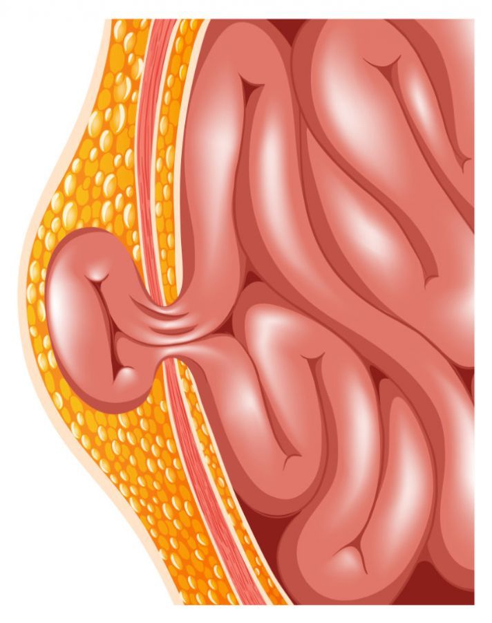
In a parastomal hernia, a protrusion occurs beneath the skin of the so-called stoma loop of the intestine. In some cases, an additional protrusion of other sections of the intestine or parts of the so-called greater omentum into the subcutaneous fatty tissue may occur. A parastomal hernia is considered a special type of incisional hernia.
It is currently estimated that up to 50 to 80 percent of patients with a colostomy also develop a parastomal hernia. Accordingly, incisional hernias are a common complication following abdominal surgery.
Symptoms of a parastomal hernia
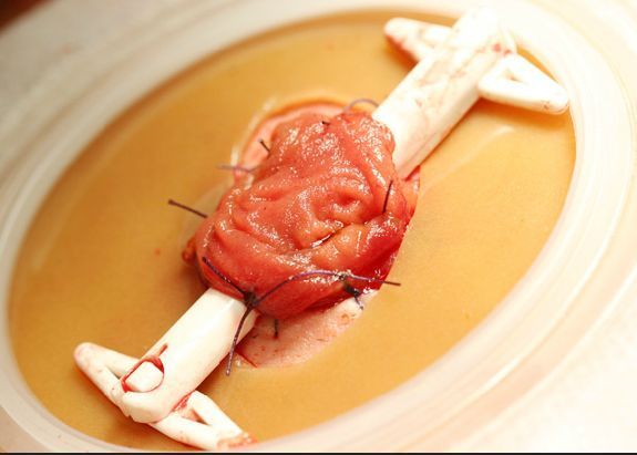
The symptoms of a parastomal hernia manifest as defects in the abdominal wall and a bulge next to the stoma. When pressure within the abdominal cavity increases, the bulge caused by the hernial sac becomes clearly visible. This occurs, for example, when coughing or straining.
Nevertheless, symptoms can vary widely, ranging from no symptoms at all to restricted movement and pain.
Pain and restricted movement occur particularly in cases of circulatory impairment, when sections of the intestine become trapped in the hernial ring. Complete obstruction of the intestine can be life-threatening. Immediate surgery is then necessary to relieve the pain and restore blood flow to the intestine.
For ostomates, the location of the stoma also plays a role: if the hernial opening is in an unfavorable location, the risk of symptoms increases. Some hernia centers now use meshes prophylactically in selected patients to strengthen the abdominal wall. An experienced team at the hospital should always decide on a case-by-case basis which measures are appropriate.
Causes and Risk Factors
A parastomal hernia mainly develops following a surgical procedure. At that point, the tissue and scar around the stoma opening have not yet sufficiently fused and stabilized.
A parastomal hernia is caused by
- a general wound-healing disorder,
- wound infections, and
- bleeding
.
- Certain medications, such as cortisone,
- disorders of collagen metabolism, and
- long-term smoking
generally increase the risk of complications following the creation of a colostomy.
General risk factors that lead to the development of a parastomal hernia include, for example,
- the patient’s age,
- obesity or being overweight,
- weak connective tissue,
- abdominal surgery,
- healed wound infections, or
- steroid therapy, such as that used to treat COPD.
All of these factors weaken the tissue. As a result, a scar may not heal properly after surgery, and the stoma may not be properly secured to the abdominal wall. Even today, a technical error by the surgeon is still considered one of the causes of parastomal hernias. However, the development of parastomal hernias shows that the surgical technique does not play a role in their cause.
This has also been demonstrated in clinical studies. In most cases, a hernia recurs when a new stoma is created. This confirms that surgical technique does not appear to play a role in the development of parastomal hernias.
Parastomal Hernia: Confirming the Diagnosis and Initiating Proper Treatment
Examination and Diagnosis of a Parastomal Hernia
In most cases, a parastomal hernia can be detected through a clinical examination from the outside. During a thorough palpation, the contents of the hernia sac may be felt.
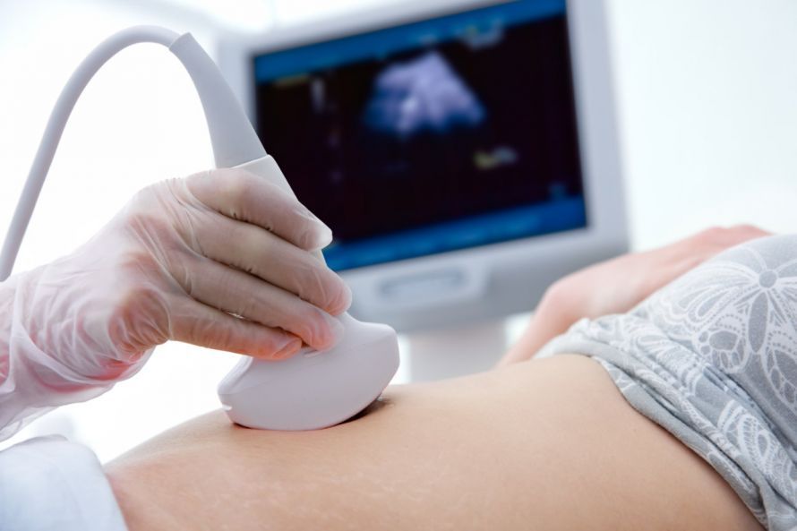
However, to establish a reliable and accurate diagnosis, imaging techniques such as ultrasound or MRI should be performed. This can also increase the detection rate of small hernias.
In the management of the hernia, a precise assessment of the diverted bowel loop plays a central role, as it provides information about the size of the abdominal wall defect and the stability of the surrounding tissue. Particularly in cases with complex findings, it is possible to detect early on whether a developing defect is enlarging and thus whether surgical planning is necessary. Postoperatively as well, close monitoring helps to detect new changes or increasing bulging in a timely manner.
Correct mesh placement is crucial for successful surgical treatment. Techniques such as the sublay technique provide stable repair of the enterostomy and significantly reduce the subsequent recurrence rate. Precise reconstruction of the abdominal wall defect permanently reinforces the hernia, ensuring that the repair remains durable over the long term.
Treatment of a parastomal hernia
Large parastomal hernias, in particular, require surgery. In these cases, the hernial orifice and hernial sac are correspondingly large and may contain a significant portion of the intestine. The goal of the surgery is to
- reposition the prolapsed abdominal contents,
- close the incision, and
- prevent a recurrence of the hernia.
A very large hernial sac often complicates the surgical conditions. The anatomical function of the abdominal wall must be restored. This is difficult because the stoma disrupts the abdominal wall’s supportive function and creates a weak spot.
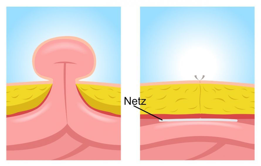
In the treatment of parastomal hernias, the use of a synthetic mesh to reinforce the hernial orifice has proven effective. This is the most reliable way to reduce the recurrence rate. Various mesh materials are available for use in different surgical procedures.
In open surgeries, lightweight polypropylene meshes with large mesh sizes are used. These come into direct contact with loops of the intestine. They are very stiff and can therefore cause irritation and fistulas. In contrast, meshes used in laparoscopic procedures must be more flexible and have a finer mesh.
Each mesh has different properties that behave differently within the body. Accordingly, the appropriate mesh must be selected for each surgical technique used to treat a parastomal hernia.
In particular, the high recurrence rates are the reason why the so-called fascial suture is no longer used for parastomal hernias. Relocating the stoma to a different site is also no longer the preferred option, as this would likely result in a new parastomal hernia. This is because the underlying conditions of the tissue and the patient remain unchanged.
Parastomal Hernia – Course, Risks, and Long-Term Considerations for Abdominal Wall Hernias
Prevention and Prognosis of a Parastomal Hernia
In general, the likelihood of developing a new parastomal hernia even after the original one has been repaired is very high. For open surgeries, the recurrence rate is estimated to be as high as 50 percent. The surgical site is exposed to a high risk of infection. This risk is lower with minimally invasive procedures, but these methods do not always yield optimal results either.
In general, patients cannot prevent a parastomal hernia on their own, especially since it results from a necessary surgical procedure.
If a patient is at high risk for developing a parastomal hernia, a synthetic mesh can be surgically implanted as a preventive measure to reinforce the abdominal wall. This strengthens the patient’s abdominal wall. The new weak spot in the abdominal wall, which was created by the formation of the artificial opening, is bypassed as effectively as possible.
A parastomal hernia often occurs where the fascia is weaker and the gap in the abdominal wall widens further due to increased pressure. For ostomates, the protrusion of intestinal segments into the resulting opening can cause additional discomfort. When structures are constricted, the risk of complications increases significantly. For this reason, the Sublay procedure is increasingly being used, as it stabilizes the fascia and reliably supports the widened fascial gap.
Guidelines and international recommendations on “the treatment of parastomal hernias” emphasize that stable reconstruction is crucial to preventing recurrent hernias. Targeted mesh placement allows for better repair of parastomal defects, thereby reducing both the recurrence rate and functional limitations in affected patients.
FAQ on Parastomal Hernias
What does the development of a parastomal hernia mean for ostomates?
In ostomates, a parastomal hernia develops due to a gap in the abdominal wall directly next to the stoma. The bulge can be bothersome in daily life and must be surgically corrected if it causes symptoms.
What role does a hernia center play in treatment?
A specialized hernia center is familiar with modern techniques such as the Sugarbaker or keyhole methods and can determine on a case-by-case basis which approach is most suitable for restoring the stability of the abdominal wall.
What distinguishes the Sugarbaker technique from the keyhole method?
In the Sugarbaker technique, the stoma is positioned laterally beneath the mesh so that the intestine cannot protrude through the fascial gap again. The Keyhole method places the mesh with an opening around the diverted stoma, but is used less frequently today because recurrence rates may be higher.
What does an enlargement of the fascial gap mean for treatment?
If the fascial gap enlarges due to strain or scar tissue, the risk increases that more sections of the intestine will enter the hernia. In such cases, the Sugarbaker or Sugarbaker-PH technique is usually preferred to permanently reinforce the abdominal wall.
What materials are used to treat a parastomal hernia?
Many centers use modern meshes, such as Rosin meshes, which are placed so that they do not constrict the stoma while simultaneously stabilizing the compromised abdominal wall.
Share this article

About the medical author
Prof. Dr. med. Ferdinand Köckerling
Medical Author · Director
View full expert profile →