An arteriovenous malformation (AVM) is a congenital vascular malformation in which arteries and veins are directly connected to one another without an intervening capillary bed. These arteriovenous malformations lead to altered blood flow within the affected blood vessels. An AVM may remain asymptomatic for a long time or become apparent through neurological symptoms. Possible symptoms include headaches, seizures, paralysis, or even a cerebral hemorrhage. A cerebral AVM in the brain, in particular, may be associated with an increased risk of hemorrhage.
Diagnosis is usually made using MRI and other imaging techniques. Treatment of an AVM is planned through an interdisciplinary approach involving specialties such as neurosurgery, neurology, and neuroradiology. Depending on the location, surgery, embolization, or radiosurgery may be considered.
What is a cerebral arteriovenous malformation?
“Cerebral” means “relating to the brain,” and the medical term “arteriovenous malformation” can be translated and explained as follows:
- Congenital, abnormal, or pathological (= “mal”)
- connections and short circuits (= formations)
- between arteries (= arterio)
- and veins (= venous).
Normally, oxygen- and nutrient-rich blood flows from the left side of the heart at high pressure (approx. 120–140 mmHg) to all organs and tissues of the body. There, in what is known as the “capillary bed,” the exchange of substances between the blood and tissue cells takes place. Capillaries are the tiniest terminal branches of the arteries with very thin walls through which substances can diffuse (pass).
On the other side of the capillary bed, tiny venous branches (venules) collect the blood, which is now low in oxygen and nutrients, and transport it at a lower pressure (approx. 10–20 mmHg) back to the right side of the heart. Here, it is supplied with oxygen via the lungs and with nutrients via the liver, before re-entering the circulatory system described above. In the presence of arteriovenous malformations, arterial blood flows directly and at high pressure into the veins via the malformations (= shunts), without undergoing gas exchange in the capillary bed.
What risks are associated with AV malformations?
The shunts described above result in the two main problems associated with AV malformations:
- pressure on the right side of the heart, as blood is rapidly returned through the shunts at high pressure and in large volumes. This can lead to chronic cardiac strain and even sudden heart failure.
- an insufficient supply of energy and oxygen to the brain tissue, since the arterial blood is diverted beforehand. This can result in tissue damage, manifesting as a stroke.
How common are arteriovenous malformations?
Approximately one in 1,000 newborns is born with an arteriovenous malformation. In principle, any organ system can be affected by such a vascular shunt. However, the anomaly occurs most frequently in the brain or facial skull.
In children under 15 years of age, arteriovenous malformations are also the most common cause of cerebral hemorrhages (intracranial hemorrhages).
How do arteriovenous malformations develop?
Arteriovenous malformations develop as early as the 4th to 8th week of pregnancy from the earliest, more primitive blood vessel structures. “Primitive” means that these structures are simple, existing before they differentiate into more complex vascular structures.
Exactly what triggers these shunts in the blood vessels is still unclear. Genetic predispositions or external factors during pregnancy may contribute to the development of arteriovenous malformations.
Blood Flow and Effects of Arteriovenous Malformations
The actual arteriovenous malformation is also referred to in medicine as a “nidus” (literally “nest”). This is the core of the anomaly. Several, often dilated, arteries supply blood to the nidus, which is then passed on to dilated veins.
The blood from the arteries is under higher blood pressure. As a result, congestion and dilation typically occur in the veins. Such venous dilations can develop into venous aneurysms or lead to stenosis (narrowing of blood vessels).
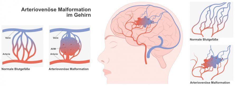
In an arteriovenous malformation, the capillary network between an artery and a vein is missing © rumruay | AdobeStock
These vascular changes eventually become visible along the course of the arteriovenous malformation.
What symptoms occur with an arteriovenous malformation?
Vascular shunts (unnatural connections between blood vessels), such as arteriovenous malformations, can remain asymptomatic for a lifetime.
However, many patients experience their first symptoms between the ages of 20 and 40, as the condition and the dilated vessels continue to expand and grow larger. Common typical symptoms include, for example:
- Brain hemorrhages: These can lead to loss of consciousness or present with signs of a stroke.
- epileptic seizures
- Headache attacks: These are sometimes initially misinterpreted as migraine attacks.
- Neurological deficits, such as numbness or paralysis
- Varicose veins: Severely dilated and externally visible veins throughout the body, particularly on the legs, may also result from an arteriovenous malformation. This should be considered, especially in cases with very pronounced findings, and cerebral manifestations should be ruled out.
How are arteriovenous malformations diagnosed?
In most patients, pulsatile vascular sounds can be heard in the cranial region. These sounds occur because blood flows at a higher velocity from the arteries into the shunted veins.
Normally, the capillary network in the organ region facilitates the exchange of substances and gases and slows down the blood flow. This capillary network is absent in an arteriovenous malformation. As a result, blood flows at high arterial pressure directly into a shunted vein. In medicine, this direct connection between an artery and a vein is also called a shunt. These higher blood flow velocities through the shunt can be visualized using transcranial Doppler or color duplex sonography.
Suspected arteriovenous shunts are then confirmed using
- magnetic resonance imaging (MRI) and
- magnetic resonance angiography (MRA)
. These two diagnostic procedures allow specialists to determine the exact location of the arteriovenous malformation.
Computed tomography (CT) is not actually significant in the imaging of vascular shunts. However, it is used in emergency diagnostics, e.g., in cases of loss of consciousness following a cerebral hemorrhage. Of crucial diagnostic and therapeutic importance is so-called invasive angiography with contrast medium. In this procedure, a catheter is advanced into the brain through a puncture in the groin, and after the administration of contrast medium, the shunts can be precisely visualized. This examination is considered invasive because the puncture site involves a breach of the body’s integrity. It should therefore only be indicated in combination with therapeutic treatment.
How is an arteriovenous malformation treated?
The treatment of vascular shunts is often difficult and carries serious risks. For this reason, each case is discussed individually at the specialized clinic by an interdisciplinary decision-making committee.
Depending on the location of the arteriovenous malformation, surgery or radiation therapy may not be feasible without risk. In these cases, conservative treatment with medication or avoidance of stressful situations is used: The goal here is to alleviate symptoms, such as epilepsy, through the use of antiepileptic drugs.
For operable arteriovenous malformations, both minimally invasive therapy and open surgery are options:
- In minimally invasive (catheter-based) procedures, specialists attempt to access the shunt system of the nidus. There, they attempt to partially or completely occlude the involved vessels using sclerosing agents or small metal coils (endovascular embolization).
- Surgical treatment involves the complete resection of the arteriovenous malformation. This means the surgeon removes the nidus entirely
The goal of any surgery is to reduce the risk of bleeding and the likelihood of recurrence of arteriovenous malformations.
Which specialists treat cerebral arteriovenous malformations?
The treatment of arteriovenous malformations in the brain involves various medical disciplines, such as specialists in
- neurosurgery,
- radiation oncology,
- neuroradiology, and
- neurology.
Conservative treatment can be provided at specialized centers and in specialist practices. Surgical procedures require treatment at a specialized clinic by specialists in neurosurgery and neuroradiology.
What is the prognosis for arteriovenous malformations?
The risk of hemorrhage is a particularly critical factor in the prognosis for arteriovenous malformations. Normally, the annual risk is about 2–3%. However, the mortality rate following a hemorrhage is quite high, at up to 10%. In addition, one in three patients continues to experience neurological deficits.
Furthermore, anyone who has experienced an initial hemorrhage due to an arteriovenous malformation has a roughly 25% chance of experiencing another hemorrhagic event in the following years.
Without treatment, one in four affected patients dies from their arteriovenous malformation within a 10-year period.
FAQ
What is an arteriovenous malformation (AVM)?
An arteriovenous malformation is a congenital abnormality of the blood vessels. In an AVM, there are direct shunts between arteries and veins, without a normal capillary bed. The so-called nidus forms the center of this vascular malformation. Arteriovenous malformations are among the most significant cerebral vascular malformations of the central nervous system (CNS).
What symptoms does an AVM cause?
Not every AVM causes symptoms. However, typical symptoms may include headaches, seizures, neurological deficits, or paralysis. An AVM in the brain can also lead to a brain hemorrhage or, in severe cases, a stroke. Symptoms often depend on the location of the AVM within the affected areas of the brain.
How is an arteriovenous malformation diagnosed?
MRI, angiography, and other imaging techniques are used for diagnosis. These examinations reveal the feeding arterial vessels, the venous drainage pathways, and the nidus of the AVM. Diagnostic and interventional neuroradiology plays a central role in this process.
What treatment options are available for an AVM?
Treatment of an AVM depends on its size, location, and risk of bleeding. Possible procedures include surgery, embolization, and radiosurgery. In endovascular embolization, a catheter is inserted through the femoral artery, and an embolization agent is precisely delivered into the vascular malformation. The goal is to eliminate the AVM or to prepare for its subsequent removal.
When is treatment necessary?
Treatment of an arteriovenous malformation is recommended if there is an increased risk of bleeding, neurological deficits, or cerebral hemorrhage. The decision regarding treatment is made through an interdisciplinary approach involving neurosurgery, neurology, and neuroradiology. Current scientific findings and individual risk factors are taken into account.
Share this article

About the medical author
Prof. Dr. med. Susanne Regus
Medical Author
Prof. Dr. med. Susanne Regus – medical author: expert articles, professional insights and medical expertise in the Leading Medicine Guide.
View full expert profile →Sources
- aerzteblatt.de
- altmeyers.org
- S1-Leitlinie „Zerebrale Gefäßmalformationen (arteriovenöse Malformationen, arteriovenöse Fisteln, Kavernome)“ (AWMF-Register-Nr. 030-088), Stand 08.08.2023: register.awmf.org/de/leitlinien/detail/030-088
- flexikon.doccheck.com
- medical-tribune.de/