An atrial septal defect (ASD) is one of the most common congenital heart defects in both adults and children. This defect involves a hole in the wall between the right and left atria, resulting in a left-to-right shunt. The ASD II, or secundum type, occurs particularly frequently in the area of the fossa ovalis. The atrial septal defect leads to volume overload of the right heart and the right ventricle.
Small ASDs are often asymptomatic, but larger defects can cause arrhythmias, pulmonary hypertension, or heart failure. Diagnosis is made using an ECG, echocardiography, or transesophageal echocardiography. Depending on the location and size of the defect, it can be closed surgically or with an occluder device via a cardiac catheterization procedure. Early treatment usually improves life expectancy and reduces long-term functional impairment of the heart and lungs.
What is an atrial septal defect?
An atrial septal defect belongs to the group of shunts. These are short circuits between the arterial and venous circulatory systems. Atrial septal defects account for approximately 10 percent of congenital heart defects.
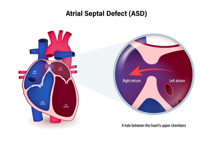 An atrial septal defect (ASD) is a hole in the wall between the heart’s atria @ Dee-sign /AdobeStock
Symptoms of an atrial septal defect
Generally speaking, the severity of symptoms associated with an atrial septal defect depends on the size of the “hole.” Smaller shunts may remain asymptomatic throughout a person’s life. For most larger defects, symptoms appear between the ages of 2 and 20.
Half of patients experience heart rhythm disturbances; signs of heart failure may develop later. Patients with an atrial septal defect usually have a pale complexion and peripheral oxygen deficiency (cyanosis). Large defects lead to exertional dyspnea (shortness of breath) and reduced physical capacity as early as infancy.
The sooner children or adults with cardiac symptoms see a doctor and receive the correct diagnosis, the better their prognosis. If the atrial septal defect remains untreated, it can lead to severe strain on the right side of the heart and heart failure. This limits the quality of life and life expectancy of those affected.
Diagnosis of an Atrial Septal Defect
Typically, the doctor detects the heart defect simply by listening to the heart (auscultation). Typical heart murmurs and a usually visible heartbeat are indicative of right-sided heart strain. Characteristic symptom complexes are also evident during the medical history (anamnesis). If symptoms are absent, the condition is usually discovered incidentally.
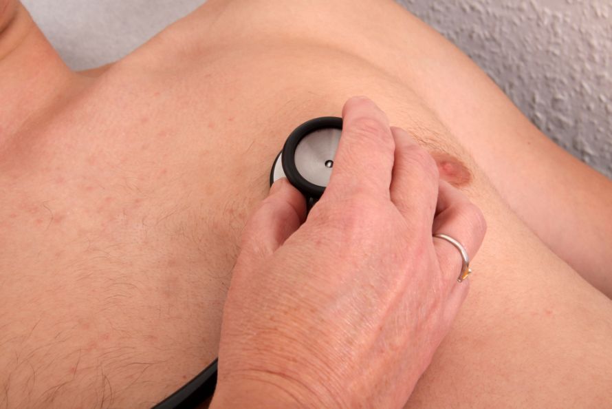 Heart examination with a stethoscope @ Klaus Eppele /AdobeStock
Imaging performed by a doctor confirms the presence of an atrial septal defect. An ultrasound examination, or echocardiography, can visualize the blood flow within the heart—particularly the flow passing through the atrial septal defect and the hole in the heart.
On an X-ray, doctors may also detect an enlarged right atrium, which results from the overload of the pulmonary circulation.
Treatment of an Atrial Septal Defect
A specialist determines on a case-by-case basis whether surgery is necessary for each patient.
The following factors are favorable for the course of the surgery and recovery:
- The patient is still in the early stages of the disease
- There is no high blood pressure in the pulmonary circulation (pulmonary hypertension) yet. A possible threshold for complications is a blood pressure of 40 mmHg or higher.
Surgical closure of an atrial septal defect before the age of 25 offers the best prospects for a normal life expectancy. The complication rate is low. In older patients who are asymptomatic, doctors have mixed views on surgical septal closure.
The surgery can be performed in two ways:
- Doctors close the atrial septal defect using a piece of tissue from the pericardium or by suturing it directly. This method is used for larger defects or when patients report significant symptoms and limitations.
- For smaller atrial septal defects, the procedure is significantly less invasive. It can be performed using a catheter that doctors guide from the groin into the heart. At the tip of the catheter is a small umbrella-like device that doctors unfold over the hole to seal it.
An atrial septal defect requires special follow-up care
After surgical repair of an atrial septal defect, patients spend some time in the intensive care unit. Here, they are placed on blood thinners and monitored.
Anticoagulation with heparin or Marcumar is necessary to prevent dangerous blood clots. These clots can enter the pulmonary circulation and cause a pulmonary embolism. The same can happen in the brain, where an arterial blockage caused by a blood clot triggers a stroke.
Which doctors treat atrial septal defects?
Specialists in atrial septal defects are experts in the fields of cardiac surgery and cardiology, as well as pediatrics (child health care) when applicable.
You can find suitable centers and specialists on this page, among other places.
FAQ
What is an atrial septal defect?
An atrial septal defect, or ASD for short, is a congenital heart defect characterized by a hole in the septum between the right and left atria. This hole in the septum causes a left-to-right shunt, resulting in increased blood flow to the pulmonary circulation. ASD type II, located in the fossa ovalis, is particularly common.
What symptoms does an ASD cause?
Small ASDs are often asymptomatic, while larger atrial septal defects can lead to shortness of breath, arrhythmias, or fatigue. The increased volume load on the right heart can cause the right ventricle to enlarge. In adulthood, pulmonary hypertension, heart failure, or dizziness are more common.
How is an atrial septal defect diagnosed?
The diagnosis is usually made using echocardiography, an ECG, and cardiac catheterization. Transesophageal echocardiography allows for precise visualization of the defect’s location and size. Additionally, hemodynamic effects on the right atrium, right ventricle, and pulmonary artery are assessed.
How is an ASD treated?
An ASD can be closed interventionally using an occluder if there is sufficient rim tissue. For more complex atrial septal defects, closure is performed surgically using a heart-lung machine. The goal is to close the defect and prevent complications such as pulmonary hypertension or stroke.
What is the life expectancy with an atrial septal defect?
After successful ASD closure, life expectancy is usually normal. Untreated large shunts, on the other hand, can lead to volume overload of the right heart, pulmonary hypertension, or functional impairment. Early treatment at a center specializing in congenital heart defects significantly improves the prognosis.
Share this article

Sources
flexikon.doccheck.com/de/Vorhofseptumdefekt
herzstiftung.de/leben-mit-angeborenem-herzfehler/angeborene-herzfehler/vorhofseptumdefekt
kinderaerzte-im-netz.de/krankheiten/vorhofseptumdefekt-atriumseptumdefekt-asd/
medlexi.de/Vorhofseptumdefekt