Craniosynostosis is a malformation of the skull in which the cranial sutures fuse prematurely. Cranial sutures are important growth zones of the skull bones. Therefore, craniosynostosis can lead to developmental disorders and permanent damage.
Below you will find more information on symptoms, causes, and treatment, as well as a list of selected craniosynostosis specialists.
In infants, the individual skull plates must be connected to one another in a flexible manner via the still-soft cranial sutures. This is the only way to ensure that the brain, which is still developing and growing, has sufficient space within the skull. Not all cranial sutures are always affected by craniosynostosis.
Furthermore, the extent of craniosynostosis can change during growth.
Craniosynostosis can lead to neurological impairments such as:
- Intellectual disability
- Vision problems
- Pain
It also leads to cosmetic symptoms such as:
- Asymmetries
- Unusual head shapes
- Facial deformities
The ossification of the cranial sutures occurs through a delicate interaction between the skull base, the meninges, and the skull bones. Brain growth sets the pace for the extent and speed of this process.

Important and well-known points here include the large fontanelle and the small fontanelle. These are two openings made of connective tissue located at the intersections of the cranial sutures.

The fontanelles provide flexibility and can be easily felt on an infant’s head. The small fontanelle closes at around 2–5 months of age, while the large fontanelle does not close until just over two years of age.
By the age of one, 90 percent of the skull’s growth is complete. By the age of two, the cranial sutures have closed, and the remaining bone growth proceeds much more slowly.
What forms of craniosynostosis are there?
Craniosynostosis is the umbrella term for premature ossification of the skull in fetuses and infants. There are specific terms depending on which cranial suture is affected:
- Frontal suture synostosis
Frontal suture synostosis refers to the ossification of the frontal suture, which runs vertically across the forehead. This prevents the forehead from developing to its normal width. Instead, this results in a spherical forehead, inadequate coverage of the lateral eye sockets, and recessed temples.
- Sagittal suture synostosis
Premature ossification of the sagittal suture, which lies between the anterior and posterior fontanelles, impairs width growth in the parietal region. To compensate, the forehead and the back of the head protrude more than average, resulting in a narrow, elongated head.
- Coronal suture synostosis
An asymmetrical forehead develops when the coronal sutures are affected. These are located on the right and left sides of the head near the hairline. Experts also refer to this as anterior plagiocephaly. The affected side is flatter in this case.
This is often accompanied by:
- A pronounced bulge of the forehead on the opposite side
- Changes in the eye socket area, making the eye appear larger. Abnormalities may also occur in the ear area or on the nose.
- Lambda suture synostosis
Asymmetries at the back of the head are usually caused by ossification of one of the two lambda sutures at the occiput. In this type of lambda suture synostosis—which usually occurs on one side—the back of the head flattens on one side.
Compensatory growth occurs in the opposite parietal region, resulting in “posterior plagiocephaly.” Due to this asymmetry, the ears usually appear to be positioned unevenly.
- Microcephaly
In microcephaly, all cranial sutures ossify prematurely. The result is an extremely small head and severely impaired brain development.
What symptoms does craniosynostosis cause?
The following symptoms occur:
- You can recognize the skull growth disorder caused by craniosynostosis by asymmetry of the head or face.
- Because the interior of the skull is too small, this leads to what is known as increased intracranial pressure. The growing brain presses from the inside against the skull, which is too small and has ossified prematurely.
- Brain damage, including visual impairments, is a typical consequence.
- Deformities in the facial area are not merely a cosmetic issue; they can also lead to functional impairments of the respiratory tract.
- In addition, reduced eye sockets can make it difficult to close the eyelids.
Diagnosis of Craniosynostosis
Craniosynostosis is often suspected simply by observing the typical changes in head shape. The cranial index provides doctors with additional guidance.
X-rays and CT scans with three-dimensional images of the skull’s structure confirm the diagnosis. This information helps the doctor plan future treatment.
Additional diagnostic procedures include an EEG to measure brain waves, further neurological examinations, and an eye exam.
Treatment of Craniosynostosis
Surgical intervention is the only treatment option for craniosynostosis.
By the third month of life, doctors can perform endoscopic surgery to remove a limited amount of bone from the ossified cranial suture.
The child then wears a special helmet until the age of 18 months. This ensures that the skull develops a normal shape. Between the fifth and 12th months of life, doctors can perform surgery to permanently establish the final, uniform shape of the head.
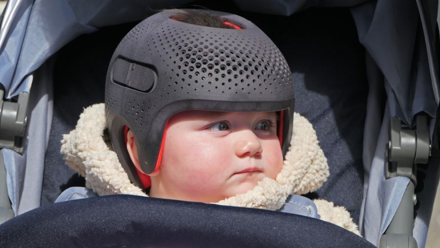 A special helmet helps shape the child’s skull into a more regular form @ peter /AdobeStock
Doctors usually treat craniosynostosis surgically within the first year of life, as the bone is more malleable at that stage.
In addition, the ability to form new bone decreases significantly. Nevertheless, older children can also undergo surgery.
These are the most common surgical methods:
- Fronto-orbital reshaping
Fronto-orbital reshaping is the most commonly performed surgery to correct deformities and functional disorders in the forehead area.
In this procedure, bone segments are removed, reshaped, and reinserted. The supporting plate used consists of absorbable material that gradually dissolves. The delicate scar is well concealed behind the hairline.
- Parieto-occipital reshaping
This surgical method is used to reshape the back of the head, for example, in cases of lambda suture synostosis. Several surgical techniques are available for this purpose.
- Decompression Surgery
Decompression surgery is performed in the cranial region of the scalp. During the procedure, the surgeon makes radial incisions in the skull bone.
The bone flaps push the growing brain apart. Later, the bone segments fuse together. Decompression surgery can be performed up until the end of the third month of life.
Recovery and Prognosis Following Treatment of Craniosynostosis
The success of surgical treatment for craniosynostosis is long-lasting. The healing process is generally good. In most cases, the noticeable skull deformity can be virtually completely corrected.
Cosmetic results in the facial area are also excellent.
Share this article