Expert Interviews
Anterior Cruciate Ligament Reconstruction

Alexandra Pfitzmann ·
Cruciate ligament tears are among the most common serious knee injuries. In Germany, approximately 70,000 to 80,000 people are affected each year—primarily while playing sports. If the anterior or posterior cruciate ligament tears completely, the knee loses its stability. In many cases, cruciate ligament reconstruction is then necessary, in which the torn ligament is replaced with a tendon from the patient’s own body to restore the natural function of the knee joint.
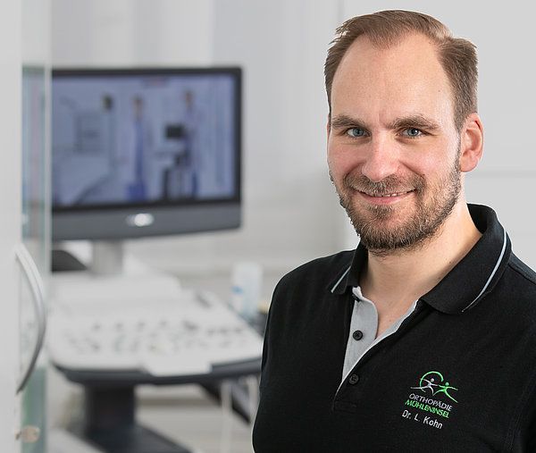
“Most accidents leading to an anterior cruciate ligament tear occur without direct physical contact. A typical cause is valgus rotational trauma: the slightly bent knee buckles inward while the thigh rotates inward and the lower leg rotates outward. Such movements often occur when decelerating in tennis, during quick changes of direction in soccer, or when landing after a jump, as the knee collapses inward and twists. This is the primary mechanism leading to a tear of the anterior cruciate ligament.
Another mechanism is hyperextension of the knee joint. This happens, for example, when someone jumps onto the knee from the front and forces it backward. This often results in additional injuries to the joint capsule. Even a normal twisting motion can cause the cruciate ligament to tear—for example, when playing soccer and your cleats get caught in the grass, your upper body continues to twist, and the knee does not follow the rotational movement.
When skiing, a caught or caught-edge ski can have the same effect; the so-called “phantom foot mechanism”—caused by the ski moving forward and the twisting motion—also leads to cruciate ligament tears. Direct impact injuries are much rarer but do occur—for example, when someone jumps into the knee from the side or from the front, as is common in American football. Unusual situations can also lead to this, such as when a dog jumps into the knee during a walk. Such cases do exist, but they remain the exception; most cruciate ligament tears occur without contact,” explains Dr. Kohn, adding regarding the symptoms:
“For those affected, this often manifests as very severe, acute pain. However, this isn’t always the case. With certain injury patterns—such as partial tears or tears very close to the bone in the thigh—the pain can be surprisingly mild. Partial tears or tears in which the synovial sheath (the lining of the joint) remains intact sometimes do not even lead to significant swelling. It sometimes happens that those affected are able to continue skiing or running despite the injury, as is occasionally observed among skiers.”
Whether cruciate ligament reconstruction is necessary or conservative treatment is sufficient depends on a combination of the injury pattern, stability, associated injuries, patient profile, and functional requirements. The decisive factor is not only the tear itself, but also the extent to which the knee’s function is impaired as a result.
“Initially, it is often possible to deduce what might have happened based on the described mechanism of the accident. This medical history provides the first indication of which structures might be affected. This is followed by a clinical examination, which, however, may be limited if the knee is severely swollen, painful, and restricted in movement. In addition, the severity of knee joint effusion is always assessed, as acute effusion following trauma is almost always a sign that a relevant structure has been injured—whether it be the meniscus, a cruciate ligament, or even bone.
In most cases, an X-ray is part of the basic diagnostic workup to rule out possible associated injuries, such as bone damage. The key diagnostic test is then an MRI, or magnetic resonance imaging, of the knee joint. It reveals the specific injury patterns, whether the cruciate ligament is torn, whether the tear is complete or partial, and the exact location of the damage. Certain cruciate ligament tears can be sutured or reattached, such as when the ligament has been torn away from the femur.
In such cases, there is a good chance of preserving the cruciate ligament. At the same time, the MRI is used to check for any associated injuries—to the meniscus, cartilage, or medial or lateral ligaments—as these influence both the treatment decision and the urgency of care,” says Dr. Kohn.
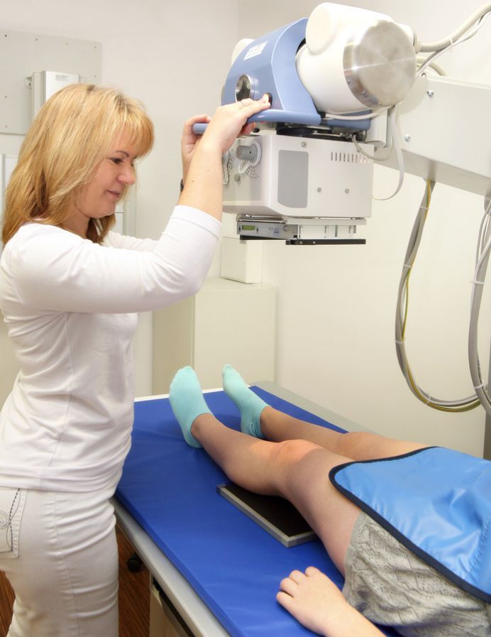
The diagnostic process includes a medical history, clinical examination, ultrasound and X-ray imaging, as well as a prompt MRI scan to determine as accurately as possible what has happened in the knee.
Conservative treatment is certainly possible in some cases; not every cruciate ligament requires surgery. The decisive factors are age, athletic and physical activity levels, and the patient’s individual needs—and, of course, any possible accompanying injuries.
“Younger, highly active individuals—especially those involved in rotational sports like soccer—are more likely to undergo surgery. Older, less active patients with lower physical demands, on the other hand, often benefit from conservative treatment. It is important that the knee remains stable over time. The muscles must be able to sufficiently compensate for the missing cruciate ligament, both in terms of strength and neuromuscular function.
If instability develops despite therapy, or if the patient reports the knee giving way, surgical treatment must be considered. Even if the patient themselves says that the instability bothers them, surgery is advisable. As a general rule: young, athletic, high physical demands—surgery is more likely; older, less active, no rotational sports—conservative treatment is more likely. Conservative therapy begins with a phase in which the swelling in the knee subsides and muscle activation is restored. As soon as mobility returns and the muscles can be controlled again, strength training, proprioceptive training, muscle-building exercises, and knee-stabilizing exercises follow.
Afterward, it becomes clear how well the patient is coping. Those who compensate well for the missing cruciate ligament and do not experience instability can be treated very successfully with conservative methods. Patients who continue to experience instability despite physical therapy often have to discontinue conservative treatment and undergo surgery after all to prevent secondary damage such as meniscus or cartilage injuries and, in the long term, osteoarthritis. “The younger the patient, the more important a stable knee joint is to prevent such secondary damage,” emphasizes Dr. Kohn.
Many patients are back to their normal daily activities relatively quickly. This often happens after just a few weeks. After four to six weeks—or two to three months at the latest—those who compensate well can once again do many things without difficulty. Patients who continue to experience instability for a longer period are usually among those for whom conservative treatment does not achieve sufficient stability.
Cruciate ligament replacement is primarily necessary when the biomechanical stability of the knee is so severely compromised by the tear that the joint can no longer reliably perform its stabilizing function.
Dr. Kohn explains: “In some cases, a cruciate ligament can actually be preserved—for example, if it has torn directly from the bone and sufficient tissue is still present. However, these are the exceptions. If the ligament tears in the middle, it has a very poor tendency to heal. In most cases, therefore, the cruciate ligament must be replaced. The body’s own tendons are available for this purpose—essentially the body’s ‘spare parts warehouse.’ The three most common grafts are the hamstring tendons on the inner side of the thigh, a portion of the patellar tendon with small bone blocks at the ends, and a portion of the quadriceps tendon. Each of these options has advantages and disadvantages, and there is no single “perfect” graft. The choice always depends on the individual patient.
The hamstring tendons are used most frequently, typically the semitendinosus tendon at the back of the thigh, which is laid in four strands. It generally fits well with the anatomy of the knee joint, has a very high tensile strength, and can be harvested through the same incision used later for fixation—which is cosmetically advantageous. Disadvantages include lower stiffness, which may allow for some elongation of the graft over time, as well as some weakness in knee flexion, since the hamstrings are involved in this movement. If the hamstring tendons have already been used—for example, in a previous surgery—or if there is a concurrent medial collateral ligament injury, the quadriceps tendon is often used instead. Its advantage is high stiffness, which helps it maintain its original length somewhat better.
However, harvesting it is technically somewhat more challenging, requires an additional skin incision, and can lead to anterior knee pain. In addition, it takes longer for quadriceps strength to fully return. The patellar tendon also provides a stable graft with good healing, but it is not the preferred choice for every patient due to potential anterior knee pain and difficulties bearing weight while kneeling.”
The choice of graft is an individualized decision based on anatomy, pre-existing conditions, athletic requirements, and any previous surgeries. All of the tendons mentioned provide stable, proven results—they differ only in their specific characteristics and respective advantages and disadvantages.
When a tendon is harvested, there is naturally a lack of tissue at that site initially, and this can lead to discomfort—and this is precisely what is referred to as “donor-site morbidity.”
“Any donor site can cause symptoms, and these vary depending on the graft. With hamstring tendons, it has been shown that the semitendinosus tendon partially regenerates in up to 70% of cases. The body also forms new tissue and restores the structure of the quadriceps tendon, similar to the patellar tendon. The patellar tendon is generally a very good graft because it is harvested with small bone blocks, allowing it to heal particularly quickly and stably within the bone canals. At the same time, however, this very harvesting technique often leads to anterior knee pain.
For people who have to kneel a lot for work—such as tile setters—this can lead to significant problems. In such cases, the patellar tendon would therefore not be a suitable graft. The harvest site thus plays an important role in deciding which tendon to use. “Depending on occupational demands, athletic activity, and individual anatomy, careful consideration must be given to which graft is the best option for each patient,” explains Dr. Kohn.
The choice of surgical technique has a significant impact on how well the knee functions after a cruciate ligament reconstruction and how stable it remains in the long term. Each technique influences the position of the graft, the restoration of natural kinematics, and the healing of the new ligament—and thus, ultimately, the functional outcome.
 Dr. Kohn with a surgical nurse
“There are various surgical options for implanting a cruciate ligament replacement, even though the core of the procedure—that is, the insertion of the new ligament—is highly standardized. The technique is adapted to the specific graft used and the patient’s individual anatomical conditions to ensure that the new cruciate ligament is positioned as anatomically correctly as possible. Differences arise primarily in the fixation of the tendon, as each type of graft has its own specific requirements.
In addition to the actual cruciate ligament reconstruction, the knee can be further stabilized. In recent years, it has become evident that reinforcing or reconstructing the anterolateral ligament (ALL) can significantly improve rotational stability and reduce the risk of re-rupture. To achieve this, an additional incision is made on the outer side of the knee, and the outer knee region is specifically reinforced. However, this additional stabilization is not necessary for every patient.
It is primarily considered for athletes who participate in rotational sports such as soccer, handball, or basketball, for patients with severe rotational instability, or in revision surgeries. When using hamstring tendons, a particularly tear-resistant suture can also be implanted. This so-called “internal bracing” acts like an internal splint, protecting the graft in the early phase while simultaneously allowing for early weight-bearing. “There are therefore several ways to optimize the outcome and further increase the stability of the knee joint,” states Dr. Kohn.
The particular challenge for the surgeon lies in fitting the new cruciate ligament as precisely as possible to the patient’s individual anatomy, since even the slightest deviations can significantly impair subsequent function.
Dr. Kohn makes this point clear: “Even though anterior cruciate ligament surgery is generally highly standardized, it remains a demanding procedure because the technique must always be adapted to the patient’s individual circumstances. The goal is not only a subjectively stable knee, but also the most accurate restoration possible of the knee’s original kinematics. To achieve this, the graft must be anchored in the anatomically correct position with millimeter precision.
The placement of the drill holes is crucial here: even small deviations can lead to instability, restricted movement, or even graft failure. Especially in tight or anatomically challenging joints, this level of precision requires a great deal of experience and a very meticulous approach. It often requires a willingness to take the time to correct positions and make adjustments until the target anatomical position is truly correct.
Added to this is the individual selection of the graft and the decision as to whether additional measures, such as ALL reconstruction (reinforcement of the anterolateral ligament), are necessary. Bone-specific characteristics must also be taken into account. All these factors make it clear that this is not a trivial operation, but rather a procedure that requires a high degree of precision, anatomical understanding, and extensive surgical experience.”
Cruciate ligament reconstruction is generally performed on an outpatient basis today. The procedure is clearly structured.
“The patient arrives at the outpatient operating room on an empty stomach, undergoes a brief preparation period, and then undergoes surgery. The procedure itself, including anesthesia and the recovery phase, takes about one hour. Afterward, the patient remains under observation for one to two hours and can then be picked up. In total, they usually spend only two to three hours in the outpatient operating room and are given an emergency number where someone can be reached at any time.
The first follow-up appointment takes place at the doctor’s office the next day. Inpatient stays are still possible under certain conditions—for example, if home care cannot be guaranteed, additional procedures are necessary, or there are specific medical reasons. In such cases, the patient generally stays in the hospital for two to three days: Many patients now feel more comfortable with outpatient care. Pain management has been so well optimized today that postoperative symptoms are usually well controlled.
In addition, many appreciate the opportunity to spend the initial period after surgery in the familiar surroundings of their own home. Nevertheless, there are people who expressly request an inpatient stay—whether due to past experiences or for personal peace of mind. “That is still possible, provided it is medically justified and the necessary conditions are met,” explains Dr. Kohn regarding the practical process for the patient.
The risks of cruciate ligament reconstruction can be divided into biological, mechanical, and functional complications—and modern procedures are specifically designed to significantly reduce these risks.
“One of the most significant risks of cruciate ligament reconstruction is graft failure—whether due to re-tearing or residual instability. This risk can be significantly reduced if the graft is positioned anatomically precise, carefully secured, and, if necessary, supported by additional measures such as an ALL reconstruction. The selection of a suitable graft and appropriate fixation material also plays a role. Another consideration is the risk of infection, which is low in cruciate ligament surgery anyway.
By soaking the graft in an antibiotic solution prior to implantation, infection rates have been significantly reduced once again in recent years. In rare cases, excessive scarring occurs, such as what is known as “Cyclops syndrome,” in which scar tissue forms in front of the cruciate ligament graft and can impair mobility. Here, too, the correct position of the drill tunnel and the proper tension of the graft are crucial—neither too tight nor too loose. The timing of the surgery can also influence the outcome.
“Despite the utmost care, some complications cannot be completely prevented; however, many risks can be significantly minimized through precise surgical technique, anatomically correct procedures, and careful postoperative care,” notes Dr. Kohn.
Cruciate ligament reconstruction is a very safe procedure, and thanks to more precise surgical techniques, improved imaging, optimized grafts, and modern rehabilitation, most risks can be significantly reduced today.
Rehabilitation following cruciate ligament reconstruction is structured, phase-oriented, and significantly more individualized than in the past. In the first few weeks, the focus is on reducing swelling, regaining knee extension, and early functional activation of the muscles.
“Rehabilitation following ACL surgery has changed significantly in recent years. In the past, it was strictly time-based—after three months you were allowed to do this, after six months that—regardless of how far along the patient actually was. Today, rehabilitation is guided much more by clearly defined criteria, even though the time required for the graft to heal continues to play a role. In the first one to two weeks, the focus is on reducing swelling, regaining full extension, and achieving initial improvement in flexion.
Equally important is the early activation of the thigh muscles to prevent prolonged inactivity. As soon as the swelling subsides, the load is increased, mobility is further improved, and the first strengthening exercises are begun. By the sixth week, the knee should be able to bear full weight, with full extension and flexion largely restored. This is followed by a phase of muscle strengthening, knee stabilization, and neuromuscular training. It is precisely during this time that many patients subjectively feel surprisingly well, even though the graft is biologically at its most vulnerable.
After four to six months, sport-specific exercises can be cautiously introduced—running training, light jumping exercises, and initial changes of direction. Whether an athlete is allowed back on the field is now determined based on various tests. These include strength measurements of the anterior and posterior thigh muscles, side-to-side comparisons, jump tests such as the single-hop or triple-hop, and an assessment of swelling and range of motion. Only when these criteria are met does the cautious return to training begin—using soccer as an example: ball control, passing, light dribbling—but no team or competitive play yet.
After another testing phase, the player can return to full team training before finally reaching the final stage: a return to competition,” says Dr. Kohn regarding the rehabilitation phase.
Each year, approximately 60 to 80 cruciate ligament surgeries are performed at Orthopädie Mühleninsel in Landshut.
“A major advantage of our practice is that all stages of treatment take place in one location. Thanks to the integrated outpatient surgical unit, procedures can be performed on an outpatient basis without having to refer patients to other facilities. However, I can also provide inpatient care at a nearby hospital. From the initial evaluation through the procedure to the entire follow-up care, the patient remains under the same care throughout. Collaboration with the physical therapists is also close and straightforward. The short distances on-site facilitate quick communication, which is particularly advantageous during follow-up care. Feedback from physical therapy reaches the practice promptly, allowing for an early response if the healing process is not progressing optimally. “This seamless care, combined with direct communication among all parties involved, ensures particularly reliable and comprehensive care,” Dr. Kohn emphasizes at the conclusion of our conversation.
- A knee specialist with a deep focus on all structures of the knee joint.
- High level of expertise in sports traumatology and complex ligament, cartilage, and meniscus injuries.
- Surgical spectrum: cruciate ligament surgeries (ACL/PCL), collateral ligament surgery, meniscus and cartilage treatments, alignment corrections, patella stabilization, and arthroscopic fracture repair.
- DKG-certified knee surgeon utilizing state-of-the-art minimally invasive techniques and 3D imaging.
- Experienced in conservative therapies such as injections, pain management, and physical therapy.
- Chief Physician for the Sports Orthopedics Division
- Two locations: Landshut-Achdorf Hospital & Orthopedics Mühleninsel.
- Prevention-oriented diagnostics
Share this article
Expert Interviews
Read next
- Expert interview
Expert interview with Professor Dr Elmar Herbst, PhD
Oct 15, 2026
Patient-centred and personalised knee surgery
Read more - Expert interview
Cardiogeriatrics: Significance and benefits in the context of hospital reform
Sep 21, 2026
Cardiogeriatrics: Significance and benefits in the context of hospital reform
Read more - Expert interview
Expert Interview with Professor Dr. Amadeus Hornemann, MPH
Sep 18, 2026
Prof. Amadeus Hornemann on Precision Medicine in Gynecology: Modern Surgery, New Techniques, Better Quality of Life
Read more