Expert Interviews
Shoulder Arthroplasty: Expert Interview with Priv.-Doz. Dr. med. Mike H. Baums

Alexandra Pfitzmann ·
Priv.-Doz. Dr. med. Mike H. Baums is the Chief of the Department of Orthopedics, Trauma Surgery, and Sports Traumatology at St. Elisabeth Hospital in Dorsten and is recognized as an expert in the field of joint surgery.
With a particular focus on shoulder, elbow, and knee joints, as well as sports traumatology, he has extensive experience in both conservative and surgical treatment of diseases and injuries of the musculoskeletal system. His practice encompasses both joint-preserving arthroscopic procedures and the implantation of artificial joints (endoprostheses), particularly in the shoulder and knee. As a certified shoulder, elbow, and knee surgeon, Dr. Baums combines the highest level of professional expertise with modern, patient-centered treatment approaches.
Treatment is always provided in accordance with current national and international standards—supported by state-of-the-art technical equipment and a highly qualified team. The clinic under his leadership offers a broad range of services, which, in addition to joint replacement, includes acute and emergency care, geriatric traumatology, as well as outpatient and inpatient procedures. The focus is on the goal of sustainably improving patients’ mobility and quality of life through precise diagnostics, individualized treatment planning, and personalized, compassionate care.
The editorial team of the Leading Medicine Guide spoke with PD Dr. Baums about shoulder joint replacement.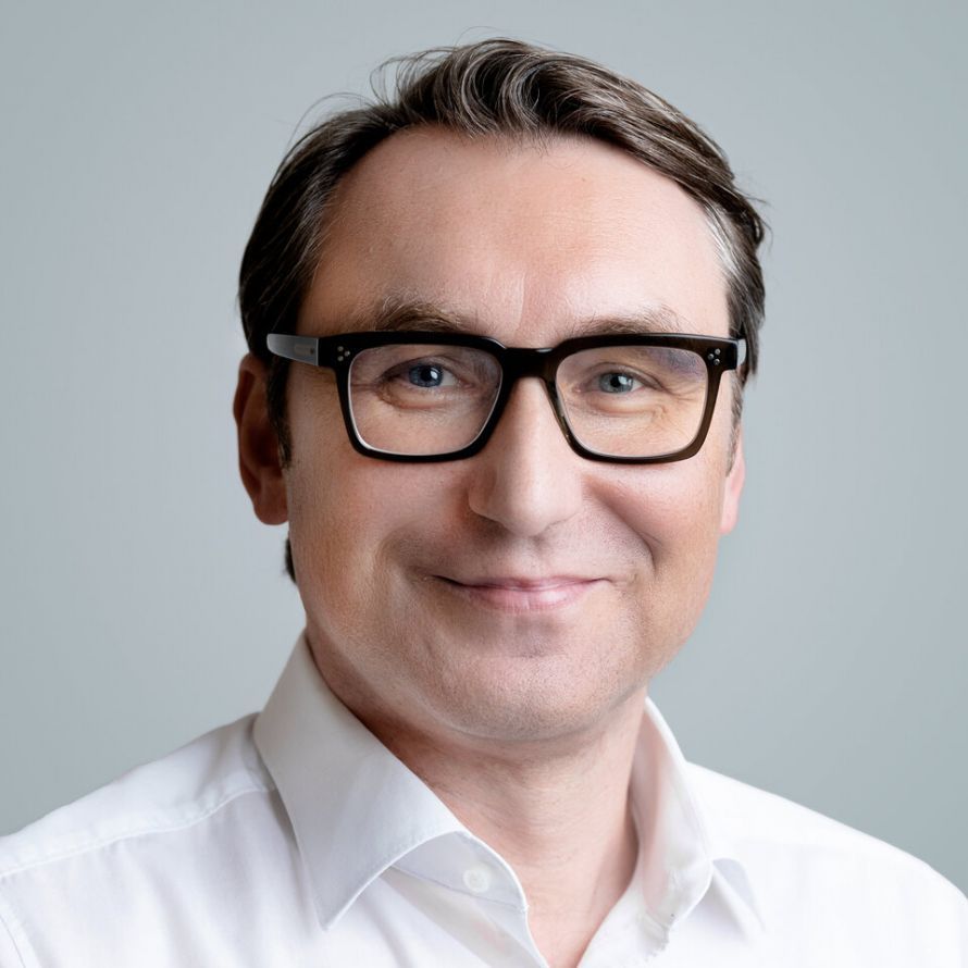
Shoulder arthroplasty has developed in recent years into an established and effective treatment method for advanced joint wear or irreparable shoulder injuries. When conservative measures are no longer sufficient, the implantation of an artificial shoulder joint can significantly improve mobility, alleviate pain, and sustainably enhance the quality of life for those affected. Depending on the findings and the individual’s specific situation, various types of prostheses are used—ranging from anatomical to reverse shoulder prostheses. Modern surgical techniques, careful assessment of indications, and targeted postoperative care ensure stable outcomes and a swift return to daily life.
Severe shoulder pain is usually caused by degenerative, inflammatory, or traumatic changes in the joint or surrounding structures.
“Shoulder pain often occurs not only locally in the shoulder but typically radiates into the upper arm—sometimes as far as the elbow. This pain pattern is initially nonspecific and can be attributed to various causes. Among the most common are osteoarthritis of the shoulder joint, rotator cuff tears, or problems with the biceps tendon. The therapeutic approach depends on the underlying condition, with conservative measures always taking priority. Physical therapy is one of the most important forms of treatment. Its primary purpose is to maintain range of motion, strengthen the muscles, and stabilize shoulder function in the long term. For certain conditions, such as tendon irritation or “calcific tendinitis,” modern procedures such as extracorporeal shock wave therapy are also used. In cases of inflammatory conditions, such as bursitis, a targeted injection under the acromion may be administered as a supplementary treatment—usually with cortisone or an anti-inflammatory medication. “Conservative therapy differs in principle very little from the treatment of other large joints, even though the biomechanical demands on the shoulder joint are particularly complex,” explains PD Dr. Baums at the beginning of our conversation, going on to elaborate further on the phenomenon of calcific tendinitis:
“Calcific tendinitis represents a specific clinical condition. In this condition, calcium crystals are deposited in the area of the rotator cuff tendon insertions. The exact cause of these deposits has not yet been fully clarified. Initially, the calcium deposits often cause no symptoms. It becomes problematic when the deposit grows large enough to cause a mechanical impingement under the acromion. The situation is particularly painful when the calcium material penetrates the bursa beneath the acromion and triggers a severe inflammatory reaction there. Conservative treatment is also the first line of defense for calcific tendinopathy. However, if symptoms persist or recur despite targeted measures, surgical removal of the calcium deposit becomes necessary. In such cases, the procedure is usually performed arthroscopically—that is, using a minimally invasive approach—to minimize trauma to the tissue and shorten recovery time. To diagnose calcific tendinitis, a conventional X-ray or ultrasound examination is often sufficient to visualize the deposits. To accurately assess the extent of the calcium deposit and evaluate the condition of the rotator cuff tendons, magnetic resonance imaging (MRI) is often recommended. This provides a detailed view of the soft tissue structures and aids in treatment decisions—particularly when considering surgical options.”
Only when conservative measures fail to produce sufficient improvement, the pain persists or even worsens, and quality of life is significantly impaired by limitations in daily activities is surgical treatment with a shoulder prosthesis considered.
“The decision on whether a shoulder prosthesis should be considered for a patient is based primarily on the imaging findings and the patient’s clinical presentation. One of the most common reasons for the need for joint replacement is osteoarthritis of the shoulder joint—a degenerative joint condition that can develop over many years, often without a specific trigger. Such degenerative changes are usually evident on conventional X-rays as a marked narrowing of the joint space and bony outgrowths, known as osteophytes. In addition to primary osteoarthritis, there are also secondary causes that can lead to premature joint wear. These include, in particular, previous fractures of the humeral head or recurrent shoulder dislocations. Such pre-existing conditions can cause long-term damage to the joint and increase the risk of developing osteoarthritis. Another important aspect in shoulder joint surgery is the integrity of the rotator cuff. Damage or complete defects in this tendon structure can permanently disrupt the balance and centering of the humeral head within the shoulder joint. In advanced cases, this leads to what is known as defect arthropathy, in which not only is the cartilage worn away, but muscular guidance is also lost. On an X-ray, this is often evident by the fact that the humeral head is no longer centered in the joint but is clearly displaced upward—an indication of decentration due to an insufficient rotator cuff. In such cases, an inverse shoulder prosthesis is often used. This specialized implant design is intended to restore joint function even in the absence of or with a severely damaged rotator cuff by shifting the force transmission to the deltoid muscle. “Imaging therefore plays a central role in distinguishing between conventional osteoarthritis and so-called rotator cuff arthropathy—and thus also in selecting the appropriate type of prosthesis,” said PD Dr. Baums.
The choice between an anatomical and an inverse shoulder prosthesis depends primarily on the structure and function of the rotator cuff, the condition of the bone, the patient’s age, and their level of physical activity.
“With an anatomical shoulder prosthesis, the damaged joint structures are replaced in accordance with the natural anatomy. The humeral head is replaced by a round prosthetic component that corresponds to the original shape. The glenoid cavity—which is normally relatively flat to slightly concave—is also fitted with a matching, form-fitting prosthesis. However, this procedure requires an intact rotator cuff. It plays a central role, as it functions, so to speak, as the ‘motor’ of the shoulder prosthesis and contributes significantly to movement control. If, on the other hand, the rotator cuff is damaged or even extensively torn, an inverse prosthesis becomes necessary. In this procedure, the anatomical structures are reversed: the spherical portion of the prosthesis is attached to the glenoid cavity, while the concave portion is placed on the humeral head. This reversal shifts the biomechanical center of the joint. As a result, the surrounding muscles are under greater tension—a measure that allows for good function even with a damaged rotator cuff. “In this case, other muscles—particularly the deltoid muscle—partially take over the functions of the damaged tendons,” explains PD Dr. Baums, adding:
“The implantation of an inverse prosthesis does not necessarily entail functional limitations; rather, it represents a different form of treatment—with adapted techniques and postoperative care. With an anatomical prosthesis, the rotator cuff must be cut to expose the joint and then carefully reconstructed at the end of the operation, which requires a longer healing phase. Postoperative care for the anatomical variant is therefore often more conservative and characterized by a longer period of immobilization. In contrast, the rehabilitation phase for an inverse prosthesis can usually be structured somewhat more flexibly, since tendon healing does not need to be taken into account. Both procedures must therefore be clearly distinguished from one another—in terms of indications, surgical technique, and postoperative care—and allow for individually tailored therapy depending on the condition of the shoulder structures.”
Surgical techniques play a central role in the management of shoulder joint disorders and significantly determine the approach, the procedure, and postoperative care. The anatomy of the shoulder allows for a relatively minimally invasive approach. There are natural muscle gaps through which the shoulder joint can be reached—particularly via the classic anterior approach. This approach utilizes the body’s existing structure and enables surgery to be performed in a gentle manner.
PD Dr. Baums comments: “However, when implanting an anatomical shoulder prosthesis, it is necessary to open the rotator cuff. This is the only way to fully expose the shoulder joint. This step is essential, as an intact rotator cuff is a prerequisite for the use of an anatomical prosthesis. After surgery, the tendon must be carefully reconstructed and reattached—which has a corresponding impact on the duration of healing. The situation is different with the inverse prosthesis. Here, the rotator cuff is often already severely damaged due to wear and tear or tears. Access to the joint is then usually gained through the existing defects, which simplifies the surgical procedure. Nevertheless, even in this case, the goal remains to work in a way that preserves as much tissue and bone as possible. Both anatomical and inverse prostheses utilize modern implant systems that enable a minimally invasive approach, resulting in minimal bone loss. Instead of traditional stem-anchored prostheses, metaphysically anchored systems are increasingly being used. These sit securely in the upper part of the humerus and preserve valuable bone substance. This is a major advantage, particularly with regard to possible future revision surgeries, as it ensures that more of the patient’s own bone remains available.”
After the surgery, the patient must be a little patient. “The patient’s arm is positioned in a special abduction pillow for six weeks. During this time, the reconstructed tendon heals, though the shoulder may still be moved passively. In addition, the patient receives a personalized post-operative rehabilitation plan, which is implemented in collaboration with the treating physical therapist. This combination of targeted immobilization and controlled mobilization promotes stable healing and ensures functional rehabilitation. Pain following shoulder surgery is generally unpleasant but can be well managed. At our clinic, patients typically receive what is known as a pain block or pain catheter from the anesthesiologists. This specifically numbs the neck and shoulder area, allowing postoperative pain to be well controlled during the first few days. This measure significantly reduces the need for pain medication and makes the particularly painful initial phase—that is, the first two to three days—much more comfortable. After the six-week period during which the shoulder is immobilized in a special splint, the rehabilitation phase begins. This typically lasts two to three weeks and can be conducted on an outpatient or inpatient basis. During this phase, the foundation is laid for further functional recovery. The goal is to improve mobility and gradually increase the shoulder’s capacity for physical activity. However, one should not expect full mobility immediately after surgery. Many patients already have a partially stiffened joint when they come in for surgery—often due to years of wear and tear and avoiding use of the shoulder. These limitations may even worsen initially due to the necessary immobilization during the first few weeks. Therefore, rehabilitation serves merely as the starting point for a longer-term process. It usually takes six to nine months for shoulder mobility to be largely restored. In some cases, full recovery is even possible—for example, for swimming or certain sports. In other cases, function remains somewhat limited—depending on how well the muscles adapt and how long the damage had been present before the procedure,” explains PD Dr. Baums.
Individual recovery depends heavily on overall physical condition—particularly muscle strength and the ability to compensate. Age, of course, plays a certain role here.
“However, what is often more decisive is how long the pre-existing damage has been present. A patient who has been living with limited shoulder mobility for ten years will take significantly longer to recover than someone with a problem that has only recently developed. As a general rule, we always work with the patient to find a personalized solution. Age alone is not a criterion for excluding a shoulder replacement. A good example is a 92-year-old female patient I operated on who, at 97, is still doing well with her prosthesis. While such cases are exceptions, they demonstrate that surgical treatment can be successful even in old age. Most patients, however, are between the ages of 60 and 70. More important than age are comorbidities—such as heart or lung conditions—as well as the patient’s subjective level of suffering. If all conservative treatment options have been exhausted and quality of life is severely impaired, a prosthesis is considered. In cases where conservative measures have not yet been attempted, this approach should be chosen first. Only when this has been exhausted is a decision made on a case-by-case basis regarding surgical treatment,” states PD Dr. Baums.
Like any major surgery, shoulder arthroplasty carries specific risks and complications that affect both the procedure itself and the postoperative period. The most common problems include infections, prosthesis loosening, dislocations, fractures, nerve injuries, and soft tissue issues.
“As with any surgery, there are certain risks associated with shoulder replacement. A key concern is the risk of infection—as with all artificial joints. Such an infection can occur not only immediately after the procedure but also in the long term and, in the worst-case scenario, lead to prosthesis loosening. Another specific risk compared to hip or knee replacements is the possibility of shoulder joint instability, which can cause the prosthesis to dislocate. Fortunately, this problem occurs only rarely, but it remains a distinctive feature of shoulder arthroplasty. In patients whose rotator cuff is intact at the outset, a tendon tear may occur over the years. In such cases, it may become necessary to convert an originally implanted anatomical prosthesis into an inverse prosthesis. This conversion is generally possible and represents a proven option when the condition of the soft tissues deteriorates over time. In terms of durability, it can be noted that shoulder prostheses have, on average, a lifespan comparable to that of hip or knee prostheses. Current registry data indicate an average lifespan of about 15 years. Of course, there are individual variations. Some prostheses last significantly longer, while others—particularly in the presence of risk factors such as diabetes mellitus or rheumatic diseases—must be replaced sooner. However, such early loosening has become rather rare. The materials used have improved significantly in recent years, so that today the majority of shoulder prostheses can be implanted cementlessly. Cemented fixation is now chosen only in exceptional cases—for example, in cases of osteoporosis or complex bone fractures where stable bony anchorage cannot be ensured. Advances in materials technology and surgical techniques thus play a key role in minimizing the risk of complications and increasing the longevity of the implants.”
St. Elisabeth Hospital in Dorsten is a highly specialized center for orthopedic and sports traumatology care. Characterized by state-of-the-art minimally invasive techniques and 3D planning, it focuses particularly on shoulder surgery and shoulder arthroplasty. Thanks to close collaboration between surgeons, physical therapists, and rehabilitation experts, patients benefit from individually tailored treatment plans that facilitate a quick return to movement and daily life.
“We perform approximately 80 shoulder replacement surgeries annually at our hospital. That is a remarkable number and attests to our high level of specialization and experience in this field. A key aspect of our work is precise and state-of-the-art surgical preparation. Before each procedure, we conduct software-assisted, three-dimensional planning. This is based on a prior computed tomography (CT) scan, which allows us to accurately map the bony anatomy. This allows us not only to precisely plan the position of the prosthesis in advance, but also to identify potential bone defects and take appropriate corrective measures into account. This three-dimensional planning also includes a simulation of the patient’s future range of motion. Although actual postoperative mobility depends largely on the condition of the soft tissues—that is, the muscles and tendons—this virtual preparation still allows for a very accurate assessment of the expected functional outcome. The use of virtual reality goggles during surgery is particularly innovative. This allows the pre-planned prosthesis to be virtually projected into the surgical field during the procedure. This facilitates the precise implementation of the plan and significantly improves the positioning of the implant components. Our organizational workflow is also well-structured. Rehabilitation measures are coordinated in advance of the surgery, ensuring a seamless transition to postoperative care for patients following their hospital stay. “In this way, we create optimal conditions for a safe surgery and a successful recovery,” explains PD Dr. Baums, and with that, we conclude our conversation.
Thank you very much, Priv.-Doz. Dr. Baums, for the valuable insight into your work!
Share this article
Expert Interviews
Read next
- Expert interview
Expert interview with Professor Dr Elmar Herbst, PhD
Oct 15, 2026
Patient-centred and personalised knee surgery
Read more - Expert interview
Cardiogeriatrics: Significance and benefits in the context of hospital reform
Sep 21, 2026
Cardiogeriatrics: Significance and benefits in the context of hospital reform
Read more - Expert interview
Expert Interview with Professor Dr. Amadeus Hornemann, MPH
Sep 18, 2026
Prof. Amadeus Hornemann on Precision Medicine in Gynecology: Modern Surgery, New Techniques, Better Quality of Life
Read more