
Expert Interviews
Expert Interview with PD Dr. Ralf Hempelmann on Spinal Canal Stenosis

Alexandra Pfitzmann ·
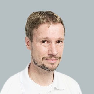
Priv.-Doz. Dr. med. Ralf Hempelmann has been a Senior Physician in Neurosurgery and Spinal Surgery at the Helios ENDO Clinic in Hamburg since 2010 and has experience in the treatment of spinal disorders. The department, which he heads together with Dr. med. Alexander Richter, a specialist in orthopedics and trauma surgery, specializes in spinal surgical procedures.
Dr. Hempelmann holds certifications in spinal neurosurgery from the German Society for Neurosurgery and a Master’s Certificate from the German Spine Society, which attest to his high level of qualification in spinal medicine. In total, he has performed more than 5,000 surgical procedures throughout his career. His work includes, among other things, disc surgeries, surgical decompression to relieve pressure on the spinal canal, stabilization and correction of spinal misalignments and segmental instabilities, the removal of tumors in the spinal canal, and peripheral nerve decompression.
In an interview with Dr. Hempelmann, the editorial team of the Leading Medicine Guide focused on spinal canal stenosis.
Spinal canal stenosis—a term that may seem unfamiliar to many at first, but which is associated with significant limitations in daily life for those affected. This narrowing of the spinal canal can lead to severe pain, numbness, and even restricted movement, all of which have a lasting impact on quality of life. What causes this narrowing? Why are some people particularly affected? And above all: What treatment options are available to improve quality of life and regain mobility?
Unlike other back conditions, which usually affect only specific nerve roots, spinal stenosis involves a structural narrowing of the entire spinal canal. This often affects multiple segments of the spine, which explains the wide range and variability of symptoms.
“In the following, we will discuss spinal stenosis of the lumbar spine: Symptoms that occur while walking are very typical of spinal stenosis, that is, the narrowing of the lumbar spinal canal. Pain often begins in the lumbar spine and radiates from there into one or both legs. The symptoms may be accompanied by sensory disturbances in the legs and, in rare cases of severe stenosis, also by temporary weakness and impaired coordination in the legs. The symptoms may also be unilateral. Typically, the symptoms subside when the person pauses, sits down, or bends forward briefly, as this causes the spinal canal to widen slightly. This intermittent need to stop is referred to as claudication. As a result, many patients can ride a bicycle for kilometers without difficulty, while they are often only able to walk a few dozen meters. The symptoms overlap with those of other spinal conditions; however, many other conditions can also cause severe pain at rest, such as certain herniated discs, spinal instability, or inflammation. Such pain at rest is rather atypical in lumbar spinal canal stenosis. Claudication caused by spinal stenosis—that is, spinal claudication— differs from intermittent claudication—which is caused by arterial circulatory disorders—in several ways, including the fact that the walking distance in spinal claudication can vary, whereas in intermittent claudication it is often quite consistent, limited to the same distance each time. “In addition, patients with circulatory disorders also experience symptoms during activities such as cycling,” says PD Dr. Hempelmann, describing the various symptoms.
The most reliable diagnosis of spinal canal stenosis requires a combination of a thorough clinical examination, imaging studies, and a detailed medical history. The goal is to confirm the narrowing of the spinal canal, determine the severity of the condition, and rule out other possible causes of the symptoms.
Regarding the diagnostic process, PD Dr. Hempelmann explains: “The most important factor is the patient’s symptoms—that is, taking a thorough medical history. No or minimal pain at rest, discomfort in the back and legs when standing for long periods or walking, and, conversely, no or minimal pain when riding a bicycle are indicative of the condition. A thorough clinical-neurological examination is then, of course, necessary. Often, the clinical findings are quite unremarkable, as patients have hardly any symptoms at rest. Many patients lack deep tendon reflexes. This examination is important for differential diagnosis (distinguishing the condition from other orthopedic, neurological, or internal medical conditions). The diagnostic imaging method of choice is magnetic resonance imaging (MRI). If this is not possible, a CT scan (computed tomography) would have to be used instead. Further measures to narrow down the diagnosis, such as local injections, are usually not necessary when there is a clear medical history and MRI findings.”
For an MRI, the patient lies relaxed inside a tube while magnetic fields generate detailed images. The examination takes about 20–40 minutes and requires no special preparation.
For a CT scan, the patient lies on a table that moves through the scanner. If a myelogram is necessary, a contrast agent is injected into the spinal canal beforehand, which is usually done under local anesthesia.
Conservative therapy is the first-line treatment for mild to moderate spinal stenosis and aims to relieve symptoms, improve mobility, and enhance quality of life.
The decision regarding which measures are most appropriate depends on the individual’s symptoms and the severity of the condition. Surgery should be considered when conservative methods have been exhausted and symptoms remain severe or worsen. Regarding this, PD Dr. Hempelmann states: “Physical therapy is effective against back pain and also against radiating pain, especially and this applies to many spinal conditions, strengthening the entire core musculature—and I am a strong advocate of isometric exercises because they do not place any strain on the spinal structures. Of course, physical therapy does not widen the spinal canal, but well-trained muscles alleviate pain for various reasons. And muscular pain is practically always a major component of back pain. And if surgery does eventually become necessary—because conservative therapy may not lead to lasting improvement—well-trained muscles are also a key factor in a favorable post-operative recovery. So the effort would not have been in vain. In general, when it comes to back pain in older adults—and this also applies to pain caused by stenosis—physical therapy must consist primarily of active exercises, with muscle training taking priority. Passive measures such as massage, heat, and others are beneficial and pleasant only as long as they are being applied. However, active exercises are the most sustainable and effective approach. - Medication can be used to support conservative treatment for a period of time, in which case common pain medications such as anti-inflammatory drugs—including ibuprofen or diclofenac—would be prescribed. If anti-inflammatory drugs are contraindicated—for example, in patients with cardiovascular conditions—metamizole can be used as an alternative.”
Imaging techniques such as magnetic resonance imaging (MRI) and computed tomography (CT) play a central role in the diagnosis and treatment planning for spinal stenosis.
MRI provides detailed insights into soft tissues and nerve structures, while CT excellently visualizes bony changes.
“MRI images are very helpful in precisely determining the location and extent of the stenosis. In certain situations—for example, when deciding how to proceed surgically in cases of additional segmental instability—additional CT scans may be indicated. Standard X-ray imaging in the standing position remains indicated as well. The patient’s medical history and clinical examination are then crucial for the surgical decision. This is because, in cases where multiple segments show significant degenerative changes, often only one segment is the primary cause of the pain. This can usually be determined through a thorough interview and examination of the patient. In such cases, only this segment should be operated on to keep the surgical intervention as minimal as possible. One should never perform more surgery than is necessary. “There is almost no indication for prophylactic—that is, preventive—surgery in cases of degenerative spinal disorders,” says PD Dr. Hempelmann.
Open microsurgery is the gold standard for spinal stenosis.
PD Dr. Hempelmann explains the differences between the surgical approaches: “The standard procedure for spinal canal stenosis is microscope-assisted decompression surgery, i.e., microsurgery. This involves making small incisions; for a single-segment procedure, the incision is about 3–4 cm long. This is currently the gold standard against which other surgical procedures must be measured. It is also possible to perform surgery percutaneously—that is, using small tubes or cannulas—which only require slight expansion of the surrounding muscles and thus result in less muscle damage; this also applies to endoscopic surgeries, which are another option for treating stenosis. Especially in the early postoperative phase, these minimally invasive procedures offer the advantage of requiring, on average, less pain medication and a slightly shorter hospital stay. To my knowledge, there is currently no evidence that these procedures offer a significant long-term advantage over open microsurgical procedures. They also have disadvantages, namely a lengthy learning curve for the surgeons. Overall, both microsurgical and minimally invasive procedures are equally recognized and are practically comparable in terms of effectiveness, which is why both approaches comply with the principles of medical practice.”
Spinal stenosis is a degenerative disease primarily influenced by our genetic predisposition and the natural aging process. Although these factors cannot be changed, acceptance of the disease plays a decisive role in how it is managed.
“So far, there’s nothing we can do about our genes or the aging process, and from my personal perspective, that’s a good thing. This condition is essentially a matter of fate, and therefore most patients can accept it as such and cope with it. This is because placing blame—not only on others but also on oneself, for example, regarding lifestyle choices—leads to a poorer recovery. Occupation and lifestyle play a minor role, if any at all. We have stenosis patients from all occupational groups and with a wide variety of lifestyles. Since degenerative disc disease also plays a role in the development of spinal stenosis, quitting smoking is recommended, as smoking and other causes of atherosclerosis are factors that accelerate disc degeneration. “In general, the best prevention for back pain is physical activity and building muscle strength. This applies both to the initial onset of back pain and to the prevention of relapses,” explains PD Dr. Hempelmann, and with that, we conclude our conversation.
Thank you very much, Dr. Hempelmann, for this informative insight into the management of spinal canal stenosis!
Share this article
About the medical author
Alexandra Pfitzmann
Editor
Alexandra Pfitzmann – medical author: expert knowledge, professional articles and medical insights in the Leading Medicine Guide.
More about the medical author →Expert Interviews
Read next
- Expert interview
Cardiogeriatrics: Significance and benefits in the context of hospital reform
Sep 21, 2026
Cardiogeriatrics: Significance and benefits in the context of hospital reform
Read more - Expert interview
Expert Interview with Professor Dr. Amadeus Hornemann, MPH
Sep 18, 2026
Prof. Amadeus Hornemann on Precision Medicine in Gynecology: Modern Surgery, New Techniques, Better Quality of Life
Read more - Expert interview
Expert Interview with Professor Dr. Konstantinos Anagnostakos
Sep 15, 2026
Prof. Anagnostakos on Painful Knee Replacements
Read more
