Expert Interviews
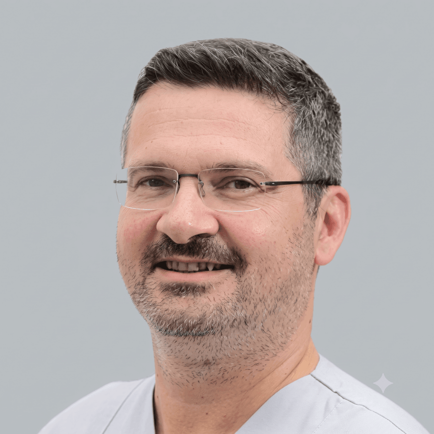
Prof. Anagnostakos on Painful Knee Replacements

Alexandra Pfitzmann · September 15, 2026
A painful knee replacement is a major burden for many patients—especially when their expectations for pain relief and mobility after surgery are not met. Pain can occur soon after the procedure or develop months or even years later. There are a wide variety of underlying causes: from mechanical problems such as a loosened or misaligned prosthesis to inflammation, infections, or issues in the surrounding soft tissue.
This is precisely why precise diagnostics and an experienced specialist like Prof. Dr. Konstantinos Anagnostakos are needed—someone who can assess the situation holistically and distinguish between harmless, temporary pain and complications that require treatment. The goal is always to clearly identify the cause of the symptoms and select a treatment that restores stable function, freedom from pain, and safe mobility.
“Pain following knee replacement is a very broad term. Many patients simply say they have ‘post-operative pain’—regardless of whether it has persisted for three months, one year, or ten years. Since they lack medical expertise, the exact cause cannot be determined from their description alone. Only diagnostic testing can clarify what type of symptoms are actually present. The term ‘painful knee replacement’ encompasses various causes: prosthesis loosening, instability, infections, and pain resulting from the progression of osteoarthritis in the parts of the joint that were not replaced.
The type of prosthesis implanted is a key factor. With partial prostheses (slide prostheses), osteoarthritis can continue to develop in the remaining compartments. With bicompartmental or tricompartmental prostheses, this affects other structures. Pain can therefore be directly related to the prosthesis or result from progressive osteoarthritis in the remaining joint compartments.
“The diagnostic process must distinguish whether the problem is caused by osteoarthritis or is prosthesis-related. Only then can the appropriate diagnostic approach and the correct treatment be determined,” explains Prof. Dr. Anagnostakos, adding:
“Pain following knee replacement can manifest as either weight-bearing pain or pain at rest—depending on the underlying cause. Processes such as the progression of osteoarthritis in unreplaced joint compartments or prosthesis loosening usually develop gradually. Symptoms increase over weeks, months, or years: initially pain on weight-bearing, and later—once a certain threshold is exceeded—also pain at rest. Acute causes follow a different pattern.
An infection can occur suddenly after a period of minimal symptoms. Instability also becomes noticeable early on. However, patients cannot directly identify instability; they merely describe the consequences, such as difficulty climbing stairs. These symptoms arise because certain ligaments—the anterior or posterior cruciate ligaments or the collateral ligaments—are replaced by the prosthesis, and the prosthesis’s degree of stabilization may not be sufficient to ensure the joint’s natural stability.
This places excessive strain on the anterior portion of the joint, which can be mistakenly interpreted as a problem with the kneecap, even though the cause lies in the instability. The challenge lies in correctly interpreting the patient’s subjective descriptions: whether the symptoms began suddenly or gradually, whether they appeared immediately after surgery or only later, and how they have changed over time. Only in combination with imaging diagnostics can the exact cause be determined and a distinction made between problems caused by osteoarthritis and those caused by the prosthesis.”

A multi-step, systematic approach is necessary to distinguish between mechanical problems, infections, and soft-tissue-related causes of a painful knee prosthesis. Each of these causes exhibits its own distinct pattern—in the clinical examination, in imaging, and in laboratory test results.
“The medical history is followed by a physical examination before imaging techniques are used. First, we verify whether the reported symptoms correspond to the clinical findings. If instability is suspected, the knee is tested in extension as well as at 30°, 60°, and 90° flexion, as instability can occur in different joint positions depending on the cause. Malalignment, rotational abnormalities, or an imbalance between the extension and flexion gaps may become apparent in certain positions. The stability of the patella is also assessed.
Further clues come from mechanical abnormalities such as crepitus, which may indicate osteoarthritis in compartments that have not been replaced or wear on the inlay. Changes in the leg axis—such as a shift to an X- or O-shape—may indicate loosening of the prosthesis and compromise stability. All this information is used to formulate an initial provisional diagnosis. Only then is a decision made regarding which imaging studies are necessary. The standard procedure is an X-ray, ideally compared with previous images, to detect changes in the prosthesis’s position.
If instability is suspected, positional X-rays can help assess the relationship between the joint surfaces. A full-leg X-ray shows the entire leg axis. “If bone defects beneath the prosthesis are suspected, a CT scan is performed,” explains Prof. Dr. Anagnostakos.
If an infection involving soft tissues is suspected, an MRI may be necessary to assess the extent of possible abscess formation. If multiple causes are possible or a distinction must be made between aseptic and septic loosening, a three-phase bone scan may be useful.
Prof. Dr. Anagnostakos comments: “Early-stage infections usually show no imaging changes, whereas late-stage infections can become visible both around the prosthesis and on the bone surface. Scintigraphy reveals contrast agent uptake in affected areas and allows for an indirect distinction between bacterial infections and nonbacterial inflammatory changes such as rheumatism or nonspecific synovitis. A structured diagnostic algorithm is crucial: The combination of medical history, clinical examination, and specifically selected imaging makes it possible to identify the exact cause of the symptoms.”
Typical signs of a loosened knee prosthesis manifest as a clear pattern involving the course of pain, response to weight-bearing, range of motion, and imaging findings. Loosening results either from mechanical wear or from an infection—both forms exhibit characteristic signs that can be clearly distinguished from one another diagnostically.
“If an infection is detected in a knee prosthesis, it is usually treated surgically, as untreated infections become chronic and can cause local symptoms such as pain and swelling, as well as spread through the bloodstream to other parts of the body. Conservative therapy is only considered if the patient cannot undergo surgery due to severe comorbidities. In such cases, long-term antibiotic therapy is used as suppression to prevent further damage without completely eliminating the infection. Prophylactic measures such as antibiotic coatings are used only in specific situations, such as with tumor prostheses featuring a silver coating or with cemented prostheses using antibiotic-impregnated bone cement.
These reduce the risk of infection but do not provide complete protection. In cases of surgical treatment, the procedure is determined preoperatively. Early infections occurring within the first few days after surgery can usually be treated in a way that preserves the prosthesis, since the pathogens have not yet become deeply entrenched. This involves removing infected soft tissues, irrigating the prosthesis, replacing interchangeable components such as the inlay, and initiating antibiotic therapy. Success rates range from about 70–80 %, depending on the study. “This prosthesis-preserving treatment is possible up to approximately three weeks after the onset of symptoms,” says Prof. Dr. Anagnostakos, adding:
“If the infection has persisted for months or years, shows changes on imaging, or has already caused loosening, it is no longer possible to preserve the prosthesis. In that case, the prosthesis must be removed. There are two procedures available for this: a one-stage and a two-stage procedure. In the one-stage procedure, the infected prosthesis is removed and a new one is implanted during the same surgery.
In the two-stage procedure, the prosthesis is first removed and a spacer containing antibiotics and made of bone cement is inserted. Once the infection has subsided, the final prosthesis is reimplanted in a second surgery. The decision between a one-stage and a two-stage procedure depends on several factors: the surgical team’s experience, the type of pathogen, the patient’s general condition, the patient’s ability to tolerate one or two anesthetic procedures, and the condition of the soft tissues. Chronic fistulas generally call for a two-stage procedure.
Internationally, the two-stage procedure is considered the gold standard, while the one-stage procedure is routinely and successfully performed only in specialized centers with many years of experience—such as the Endo-Klinik Hamburg.”
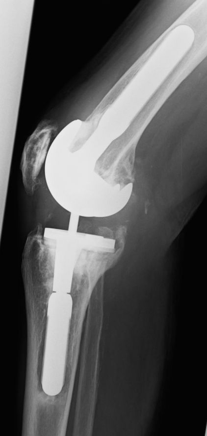
Photo: Loosening of a partially-coupled prosthesis
Increasing load-dependent pain, a feeling of instability, swelling or signs of inflammation, abnormal laboratory values, and radiological signs of loosening indicate that a knee prosthesis is no longer securely anchored. In such cases, revision surgery is often necessary to restore joint function and prevent further damage.
Revision surgery on a painful knee prosthesis is among the most demanding procedures in joint surgery. The challenges lie both in the technical execution and in strategic planning, because every revision presents unique anatomical, mechanical, and infectious disease-related characteristics. Centralization in endoprosthetics is an important aspect because patients benefit from a high level of specialization during complex procedures—especially revision surgeries.
Prof. Dr. Anagnostakos comments: “Primary surgeries are performed in large numbers at many hospitals, but revision surgeries pose significantly greater challenges. They often require additional specialties such as intensive care medicine, internal medicine, microbiology, or infectious diseases. As a result, some hospitals cannot offer revision surgeries despite having a high level of expertise in primary joint replacement. This does not mean that these hospitals are subpar, but rather that the demands of revision surgery require a different organizational structure.
Certified joint replacement centers provide a point of reference, but certification alone says nothing about how many revision surgeries are actually performed. The minimum numbers for certification are clearly defined, but there is no upper limit. Two hospitals may each perform 1,000 knee surgeries per year, but one has 950 primary procedures and 50 revision surgeries, while the other has 700 primary procedures and 300 revision surgeries. Both are considered maximum-care joint replacement centers, yet the second clinic has significantly more experience with revision surgeries.
These differences are not transparent to patients. That is why it is important to inquire individually—either directly at the hospital or through private orthopedic surgeons and primary care physicians—which facility is suitable for a revision surgery. The number of cases varies greatly between hospitals and federal states. The Nardini Clinic performs a comparatively high number of revision surgeries. According to EndoCert guidelines, a maximum-care center should perform at least 50 revision surgeries per year. This figure was already reached there by mid-year, which underscores the high level of specialization in the field of revision arthroplasty,” he adds:
“Patients come from both the immediate vicinity and from farther away. Some travel specifically to the hospital because they have chosen a specialized clinic based on their own research. Others come from regions where the treating physician previously practiced and are willing to travel longer distances of 80 to 100 kilometers to do so. For complex procedures such as revision surgeries, this effort is worthwhile, as proper treatment is crucial for the long-term outcome.
Today, joint replacements have a long lifespan of about 20 years or more. Therefore, it makes sense for many patients to travel a greater distance for a short period of treatment if this ensures the long-term functionality of the prosthesis. Past examples show that patients deliberately choose hospitals with a particularly good reputation and thus achieve excellent results.
From the perspective of healthcare providers as well, it makes sense that, if they were to undergo surgery themselves, they would want to be thoroughly informed about the healthcare landscape. The choice of clinic depends not only on one’s own practice but also on the general health policy situation and regional structure.”
Technically, the removal of an old prosthesis is one of the most difficult steps. Components are often firmly fused to the bone or securely anchored with cement. The implant must be removed completely and in a way that minimizes tissue damage, without further damaging the already compromised bone. The situation is particularly challenging when loosening or an infection is already present, as the bone structures are often weakened, thinned, or irregular in such cases.
The healing process following knee replacement revision surgery is significantly more complex than after a primary implantation, because the tissue is already damaged, bone may be missing, and the stability of the joint must be rebuilt. Nevertheless, recovery follows a clear pattern divided into several phases.
“Aseptic revisions—that is, replacements without infection—usually proceed similarly to primary surgeries: Modern implants allow for immediate full weight-bearing, and mobilization begins right after surgery. Nevertheless, the healing process is often somewhat slower, as the soft tissues are subjected to stress once again and patients undergoing revision surgery are usually older than those undergoing primary implantation. As a rule, they regain good mobility after six to twelve weeks, walk without assistive devices, and report a significant improvement in their quality of life.
It is important to have realistic expectations: A replacement surgery restores function but does not result in “rejuvenation.” “Very strenuous activities—such as long mountain hikes—are possible, but they are physically demanding and come with corresponding muscular and fitness limitations,” says Prof. Dr. Anagnostakos, concluding our conversation:
“In the case of septic revision surgeries, particularly with a two-stage procedure, the recovery process is significantly longer. Depending on the type of bacteria and the patient’s overall condition, there is a two- to eight-week interval between the removal of the infected prosthesis and the reimplantation of the new prosthesis, during which an interim prosthesis made of antibiotic-impregnated bone cement is used.
It often takes three to four months to fully recover after the second surgery. These longer interim phases and the overall more complex course of treatment must be discussed in detail as part of the individual consultation so that patients know what challenges and timeframes they can expect.”
- Specialist in hip and knee arthroplasty — extensive experience in the implantation and revision of hip and knee prostheses
- Expert in revision surgery — treatment of loosened, infected, or defective prostheses, including complex replacement surgeries
- Chief of Joint Surgery & Sports Traumatology, Nardini Klinikum Zweibrücken — Director of a certified Endoprosthetics Center with the highest quality standards
- Minimally invasive and navigation-assisted techniques — precise prosthesis placement, tissue-sparing procedures, faster rehabilitation
- Broad surgical spectrum — modern endoprosthetics, arthroscopic procedures on the knee, shoulder, and ankle, treatment of acute and chronic sports injuries
- International Recognition — in-depth training, scientific expertise, establishment of a center of excellence for hip replacement arthroplasty
- Focus on mobility and quality of life — precise planning, modern technology, and extensive surgical experience for optimal results in cases of painful knee or hip prostheses
Share this article
About the medical author
Alexandra Pfitzmann
Editor
Alexandra Pfitzmann – medical author: expert knowledge, professional articles and medical insights in the Leading Medicine Guide.
More about the medical author →Expert Interviews
Read next
- Expert Interviews
Expert Interview with Professor Karl Philipp Kutzner, M.D.
Sep 11, 2026
Prof. Kutzner on Cutting-Edge Endoprosthetics at ENDOPROTHETICUM Mainz
Read more - Expert Interviews
Expert Interview with Dr. Emanuel Stutz, M.D.
Sep 9, 2026
Hyperthermia Treatment for Cancer
Read more - Expert Interviews
Expert Interview with Dr. Moustafa Elshafei, FACS
Sep 7, 2026
Focus on Colorectal Cancer: Early Detection, Better Treatment—On Opportunities and Modern Medicine
Read more