Spinal canal stenosis—also known as spinal canal narrowing—is a condition of the spine in which the bony canal through which the spinal cord and nerve roots pass becomes narrowed. This narrowing of the spinal canal puts pressure on the nerves, which can cause back pain and leg pain when walking. Lumbar spinal stenosis occurs particularly frequently in the lumbar spine (LS), while the cervical form is less common in the cervical spine (CS).
Symptoms usually develop gradually and worsen with age, as degenerative changes such as herniated discs, osteoarthritis, or thickening of the ligamentous structures can narrow the spinal canal.
Depending on the severity of the condition, various treatment options are available: ranging from conservative therapy with physical therapy and medication to surgical decompression. An early diagnosis of spinal stenosis helps maintain quality of life and prevent potential neurological deficits.
Definition: What is spinal stenosis?
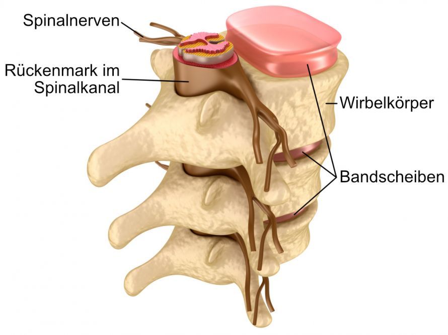
The spinal canal is the bony canal within the spine that protects the spinal cord and nerve roots. It is formed by the interaction of the individual vertebral bodies, their arches, and ligaments. The spinal nerves branch off from here to supply the muscles and skin.
Spinal stenosis occurs when this canal narrows, causing pressure on the spinal cord or the nerve roots. This narrowing can occur in various sections of the spine—most commonly in the lumbar spine (lumbar spinal stenosis) and less commonly in the cervical spine (cervical spinal stenosis).
Typically, the narrowing does not affect the entire spinal canal, but only individual segments. This leads to reduced nerve supply and symptoms that are particularly noticeable when walking, standing, or maintaining an upright posture for extended periods.
Spinal canal stenosis can have the following effects:
- A feeling of pressure or pain in the back
- Numbness or tingling in the legs or arms
- Weakness in the muscles
- Increasing limitation in walking distance
The condition primarily affects people over the age of 60 and is one of the most common causes of chronic back pain in older adults. Degenerative changes, an age-related decrease in intervertebral disc height, or thickening of the ligamentous structures within the spinal canal contribute significantly to its development.
Primary and Secondary Spinal Canal Stenosis – Differences
Spinal canal stenosis is generally classified into two forms: the primary (congenital) and the secondary (acquired) form. Both lead to a narrowing of the spinal canal but differ in their cause.
Primary Spinal Canal Stenosis
This form is congenital and occurs relatively rarely. It is usually caused by a genetically determined, abnormally narrow spinal canal. Affected individuals have a narrower spinal canal from an early age.
Symptoms often do not appear until later in life, when additional degenerative changes such as disc degeneration or osteoarthritis occur.
Typical characteristics:
- Congenital, structural narrowing of the spinal canal
- Symptoms often do not appear until adulthood
- symptoms are usually symmetrical
Secondary spinal canal stenosis
The secondary or acquired form is significantly more common. It develops over the course of a person’s life due to signs of wear and tear on the spine. Common causes include:
- age-related flattening of the intervertebral discs
- Formation of bone spurs (osteophytes) on the vertebral bodies
- Thickening of the ligamentous structures (ligamentum flavum)
- Osteoarthritis of the small vertebral joints (spondylarthrosis)
- spondylolisthesis or previous surgeries
These changes cause the spinal canal to narrow progressively, exerting pressure on the nerve roots or the spinal cord. Lumbar spinal stenosis is particularly common in the lumbar spine, as it bears the majority of the body’s weight.
The secondary form is one of the typical degenerative diseases of old age and primarily affects people over the age of 60.
Causes of Spinal Canal Stenosis
Causes: Degeneration, Herniated Discs, and Poor Posture
The causes of spinal stenosis are varied and depend on whether it is a congenital (primary) or acquired (secondary) form. Most commonly, the narrowing of the spinal canal results from degenerative changes in the spine that increase with age.
The most common causes of acquired spinal canal stenosis include:
- Intervertebral disc damage: Due to age-related wear and tear, intervertebral discs lose height and elasticity. This can lead to disc bulging or a herniated disc, which further narrows the spinal canal.
- Thickening of ligamentous structures: The so-called ligamentum flavum, an elastic ligament within the spine, thickens over the years and presses on the spinal cord or nerve roots.
- Bone spurs (osteophytes): Arthritic changes in the small vertebral joints (facet joints) can lead to bony outgrowths that narrow the spinal canal.
- Spondylolisthesis: Due to instability, a vertebral body can slip forward and exert pressure on the spinal canal.
- Postural abnormalities and degenerative scoliosis: A lateral curvature of the spine alters the distribution of stress and asymmetrically narrows the spinal canal.
- Scar tissue following surgery: Previous spinal surgeries can lead to adhesions that contribute to secondary spinal canal stenosis.
As the intervertebral discs lose their height and cushioning effect due to wear and tear, the vertebral bodies tend to ossify. The video shows an example of disc degeneration and its consequences:
Congenital abnormalities, such as an overly narrow vertebral arch or a short pedicle, can also cause primary spinal canal stenosis at a young age.
The lumbar spine is particularly commonly affected, as it bears the weight of the upper body and is therefore especially susceptible to degenerative changes.
The symptoms of spinal stenosis depend on which section of the spine is affected and how severe the narrowing is. In most cases, symptoms begin gradually and worsen over months or years.
Typically, the pain worsens when walking or standing and improves when sitting or bending forward. This phenomenon is called claudicatio spinalis—leg weakness and pain that depends on the distance walked, resulting from nerve compression.
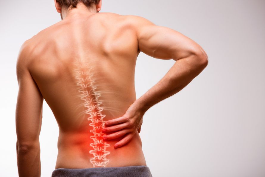
Common Symptoms of Lumbar Spinal Canal Stenosis (LWS)
Lumbar spinal stenosis is the most common form of the condition. The narrowing of the spinal canal in the lumbar spine leads to compression of the nerve roots that supply the legs.
Typical symptoms:
- Back pain that radiates into the buttocks, thighs, or calves
- Leg pain or tingling when walking or standing
- Numbness or weakness in the legs
- Improvement when sitting or leaning forward (e.g., over a shopping cart)
- Reduced walking distance, as pain increases after a few minutes
- In severe cases: unsteadiness while walking or occasional urinary dysfunction
These symptoms result from pressure on the nerve roots in the lumbar spine, which impairs signal transmission to the legs.
Symptoms of cervical spinal stenosis (cervical spine)
In cervical spinal stenosis—that is, a narrowing of the spinal canal in the cervical spine—other symptoms are more prominent, as the spinal cord is directly affected here.
Possible signs:
- Neck pain radiating into the shoulders and arms
- Tingling, numbness, or a sensation of coldness in the hands and fingers
- Coordination problems when walking (unsteady gait)
- Weakness in the arms or legs
- In advanced cases: myelopathy with paralysis and impaired fine motor skills
The cervical form is considered potentially more dangerous because it exerts pressure on the spinal cord itself—which, if left untreated, can cause permanent neurological damage.
General accompanying symptoms and complications
Regardless of the location of the narrowing, additional symptoms may occur that are attributable to the restricted nerve supply:
- Muscle tension along the spine
- Unsteady standing or walking during prolonged activity
- Numbness or cramps in the legs
- Chronic back pain of varying intensity
In advanced stages, spinal stenosis can lead to neurological deficits. These include gait disturbances, sensory disturbances, and, rarely, incontinence. Early diagnosis and appropriate treatment are therefore crucial to prevent permanent damage.
Diagnosis of Spinal Canal Stenosis
The diagnosis of spinal stenosis involves several steps and aims to determine the exact location, cause, and extent of the narrowing. The goal is to distinguish between primary and secondary forms and to rule out other causes of back pain.
Physical Examination and Medical History
The process begins with a detailed medical history. The doctor asks the patient about
- the nature, duration, and intensity of the back pain,
- limitations when walking or standing,
- neurological symptoms such as numbness or weakness.
This is followed by a physical examination to assess:
- spinal mobility
- Muscle strength and reflexes in the arms and legs
- Sensory disturbances along the nerve pathways
Based on these findings, it is often possible to determine whether the patient has lumbar spinal stenosis or cervical myelopathy (pressure on the spinal cord in the cervical spine).
Imaging procedures (MRI, CT, X-ray)
Imaging techniques are used to confirm the diagnosis.
- Magnetic resonance imaging (MRI): The most important method for visualizing the spinal cord, nerve roots, and soft tissue structures. It shows exactly how severely the spinal canal is narrowed and which structures are causing the pressure.
- Computed tomography (CT): Provides detailed images of the bones and can be particularly helpful in cases of bony narrowing of the spinal canal.
- X-rays: These reveal misalignments, spondylolisthesis, or degenerative changes in the spine.
In some cases, a myelogram (contrast-enhanced examination of the spinal cord) is performed to assess the flow of cerebrospinal fluid.
Neurological Tests and Functional Diagnostics
Supplementary tests are used to assess nerve function:
- Electromyography (EMG): measures the electrical activity of the muscles
- Nerve conduction velocity (NCV): shows how well nerve impulses are transmitted
- Walking and stress tests: used to objectively assess typical spinal claudication
These examinations help to better assess the extent of nerve compression and the need for surgical treatment.
Differential Diagnosis
Since back pain and leg symptoms can also occur in other conditions, accurate differentiation is important. Common differential diagnoses include:
- Herniated or bulging disc
- Peripheral arterial disease (PAD) – impaired blood flow to the legs
- Osteoarthritis of the hip or knee joints
- Diabetic polyneuropathy
To distinguish between these conditions, blood flow to the legs is assessed, among other things. While pain in PAD worsens with leg exertion, in spinal stenosis it improves when sitting or leaning forward.
Conservative Treatment of Spinal Canal Stenosis
In cases of spinal canal stenosis, conservative treatment is the initial priority. This form of therapy is particularly suitable for mild to moderate symptoms and aims to relieve pain, maintain mobility, and avoid surgery whenever possible.
Conservative treatment can combine various measures—ranging from targeted physical therapy to medication and minimally invasive injections.
Conservative Treatment: Physical Therapy, Medications, and Injections
Physical Therapy and Exercise Training
Physical therapy is a central component of conservative treatment for spinal stenosis. Targeted exercises improve posture, mobility, and muscle strength, particularly in the deep back muscles (autochthonous muscles).
Goals of physical therapy:
- Stabilization of the spine
- Improvement of posture
- Relieving pressure on the spinal canal by flexing the lumbar spine
- Pain relief through stretching and mobilization techniques
Typical interventions:
- Manual therapy to release blockages and tension
- Strengthening exercises for the abdominal and back muscles
- Exercise therapy with a focus on back-friendly activities (e.g., swimming, cycling)
- Heat therapy to relax muscles
Regular exercise promotes blood circulation and can help counteract degenerative processes in the spine.
Medication
Various medications are used to relieve pain. The choice depends on the severity of the symptoms.
Commonly used active ingredients:
- Nonsteroidal anti-inflammatory drugs (NSAIDs) such as ibuprofen or diclofenac for pain relief and to reduce inflammation
- Muscle relaxants for severe muscle tension
- Corticosteroid medications (oral or injected) to reduce nerve inflammation
- Opioids (e.g., tramadol or morphine) for severe pain, usually only for short-term use
Antineuropathic medications such as pregabalin or gabapentin can also help reduce burning nerve pain.
Important: Medication should always be monitored by a doctor to avoid side effects or drug interactions.
Infiltrations and injection therapies
If conservative measures alone are not sufficient, invasive but minimally invasive procedures can help. In these procedures, medications are injected directly into the affected areas of the spine.
Typical procedures:
- Periradicular therapy (PRT): Injection of a corticosteroid directly into the irritated nerve root under CT guidance
- Epidural infiltration: Administration of medication into the epidural space to reduce swelling and inflammation
- Facet joint infiltration: Injection into the small vertebral joints in cases of concomitant osteoarthritis (spondyloarthrosis)
These treatments have anti-inflammatory, decongestant, and pain-relieving effects.
They can often delay or even prevent the need for surgery.
Surgical Treatment of Spinal Canal Stenosis
If conservative measures do not provide sufficient relief or if neurological deficits occur, surgical treatment may be necessary. The goal of the surgery is to eliminate the narrowing of the spinal canal and reduce pressure on the nerves and spinal cord—a procedure known as decompression.
Today, surgical treatment is usually performed using microsurgical or minimally invasive techniques to preserve tissue and speed up rehabilitation.
Microsurgical Decompression (Decompression Surgery)
Microsurgical decompression is considered the standard procedure for severe spinal canal stenosis. Under a surgical microscope, the structures narrowing the spinal canal are selectively removed—such as thickened portions of the ligamentum flavum, bony spurs, or parts of the vertebral arches.
Procedure:
-
The procedure is performed under general anesthesia.
-
The surgeon makes a small incision in the skin over the affected section of the spine.
-
Using special microsurgical instruments, the narrowed area is widened.
-
The spinal cord and nerve roots are relieved of pressure.
After the surgery, the pressure on the nerves is immediately relieved, resulting in a significant reduction in the typical back and leg pain.
Advantages:
- High precision thanks to microscopic techniques
- Minimal tissue damage
- Short recovery time
- Very high success rate when indicated correctly
Stabilization (spondylodesis) in cases of instability
If the spine becomes unstable as a result of decompression—for example, in cases of spondylolisthesis or severe degenerative changes—additional spinal fusion (spondylodesis) may be necessary.
In this procedure, the affected vertebral bodies are fixed in place using screws and plates to ensure stability.
This method prevents the spinal canal from narrowing again, but it does limit the mobility of the operated segment.
Alternatively, dynamic stabilization systems are available that preserve a certain degree of mobility and relieve pressure on the adjacent segments of the spine.
Minimally Invasive Procedures
In recent years, minimally invasive surgical techniques have become established. They allow for treatment that is particularly gentle on the tissues, with smaller incisions and a faster recovery.
Examples:
- Endoscopic decompression: Access via an endoscope with a camera; minimal blood loss, short surgery time.
- Interspinous spacers: small implants placed between the spinous processes to widen the spinal canal (usually performed on an outpatient basis).
These procedures are particularly suitable for patients with localized narrowings and no significant instability.
Risks and Prospects for Success
As with any surgery, there are potential risks associated with decompression, though these are rare.
Possible complications:
- Postoperative bleeding or infection
- Temporary nerve irritation (e.g., numbness or tingling)
- Rarely: spinal instability
Overall, the prognosis is very good:
About 80–90% of patients report significant pain relief and improved walking ability.
In the long term, targeted physical therapy can help ensure spinal stability and prevent relapses.
Prognosis and Quality of Life After Spinal Stenosis
The prognosis for spinal canal stenosis is very good in most cases.
With a timely diagnosis and appropriate treatment—whether conservative or surgical—pain can be significantly alleviated and mobility maintained in the long term.
Patients who regularly strengthen their back muscles and continue with physical therapy exercises can effectively prevent relapses.
Quality of life usually improves significantly just a few weeks after treatment begins.
FAQ – Frequently Asked Questions About Spinal Canal Stenosis
What is spinal stenosis?
What causes spinal canal stenosis?
In most cases, spinal canal stenosis is a degenerative change in the spine that develops with age.
Possible causes include:
- Wear and tear of the intervertebral discs
- Thickening of the ligaments in the spinal canal
- Osteoarthritis of the small vertebral joints
- Spondylolisthesis or poor posture
These processes cause the spinal canal to gradually narrow, putting pressure on the nerve roots.
What symptoms does narrowing of the spinal canal cause?
The cause of the symptoms usually lies in pressure on the spinal cord or the nerves exiting it. Typical symptoms include back pain, leg pain, tingling, or numbness.
A characteristic feature is that the pain worsens when walking but improves significantly when sitting or bending forward—a sign of lumbar stenosis.
How is spinal canal stenosis treated?
Treatment for spinal canal stenosis depends on the severity of the symptoms.
For spinal canal stenosis, conservative therapies such as physical therapy, heat therapy, and exercise therapy, as well as anti-inflammatory medications, are used initially.
If these measures are insufficient, microsurgical decompression or minimally invasive surgery can remove the narrowing and widen the spinal canal.
Does spinal canal stenosis always require surgery?
No, surgery is only necessary if conservative methods do not bring about improvement or if neurological deficits occur.
In most cases, however, symptoms can be managed through targeted exercise, back strengthening, and pain management.
What is the prognosis after treatment for spinal canal stenosis?
Most patients benefit from the treatment in the long term. Through consistent follow-up care, strengthening the back muscles, and avoiding poor posture, mobility can be maintained and the risk of a relapse significantly reduced.
Share this article

About the medical author
Prof. Dr. med. OliverP. Gautschi
Medical Editor · In-patientphysician, St. Anna Hospital Neurology and Spinal Centre · Lucerne
View full expert profile →