Expert Interviews
Glioblastoma: Challenges, Treatments, and Glimmers of Hope—An Expert Interview with Prof. Gousias

Alexandra Pfitzmann ·
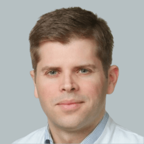
Professor Dr. Dr. med. Konstantinos Gousias is undoubtedly a leading figure in the field of neurosurgery and a valuable asset to St. Marien Hospital in Lünen, North Rhine-Westphalia. His impressive professional career and his tireless dedication to his patients set him apart as one of the leading experts in his field. With extensive medical training and an impressive career—including leadership roles at renowned tumor and spine centers—Prof. Dr. Dr. Gousias brings an unparalleled level of expertise and experience to patient care. His ongoing service on German and international commissions and advisory boards underscores his outstanding expertise. A remarkable specialization in brain and spinal cord tumor surgery, skull base surgery, and complex spinal procedures makes Prof. Dr. Dr. Gousias a highly sought-after specialist in this field.
The state-of-the-art neurosurgical operating rooms in Lünen enable his team to perform innovative minimally invasive procedures, which undoubtedly improve his patients’ quality of life. One of the greatest strengths of the hospital in Lünen is undoubtedly the interdisciplinary neurocenter INZ, which facilitates close collaboration between various specialized departments. This ensures precise and comprehensive care for patients with neurological and neurosurgical conditions around the clock. Precise and personalized care is of the utmost importance, and this is a cornerstone of Prof. Dr. Dr. Gousias’ treatment philosophy. He and his team take the time to thoroughly examine each patient, make a diagnosis, and work with the patient to develop a customized treatment plan.
In addition, Prof. Dr. Dr. Gousias is extremely active not only in clinical practice but also in research and teaching. His numerous scientific publications and contributions to renowned journals underscore his commitment to advancing medical care. Prof. Dr. Dr. Gousias is certified in the fields of neurosurgical oncology and spinal surgery by the German Academy of Neurosurgery as well as the German and European Spine Societies. These certifications attest to his professional competence, expertise, and experience in the care of neurosurgical patients. The editorial team of the Leading Medicine Guide spoke with him specifically about glioblastoma—a particularly aggressive form of brain tumor.
Glioblastomas are one of the most insidious forms of brain tumors, posing immense challenges for both medical professionals and patients. These highly aggressive brain tumors are known for their rapid progression and the difficulty in treating them. Their complex characteristics and their infiltration into surrounding tissue make them a unique medical challenge. The prognosis is often grim, but there are ongoing efforts in science and research to develop better therapies and treatments that could improve survival rates and quality of life for those affected.
The exact causes of glioblastoma are not fully understood.
“The number of new glioblastoma cases per 100,000 people per year is 4–5. The risk of glioblastoma increases with age, and they most commonly occur between the ages of 55 and 65. There are few established risk factors. Radiation, particularly ionizing radiation, has been identified as a potential risk factor for the development of brain tumors such as glioblastomas. People who have been exposed to certain sources of radiation—such as ionizing radiation—either occupationally or medically (e.g., during radiation therapy for cancer treatment) may have an increased risk of developing this type of tumor. Then there is genetic predisposition, such as neurofibromatosis, a genetic disorder that affects the nervous system. However, these risk factors do not apply to most patients.
Current research is focusing on familial predisposition—that is, the tendency of a family or certain family members to develop a specific disease or disorder. This tendency is influenced by genetic factors and can lead to family members having an increased risk of developing a specific disease, based on genetic predispositions or shared environmental factors. More than 50 genes have already been identified in this context. Even this etiology accounts for only about 4% of cases. Therefore, the cause of the tumor remains unclear for the vast majority of patients. If one absolutely wants to have their genetic makeup analyzed to calculate the risk of a brain tumor, this could potentially lead to unpleasant and stressful situations. After all, we can’t test every two months to see whether a tumor is forming or not. An annual MRI checkup is not sufficient, because a glioblastoma grows extremely rapidly—within just two to three months. In laboratory studies, we have found that tumor cells can double in number within 36–48 hours. The tumor is therefore incredibly aggressive; it grows diffusely in the brain through uncontrolled cell division, with the malignant cells dividing even more rapidly. Unfortunately, we haven’t made much progress yet in prevention,” explains Prof. Dr. Dr. Gousias regarding the development of this malignant tumor. There are therefore no specific measures to prevent the development of these tumors. Instead, research is focused on developing better diagnostic methods and treatment options.
Ionizing radiation is a form of radiation that has enough energy to remove electrons from atoms or displace them from their orbits, thereby creating ions. It encompasses various types of radiation, including X-rays, gamma rays, and high-energy particles such as alpha and beta radiation.
The diagnosis of glioblastoma involves a series of steps and imaging procedures.
“The tumor is very rarely discovered incidentally. This is because the patient typically experiences various neurological symptoms, which usually occur suddenly and vary in severity depending on the size and location of the tumor. Typical complaints in the form of specific symptoms include, for example, epileptic seizures, as well as paralysis or visual disturbances. However, nonspecific symptoms such as headaches or dizziness are also possible. Of course, you shouldn’t panic just because you have a headache once in a while! Imaging techniques such as a CT scan or an MRI are used for diagnosis. The latter is the diagnostic method of choice. This is where a suspicion of glioblastoma can be confirmed. The final diagnosis is then made through histological and molecular testing. “This means the patient undergoes surgery, since the tumor must be removed in any case. The resected tissue is then examined, and only then can it be determined with certainty whether it is a glioblastoma,” explains Prof. Dr. Dr. Gousias.
Diagnosing a brain tumor—especially a glioblastoma—is extremely challenging for doctors and medical professionals.
It is an emotional and ethical burden to have to inform a patient of such a serious and potentially life-threatening diagnosis. It requires a great deal of empathy, sensitivity, and strong communication skills to convey this news appropriately. Prof. Dr. Dr. Gousias describes the cautious approach: “As a doctor, you need a great deal of experience here. The patient and the patient’s family know that something is growing in the brain that doesn’t belong there. Before the surgery, there is only a suspicion of glioblastoma. The final diagnosis isn’t confirmed until 2–3 days after the surgery. However, the surgery means the first battle has already been won. After all, the tumor has been removed. Then, together with the patient, the family, and fellow physicians, we can discuss how to plan the next steps in the battle; in that sense, the conversation remains positive. A climate of trust must be established, and the patient must know that the treating physician is always available.” The diagnosis and classification of glioblastomas are therefore very complex, and the exact treatment plan depends on various factors, including the tumor grade and the patient’s overall health. Collaboration between neurosurgeons, oncologists, and other specialists is crucial for developing an appropriate treatment plan.
The main treatment options for patients with glioblastomas.
Surgical removal of the tumor is almost always the first step in treating glioblastomas. The goal is to remove as many tumor cells as possible without damaging healthy brain tissue. In some cases, however, complete removal is not possible because glioblastomas grow invasively into the surrounding tissue. “Once a glioblastoma is suspected, we aim to perform surgery promptly, usually 2–3 days after the suspicion is raised. To do this, we need thorough preparation—that is, a large amount of imaging data, not only of the tumor itself but also of the surrounding regions, the nerve fibers, and the pathways of the nervous system in the head—so that we can determine the exact location of the tumor and its impact on brain functions. This is crucial for the surgical technique and the entire procedure. For example, there are superficially located tumors that can simply be completely and radically removed. And then there are those located near motor and language centers. We cannot simply remove those. But during awake surgery, critical brain regions can be identified so that the best possible resection can be performed,” says Prof. Dr. Dr. Gousias.
The choice of the optimal treatment depends on the patient’s individual situation and the recommendations of the treatment team. The combination of surgery, radiation therapy, and chemotherapy is the standard treatment for glioblastomas, but other treatment options may also be considered. It is important to work closely with the medical team to develop a personalized treatment plan and to take the latest research findings and clinical trials into account.
Glioblastoma research focuses on various aspects.
The identification of genetic alterations and molecular markers in glioblastomas has helped deepen our understanding of the disease and develop potential targeted treatment approaches. Research into immunotherapy and targeted therapies for glioblastomas is in full swing, and new treatment approaches are being developed. Prof. Dr. Dr. Gousias is optimistic about this: “The fact is that after tumor resection, so-called satellite tumor cells always remain. The goal is to target and destroy these cells. I am a big advocate of immunotherapy, which aims to activate the body’s immune system to fight tumor cells. We do this here at Marien Hospital as well, in collaboration with colleagues from Düsseldorf, using what are known as dendritic cells. For this, cells are extracted from the patient’s blood, which are then loaded in the lab with antigens from the tumor removed in our operating room. These cells are then reinfused into the patient, where they fight the remaining tumor cells in the body.”
A glioblastoma can significantly impact the lives of patients and their families, as it is an aggressive and life-threatening form of brain tumor. The effects are varied and can be emotional, physical, social, and financial.
Glioblastomas can cause neurological symptoms that impair mobility, speech, perception, and other functions. Patients may face functional limitations and side effects from treatments, such as fatigue, nausea, and pain. Furthermore, a diagnosis of glioblastoma is extremely distressing for patients and their families. The emotional strain associated with the disease is significant, and anxiety, depression, and grief are common reactions. “The patient’s (and the family’s) mental stability is severely strained, and the patient goes through various phases. There are times when they feel afraid and times when they feel angry. And, of course, the patient always has questions. Psycho-oncological therapy can help them develop new perspectives. The goal is to motivate them. Patients never give up at this point because they’ve already come so far. Together, we always look to the future with optimism, since quality of life plays a very significant role—sometimes even a greater one than life expectancy itself,” explains Prof. Dr. Dr. Gousias.
Support from psychologists, social workers, or counselors can help patients and their families cope with the emotional and social challenges associated with the disease. Support groups offer those affected an opportunity to share experiences, find support, and connect with people in similar situations. In advanced stages of the disease, palliative care and hospice care can improve quality of life and provide support to patients and their families. Support from friends and loved ones, open communication with the medical team, and access to information about the disease are also crucial. Every individual situation is unique, so it is important to consider the needs and preferences of the patient and their family in order to find the most appropriate support options.
A Look to the Future.
Research is advancing by leaps and bounds and has led to significant improvements in the successful treatment of brain tumors in recent years. “I hope for improvements every day. Surgeries usually go well, and we have many excellent tools, so we can successfully remove even the most difficult tumors. But surgery is followed by systemic therapy in the form of radiation and/or chemotherapy. The last drug established for this purpose dates back to 2005. We definitely need a newer and more effective systemic therapy. But I’m very optimistic about this, since immunotherapy—which is currently still considered experimental—is a promising therapeutic approach for the future. Why? Because we can use the body’s own cells to specifically target the tumor. This also means we don’t have any problems with toxicity. I myself have been conducting research in immunology for more than 20 years and am very optimistic about this field.”
Prof. Dr. Dr. Gousias will leave Marien Hospital at the end of 2023 and return to his homeland, Greece. “In Athens, I will become chief physician at a private academic clinic and will continue my fight against tumors there!” explains Prof. Dr. Dr. Gousias enthusiastically, bringing our conversation to a close.
Prof. Dr. Dr. Gousias, Director of the Department of Neurosurgery
Athens Medical Center
Distomou 5-7, Marousi
Athens, Greece
Share this article
About the medical author
Alexandra Pfitzmann
Editor
Alexandra Pfitzmann – medical author: expert knowledge, professional articles and medical insights in the Leading Medicine Guide.
More about the medical author →Expert Interviews
Read next
- Expert interview
Cardiogeriatrics: Significance and benefits in the context of hospital reform
Sep 21, 2026
Cardiogeriatrics: Significance and benefits in the context of hospital reform
Read more - Expert interview
Expert Interview with Professor Dr. Amadeus Hornemann, MPH
Sep 18, 2026
Prof. Amadeus Hornemann on Precision Medicine in Gynecology: Modern Surgery, New Techniques, Better Quality of Life
Read more - Expert interview
Expert Interview with Professor Dr. Konstantinos Anagnostakos
Sep 15, 2026
Prof. Anagnostakos on Painful Knee Replacements
Read more