Glioblastoma is the most common malignant brain tumor in adults and is one of the most aggressive brain tumors. Glioblastomas arise from glial cells and are characterized by rapid tumor cell growth. Because of their diffuse spread throughout brain tissue, treating glioblastoma is often challenging. The most common symptoms include headaches, neurological deficits, and epileptic seizures.
The diagnosis is made using modern diagnostic methods such as MRI (magnetic resonance imaging) and histological examination of the tumor tissue. Treatment typically involves surgery, radiation therapy, and chemotherapy. Despite modern treatment, the prognosis for glioblastoma remains challenging. However, advances in neurosurgery, neuro-oncology, and molecular research are continuously improving treatment options.
Definition: What is a glioblastoma?
Glioblastomas are classified as diffuse-infiltrating brain tumors. This means they grow into healthy brain tissue rather than displacing it. The term “glioma” reflects the now-outdated assumption that glioblastomas originate in the nervous system’s supporting tissue, known as glial cells.
Based on its characteristics, the World Health Organization (WHO) classifies glioblastomas as Grade IV and thus as aggressive tumors. In the majority of cases, the glioblastoma first develops in one of the two cerebral hemispheres (Fig. 1)
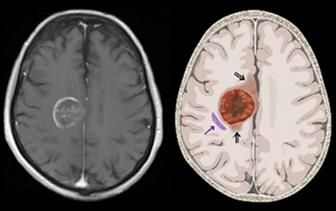
Fig. 1: Glioblastoma on MRI (left) showing the infiltration zone (double arrows) and path of spread (arrow) not visible on the left MRI image. [Source: Prof. Dr. Andreas Raabe, Inselspital Bern]
Symptoms of Glioblastoma
The intensity and severity of glioblastoma symptoms depend on the exact location of the brain tumor. Depending on the affected region of the brain, symptoms can vary widely, which often makes diagnosis difficult. Generally, symptoms appear within a few weeks.
The brain cannot move out of the way of the space-occupying tumor inside the hard skull. Nor can it adapt to the changed pressure conditions. As a result, patients primarily suffer from headaches—especially at night or in the early morning hours.
Patients report that the pain initially subsides on its own over the course of the day. Unlike other headaches, however, glioblastoma-related headaches recur at regular intervals. Over-the-counter medications, such as those available at drugstores and pharmacies, become ineffective over time.
In addition, patients with glioblastoma also frequently experience the following symptoms, which are common to all brain tumors:
- dizziness,
- coordination problems,
- visual disturbances,
- seizures,
- changes in personality,
- nausea, and
- fatigue and general exhaustion.
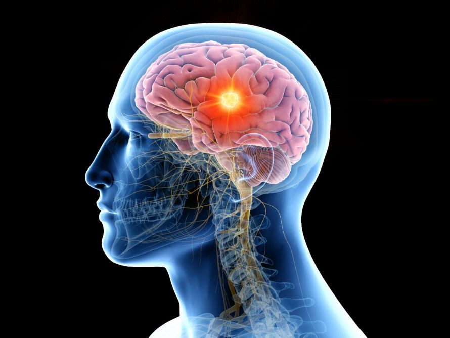
Anatomically accurate depiction of skull structures with a brain tumor © SciePro | AdobeStock
Causes and Risk Factors
The exact causes of glioblastoma remain unknown. Nevertheless, these tumors are among the most common types of brain tumors. The majority of patients are diagnosed between the ages of 60 and 70. The average age at diagnosis is 64, though this does not rule out the possibility that children can also develop glioblastomas.
It is interesting to note that men are about 1.7 times more likely to be affected by glioblastoma than women. Data from the U.S. Brain Tumor Registry also show that people of white ethnicity are particularly prone to developing glioblastoma. Based on their origin, a distinction is now made between primary and secondary glioblastomas. A primary glioblastoma, for example, arises from astrocytes—important support cells of the central nervous system. Since these astrocytes are regularly renewed, errors can occur during cell renewal. The cells then begin to grow uncontrollably and eventually form a glioblastoma.
Secondary glioblastomas, on the other hand, develop from pre-existing tumors. They thus represent the final stage of a disease that has been ongoing for some time. Ionizing radiation is also discussed as a possible factor in the development of glioblastoma. As a result, the internet in particular is full of theories and opinions regarding the influence of cell phones on glioblastomas and their development. But do cell phones or smartphones really cause brain tumors?
Based on the current state of research, experts say: Even large-scale epidemiological studies in humans have so far found no evidence that the use of cell phones leads to an increased risk of developing a brain tumor.
In contrast, extensive animal studies suggest an increased risk of tumors caused by mobile phone radiation in male rats and mice. However, they cannot demonstrate a dose-response relationship, nor can they explain the absence of this effect in female animals. Various authorities therefore classify the potential risk very differently: ranging from “harmless” to “possibly slightly carcinogenic—cannot be ruled out.”
Examinations and Diagnosis
Since the symptoms of glioblastoma usually appear suddenly, some patients first consult a neurologist. The neurologist begins by taking a medical history (anamnesis).
The most important diagnostic tool for reliably detecting glioblastoma is and remains magnetic resonance imaging (MRI). This imaging technique allows doctors to visualize the tumor (Fig. 2).
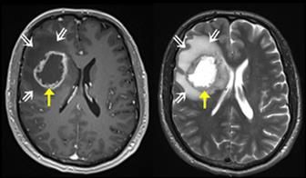
Fig. 2: Axial MRI T1 sequence with contrast agent (left) and T2 sequence (right). A glioblastoma typically appears as an irregular, ring-shaped mass that takes up contrast agent. The yellow arrow points to the tumor and its compartments, and the white double arrows indicate the surrounding cerebral edema. [Source: Prof. Dr. Andreas Raabe, Inselspital Bern]
An area in the brain suspected of being a tumor is, for example, a bright, ring-shaped structure visible on MRI images. If glioblastoma is suspected, doctors sometimes confirm the diagnosis using a tissue sample, known as a biopsy. In most cases, however, the tumor is operated on immediately without a prior biopsy.
General Information on Treatment
Due to the rapid growth of a glioblastoma, surgery should be performed as soon as possible. As time passes and the tumor grows, more cells spread into the surrounding tissue. This causes the tumor to continue expanding. At the same time, the risks associated with surgery increase. Surgery should therefore ideally be performed within 1–2 weeks of diagnosis.
For glioblastomas, the current “standard of care” consists of a combination of
- microsurgical tumor resection,
- radiation therapy, and
- chemotherapy.
Experts currently estimate that at least 80% of the tumor must be surgically removed. In such cases, surgery provides patients with a survival benefit. However, only MRI-confirmed complete resection results in a significant survival benefit.
Surgery
To ensure optimal preparation for surgery, a specialized MRI is required to provide the most accurate assessment and surgical planning possible. Depending on the tumor’s location, patients may also undergo additional tests.
Goal of Surgery
Surgical removal (resection) is now an integral part of the treatment plan and its first step. A greater extent of resection has a favorable effect on the course of the disease; therefore, MRI-complete tumor resection is the goal of the surgery. Furthermore, removal of the tumor reduces the mass effect and, consequently, the symptoms.
Surgical removal of the tumor tissue also enables histological and molecular biological analysis of the glioblastoma in question.
In addition, the more tumor tissue that is removed, the greater the survival benefit from the surgery. However, distinguishing between tumor and brain tissue is often difficult, especially at the margins.
Modern Technologies
For this reason, various modern technologies are used here. These include, for example, microscope-guided image navigation—known as neuronavigation—which utilizes a GPS-like system to enable surgeries with millimeter precision.
The high-performance surgical microscope allows for truly precise work on the brain. On the surgical microscope,
- fiber tracts marked prior to surgery,
- the tumor itself, and
- other important centers
can be virtually overlaid and projected onto the surface of the head (Fig. 3). This “augmented reality” allows the surgeon to better orient themselves and plan a customized approach to the tumor in advance. In doing so, they follow the principle of “as small as possible, but as large as necessary.”
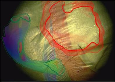
Fig. 3: Virtual reality achieved by overlaying the marked tumor and nerve tracts on the surgical microscope. A strip of skin is visible in the center; this was shaved, disinfected, and covered with plastic film before the operation began. Superimposed on this are the tumor in red and the important nerve pathways in color. [Source: Prof. Dr. Andreas Raabe, Inselspital Bern]
Postoperative Quality Control
An MRI scan is performed 24 to 48 hours after the procedure for quality control. Glioblastomas invade brain tissue in a very diffuse manner. Therefore, even after a resection that appears “complete” on MRI, a few tumor cells always remain. These must be destroyed through subsequent radiation therapy and chemotherapy. This means that, regardless of the MRI image, radiochemotherapy is always administered.
If residual tumor is visible on the early 24–48-hour follow-up MRI, it should be removed in a second surgery, provided its location allows for it. This surgery often takes place as early as the next day.
Experience shows that, despite all technical aids, removable residual tumor tissue remains in 5–10% of patients. A second surgery is advisable in these cases. Experts at Inselspital Bern (Switzerland), for example, were able to demonstrate in a study of their own patients that
- this almost always leads to complete removal,
- is well tolerated, and
- is associated with only minimal risk to patients.
This extended the hospital stay by only about two additional days.
Course and Prognosis
Glioblastomas belong to the group of Grade IV tumors. They are thus classified in the highest grade of the WHO tumor classification system.
The prognosis is significantly improved by prompt treatment and, above all, surgical removal of the tumor. The two pillars of glioblastoma treatment are
- the controlled and safe removal of the tumor and
- the combined use of radiation therapy and chemotherapy.
Regarding life expectancy, current research already indicates an increase in the number of patients achieving long-term survival. On average, with optimal and individually tailored treatment, life expectancy of 2 years or more can now be achieved.
The individual prognosis depends on a number of factors. The following list shows some of these factors that are believed to have a positive effect on the prognosis, such as:
- younger age,
- good general health or physical condition,
- no loss of neurological function prior to surgery,
- short time to surgery, i.e., earliest possible tumor removal,
- minimal or no steroid administration (dexamethasone) before and after surgery,
- no neurological deficits after surgery, in particular no paralysis or partial paralysis resulting from the surgery,
- Complete tumor removal as confirmed by T1-weighted contrast-enhanced MRI,
- no complications during or after surgery.
For individual patients, only a rough estimate of the timeline is possible, but no exact prediction can be made. All figures mentioned here are ultimately based on the “average” of a large number of patients and do not allow for conclusions to be drawn about individual patients.
A Note on Methadone
In recent years, there has been increasing discussion of methadone in connection with glioblastoma. Many posts on this topic can be found in online forums, while standard therapies are criticized in the same context.
However, the assumption that methadone is helpful is not based on systematic, scientifically collected, and publicly available data. To date, there is no evidence of the effectiveness of methadone therapy for glioblastomas.
Furthermore, methadone is not without side effects and can significantly impair quality of life if used improperly. In this context, we also refer to the statement issued by the German Neuro-Oncology Working Group.
Summary
Glioblastoma is one of the most malignant brain tumors. It develops relatively quickly. It primarily affects middle-aged people. The risk factors are largely unknown. The medical literature is limited to mere conjecture that does not go beyond the theoretical stage.
Despite intensive treatment methods, life expectancy for half of those affected is only 1–2 years after diagnosis. The other half of patients live longer. However, the disease remains incurable, despite advances in modern medicine and research.
FAQ
What is a glioblastoma?
A glioblastoma is a malignant brain tumor that originates from glial cells, specifically astrocytes. Glioblastoma multiforme is classified as WHO Grade 4 and is considered particularly aggressive. Glioblastomas invade brain tissue diffusely and therefore usually cannot be completely removed. Glioblastoma is the most common primary brain tumor in adults and is also referred to as GBM.
What symptoms does a glioblastoma cause?
The symptoms of a glioblastoma depend on the tumor’s location in the brain. Common symptoms include headaches, neurological deficits, epileptic seizures, paralysis, or other complaints. Other symptoms may include speech difficulties, memory problems, or personality changes. With glioblastoma, symptoms often develop rapidly due to the fast growth of the tumor cells.
How is glioblastoma diagnosed?
Glioblastoma is diagnosed using imaging techniques such as MRI (magnetic resonance imaging) or CT (computed tomography) with contrast dye. Tissue is then collected and subjected to histological and molecular analyses. This confirms the diagnosis of glioblastoma and enables the development of a personalized treatment plan. Neurosurgery, neuro-oncology, and other specialties work together in an interdisciplinary manner to establish the diagnosis.
What does the treatment for glioblastoma involve?
Treatment for glioblastoma usually begins with surgical removal of as much of the tumor as possible. This is followed by standard therapy consisting of radiation therapy and chemotherapy with temozolomide. Tumor-treating fields (electrical fields) may also be used as an adjunct. Treatment is tailored individually to the disease, genetic characteristics, and the extent of the tumor.
What is the prognosis and life expectancy for glioblastoma?
The prognosis for glioblastoma depends on various factors such as age, overall health, molecular characteristics, and the success of treatment. Despite modern treatment, the tumor often recurs. Clinical trials are continuously exploring new approaches to improve treatment. Patients with glioblastoma benefit from specialized care at neuro-oncology centers to receive the best possible treatment.
Share this article
Sources
- hirntumorhilfe.de/hirntumor/tumorarten/glioblastom/ [zuletzt aufgerufen am 02.09.2019]
- Inskip PD et al., Cellular-telephone use and brain tumors. N Engl J Med 2001; 344: 79–86
- Jakola AS et al., Surgical resection versus watchful waiting in low-grade gliomas. Ann Oncol 2017; 28: 1942–1948
- onko-portal.de/basis-informationen-krebs/krebsarten/hirntumor/astrozytome-und-glioblastome.html [zuletzt aufgerufen am 02.09.2019]
- krebsinformationsdienst.de/vorbeugung/risiken/krebs-durch-handys-mobilfunk-elektrosmog.php
- Lahkola A et al., Mobile phone use and risk of glioma in 5 North European countries. Int J Cancer 2007; 120: 1769–1775
- S2k-Leitlinie „Gliome“ (AWMF: 030/099) [derzeit in Überarbeitung voraussichtl. bis Ende 2019]
- Schucht P et al., Early re-do surgery for glioblastoma is a feasible and safe strategy to achieve complete resection of enhancing tumor. PLoS One 2013; 8 :e79846
- Fachliche Unterstützung und Beratung: Prof. Dr. Andreas Raabe und Kollegen vom Inselspital Bern (Schweiz)