A brain tumor requires individualized treatment based on the type, size, and location of the tumor in the brain. Brain tumor surgery is often the most important step in the treatment of brain tumors and is performed with high precision in modern neurosurgery. The goal of the surgery is to remove the tumor as completely as possible while preserving healthy brain tissue.
Before each procedure, specialists use MRI, CT, and other imaging techniques to create a detailed surgical plan. Depending on the findings, surgery, radiation therapy, chemotherapy, or other forms of therapy are combined in an interdisciplinary approach. Modern techniques such as neuronavigation, neuromonitoring, and intraoperative fluorescence enhance safety during surgery. As a result, the quality of life and neurological functions of many patients can be improved in the long term.
The term "brain tumor" is a general term for all benign and malignant tumors located within the skull (intracranial tumors).
Generally, the treatment options for a brain tumor
- surgery,
- radiation therapy, or
- chemotherapy
or a combination of these options. In most cases, the goal is to surgically remove the tumor.
Definition of Brain Tumors
In the strict sense, brain tumors are defined as all tumors of the neuroectodermal tissue. These are tumors that originate, for example,
- the nervous tissue of the brain,
- the hormone-producing glands in the brain, and
- the supporting and connective tissue
. Examples of these neuroepithelial tumors include
- astrocytomas (usually arising from astrocytes, which are part of the supporting tissue),
- gliomas (often arising from glial cells, which are part of the supporting and nutritive tissue), and
- medulloblastoma (a tumor of the cerebellum).
In a broader sense, meningeal tumors are also classified as brain tumors. These develop, for example, in the meninges (meningiomas) or in adipose tissue cells (lipomas). Brain metastases, which originate from a primary tumor outside the brain, are also classified as brain tumors. For example, cells from a breast tumor can enter the brain via the bloodstream and establish themselves there. This is referred to as a brain metastasis or a secondary tumor.
Brain tumors account for a relatively small proportion of cancers in adults compared to other types of cancer, such as breast cancer, colorectal cancer, and others. In children, however, they are among the more common types of tumors. The most common brain tumors include meningiomas and gliomas.
Causes of Brain Tumors
The reasons why brain tumors develop are not yet fully understood. Brain tumors also occur more frequently in association with certain genetic defects (e.g., hereditary neurofibromatosis). Therefore, researchers also assume that a genetic predisposition plays a role in the development of brain tumors.
However, exposure to carcinogenic substances also increases the risk of developing a brain tumor. These include, for example, certain pesticides as well as radiation therapy.
Symptoms of a Brain Tumor
The first symptom of a brain tumor is usually a headache. Lying down increases blood volume in the head, leading to a rise in intracranial pressure at night. This results in nighttime headaches.
These headaches improve during the day but are often accompanied by nausea and vomiting. Within a short time, the frequency of headaches caused by the brain tumor increases.
Other symptoms also depend on the area of the brain where the tumor is located. Depending on which nerves it presses on and into which areas it grows, additional symptoms—some more typical than others—may appear. These often include
- paralysis of the arms and legs,
- speech difficulties,
- visual disturbances, and
- seizures (epileptic seizures).
As the brain tumor grows, disturbances in consciousness and changes in personality also increasingly develop. Some symptoms provide clues as to which area of the brain is affected by the tumor. For example, if paralysis occurs in the right leg, this is a sign that the tumor is located in the left hemisphere of the brain. Certain neurological deficits can also provide clues as to the location of the brain tumor within the brain.
Diagnosis of Brain Tumors
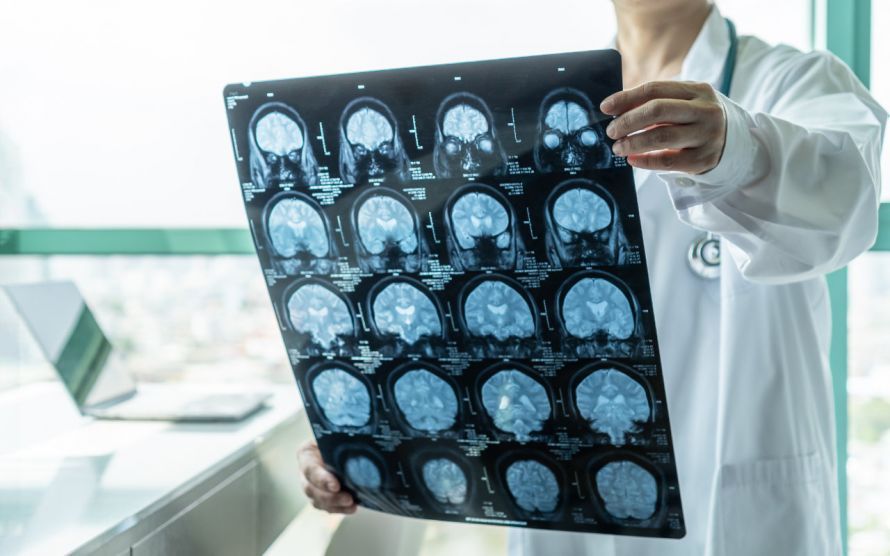
The diagnostic process begins with a medical history interview and a physical examination. If a brain tumor is suspected, imaging tests are crucial. In particular,
- magnetic resonance imaging (MRI) and
- computed tomography (CT)
are particularly important here. These allow physicians to determine the exact location and extent of the brain tumor.
Depending on the findings and symptoms, further diagnostic measures may be necessary to obtain more precise information. These include, for example,
- a lumbar puncture, in which cerebrospinal fluid is examined,
- an electroencephalogram (which measures brain waves), and
- angiography (imaging of the blood vessels in the brain using a contrast agent in an X-ray).
Occasionally, the back of the eye is also examined (ophthalmoscopy).
Surgical Treatment of Brain Tumors
Generally, the following options are available for treating a brain tumor:
- surgery,
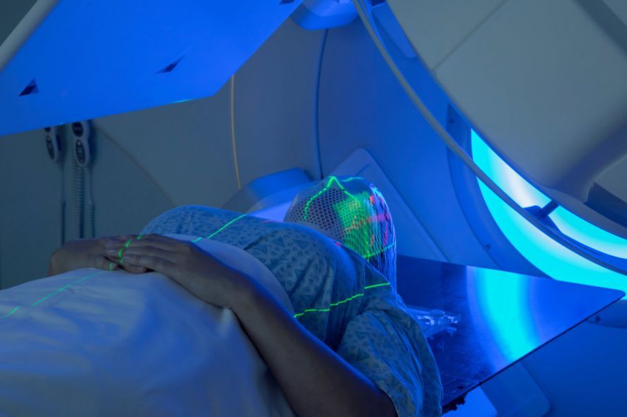
- radiation therapy, and
- chemotherapy (which is rarely used) or
- a combination of these
. The ideal treatment depends, among other factors, on the type of tumor, its location in the brain, and its size. If the size and location of the brain tumor allow it, the goal is to completely remove the tumor.
If functionally important areas of the brain are already affected, tumor removal could carry an increased risk of neurological deficits, such as
- speech disorders or
- paralysis
In these cases, often only part of the brain tumor is removed. This is followed by chemotherapy and/or radiation therapy to further combat the remaining tumor.
The primary goals of brain tumor surgery are
- to obtain a tissue sample for subsequent histological examination,
- relieving pressure on the brain, and
- removing as much of the tumor mass as possible.
It is crucial that the surgery does not endanger the patient and that no healthy brain tissue is destroyed. For benign brain tumors, surgical removal of the tumor is usually sufficient. For malignant tumors, radiation therapy and/or chemotherapy often follow.
Information on Brain Tumor Surgery
Brain tumor surgery is indicated (i.e., brain tumor surgery should be performed) if
- the brain tumor is growing rapidly and is already very large,
- the brain tumor is easily accessible and removable, and
- the patient’s overall condition and age permit surgical intervention.
In addition to removing the tumor, the primary goals of brain tumor surgery are, in particular,
- to improve quality of life,
- to delay deterioration, and
- to create better conditions for subsequent radiation therapy and/or chemotherapy.
Radiation therapy for a brain tumor © Mark Kostich | AdobeStock
Brain Tumor Surgery Under General Anesthesia or as Awake Surgery
Brain tumor surgery is usually performed under general anesthesia. If the brain tumor is located very close to the speech center or other important functional centers, the procedure is usually performed as awake surgery (awake craniotomy).
In this procedure, the patient is briefly awakened from anesthesia after the skull has been opened. Using various tests, the medical team determines how much tumor tissue can be removed without causing functional deficits.
By having the patient read, speak, or name objects during the operation, serious speech impairments, for example, can be avoided.
Risks and Complications of Brain Tumor Surgery
The risks of brain tumor surgery depend mainly on the tumor’s size and its location in the brain. In addition, the involvement of cerebral blood vessels, the patient’s age, and their overall health also play a role.
If the brain tumors are still small and located in favorable areas of the brain, the risk of brain tumor surgery is very low. With very large tumors, however, the risk increases significantly. Modern techniques have significantly reduced the risk of brain tumor surgery. These include, for example,
- endoscopic and microsurgical techniques,
- neuronavigation,
- intraoperative and functional imaging,
- intraoperative monitoring of brain function (neuromonitoring).
Nevertheless, an individualized consultation with the patient prior to brain tumor surgery is very important. Complications may arise during brain tumor surgery that temporarily or permanently impair a patient’s ability to drive. Therefore, a driving ban remains in effect until the doctor determines that it is safe to drive.
Other possible complications include, for example,
- visual disturbances,
- epileptic seizures, and
- impaired cognitive function.
Intraoperative Neuronavigation
Intraoperative neuronavigation is a computer-assisted surgical procedure used in neurosurgery. It enables the planning of brain tumor surgeries and facilitates spatial orientation during the procedure.
This requires three-dimensional image data of the brain. These images are acquired during surgery using
- computed tomography (CT),
- magnetic resonance imaging (MRI), and
- ultrasound
and displayed as a 3D image. The position of the surgical instruments is combined with the 3D images and also displayed. This allows the neurosurgeon to know exactly where in the brain he is located during the operation, without any delay. If necessary, a CT scan can be performed during brain tumor surgery, and the image data can then be updated.
In preparation for the surgery, the brain structures can be studied in detail, allowing the surgeon to choose the ideal approach to the brain tumor. The surgeon also assesses the blood supply in the surgical area and identifies functionally important brain regions.
Thanks to the planning of the brain tumor surgery and the extreme precision during the procedure,
- functionally important areas of the brain (e.g., the speech center) are spared,
- the skull incision kept smaller, and
- accidental damage to blood vessels is avoided.
Intraoperative Neuromonitoring
Intraoperative neuromonitoring allows for the electrophysiological monitoring of important neurological functions during surgery. This enables the timely detection of damage to nerve tissue and the localization of functionally important areas prior to brain tumor surgery.
Through intraoperative monitoring, the neurosurgeon receives continuous information about the function of the brain area currently being operated on. This ensures that important brain structures are spared to the greatest extent possible.
Minimally Invasive Tissue Biopsy
Removal of a brain tumor via open brain surgery is not always possible in the following cases:
- Patients with diffusely growing (i.e., invasively spreading into the brain) or large cystic tumors,
- Patients with small tumors in unfavorable locations (e.g., in the basal ganglia or the brainstem),
- Elderly patients or patients in poor general health.
However, the tumor type must be known in order to determine an effective treatment (chemotherapy, radiation therapy). The tumor sample for histological examination is obtained using a stereotactic, computer-guided technique. This is a minimally invasive neurosurgical procedure.
To do this, the patient’s head and the medical instruments are secured in a firmly bolted frame. Stereotactic tissue sampling is often combined with real-time imaging using neuronavigation. This allows for precise, largely injury-free movement within the brain, enabling the tissue sample to be safely extracted.
Brain Tumor Surgery
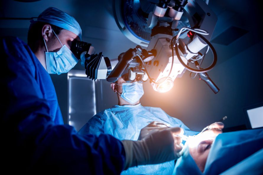
Open surgery refers to the opening of the skull followed by the removal of the tumor, as completely as possible. Brain tumor surgery is usually performed under general anesthesia or as awake surgery. For this, the head is secured in a three-point head clamp, and the skin is incised in a straight or curved line. Next, a section of the skull is sawed away, and the dura mater is opened.
Using neuronavigation and under microsurgical conditions, the surgeon guides the instruments to the tumor and removes it. Occasionally—especially with larger tumors—ultrasonic aspirators are also used. Fluorescence-guided resection makes it easier to identify the tumor. To achieve this, the patient ingests a substance beforehand that accumulates in the tumor and is made visible using blue light. During the surgery, the surgeon removes as much of the tumor mass as possible, taking care not to damage adjacent functional areas.
Brain tumor surgery may take place near functional centers, nerve pathways, and cranial nerves that control, for example,
- sensory and motor functions,
- hearing, and
- the facial and tongue muscles
. In such cases, intraoperative neurophysiological monitoring is performed to monitor the function of these areas during the procedure.
After the tumor is removed, bleeding is controlled, and if necessary, the results are verified intraoperatively using CT or MRI. The surgeon closes the dura mater and the incision. The patient wakes up in the neurosurgical recovery unit after brain tumor surgery.
To assess the outcome of the brain tumor surgery, a CT or MRI follow-up scan is performed one or two days after the procedure. If there are no complications and with the support of physical therapy, the patient can be discharged from the hospital after about 7 to 10 days. If necessary, the patient will continue treatment at their local hospital or a rehabilitation clinic.
Prognosis for a Brain Tumor
The prognosis for a brain tumor depends on numerous factors, including
- the growth pattern of the tumor cells,
- the tumor’s location in the brain,
- the amount of tumor mass remaining after brain tumor surgery, the
- type of tumor, and
- the available treatment options.
It can therefore vary greatly from patient to patient. A recurrence of the tumor also has a negative impact on the long-term prognosis.
A benign, slow-growing brain tumor that is easy to remove and does not recur has a favorable prognosis. In contrast, brain metastases worsen the prognosis of the primary tumor.
Conclusion
Modern brain tumor surgery is now a highly precise and often minimally invasive surgical procedure for treating brain tumors. Whether benign tumors, gliomas, or a glioblastoma—the choice of treatment depends on the type of tumor, its location within the central nervous system, and the affected area. The goal of any brain tumor surgery is the complete removal of the tumor, provided this can be done without damaging vital brain functions or cranial nerves. If complete resection is not possible, residual tumor tissue can be treated with radiation therapy, chemotherapy, or modern immunotherapies.
During neurosurgical procedures, neuromonitoring, navigation systems, surgical microscopes, 5-ALA fluorescence, intraoperative techniques, and three-dimensionally processed image data from imaging modalities all support the safe removal of the tumor. These technologies are particularly crucial for tumors near the base of the skull or sensitive brain tissue. Prior to the procedure, a biopsy and discussion at a tumor board meeting can help plan the optimal treatment.
Depending on the findings, the tumor is removed surgically, with the focus on complete removal, preservation of the meninges, and achieving the most successful outcome possible. For patients, the most important goal—in addition to treating a brain tumor—is, above all, improving their quality of life.
FAQ
When is brain tumor surgery necessary?
Brain tumor surgery is recommended when a tumor can be safely removed or when a tissue sample is needed for diagnosis. Brain tumor surgery is often performed to remove the tumor as completely as possible, alleviate symptoms, and lay the groundwork for further treatment of brain tumors.
How is brain tumor surgery performed?
Before the surgery, MRI, CT, and other imaging techniques are evaluated. During the procedure, neuronavigation, a surgical microscope, intraoperative fluorescence with 5-ALA, and neuromonitoring are frequently used. The surgeon continuously monitors vital brain functions to protect healthy brain tissue as effectively as possible.
Can a brain tumor be completely removed?
Whether a brain tumor can be completely removed depends on the type of tumor, its location, and its proximity to important areas of the brain. Benign tumors can often be completely removed, whereas in the case of gliomas or glioblastomas, residual tumor tissue must sometimes be intentionally left behind to prevent neurological deficits.
What treatment follows brain tumor surgery?
After a successful surgery, an interdisciplinary tumor conference determines the course of further treatment. Depending on the type of tumor, this may include radiation therapy, chemotherapy, and modern immunotherapies. The goal is to treat any remaining tumor cells and improve patients’ quality of life.
What are the risks of brain tumor surgery?
Like any neurosurgical procedure, brain tumor surgery carries risks such as bleeding, infection, or neurological deficits. However, thanks to modern surgical techniques, precision, monitoring, and specialized neurosurgeons, these risks have been significantly reduced. The treating specialist performs an individual risk assessment before each surgery.

About the medical author
Dr. Claus Puhlmann
Medical Author · Medical journalist
More about the medical author →