Expert Interviews
Modern Techniques in Interventional Endoscopy

Alexandra Pfitzmann ·
The editorial team of the Leading Medicine Guide learned more about modern techniques in interventional endoscopy during a conversation with Prof. Dr. Weismüller.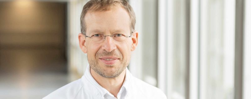
Modern interventional endoscopy techniques are revolutionizing the diagnosis and treatment of diseases of the digestive tract. Through the use of innovative endoscopic techniques, numerous diseases can be treated in a minimally invasive, precise, and gentle manner. These advanced procedures enable, for example, the targeted removal of tumors, the treatment of strictures, the placement of stents, as well as preoperative diagnosis and therapy for complex clinical conditions.
Thanks to continuous technical advancements, interventional endoscopic methods not only improve treatment outcomes but also significantly enhance patients’ quality of life. They thus represent a key pillar of modern medicine, where interdisciplinary collaboration and the use of the latest technologies are of paramount importance.
“Many people primarily associate the term ‘gastroenterologist’ with gastroscopies and colonoscopies. In fact, about 30 years ago, endoscopy was also predominantly a diagnostic procedure—and in the public perception, this is often still the case. In private practices, diagnostic endoscopy remains the primary focus, even though small polyps are removed or tissue samples (biopsies) are taken there.
In hospitals, however, the picture has changed significantly. Thanks to technical innovations in recent years, the focus has shifted markedly from purely diagnostic procedures to interventional—that is, therapeutic—procedures. Today, gastroenterologists treat not only the esophagus, stomach, duodenum, small intestine, and large intestine, but also the liver, bile ducts, and pancreas. In doing so, they work closely with colleagues from visceral surgery—that is, abdominal surgery.
This close collaboration has led to a modern understanding of what is known as visceral medicine: gastroenterology and surgery approach the same clinical pictures from different perspectives in order to jointly determine the best possible individualized treatment for their patients.
“While the miniaturization of surgical procedures has brought about major advances in surgery, modern endoscopes in the field of gastroenterology now make it possible to perform operations from the inside—without any abdominal incision at all,” said Prof. Dr. Weismüller at the start of our conversation, adding:
“The advantages of these minimally invasive therapies are significant: In the case of early-stage tumors, for example, growths can be removed endoscopically without the need for surgery. The affected organ—such as the rectum or the esophagus—is preserved, which is a crucial factor for the quality of life of those affected. At the same time, it is important to understand the limitations of these procedures.
That is why all patients are discussed in an interdisciplinary setting—in tumor boards or special case conferences—to determine the best individual treatment. Modern endoscopic methods offer the major advantages of reduced trauma, organ preservation, and a significantly shorter hospital stay.”
In recent years, numerous technical innovations have significantly advanced the development of modern interventional endoscopy and expanded its range of applications.
One of the most significant developments is the improvement of the endoscopes themselves, particularly the introduction of high-resolution 4K and even 8K cameras, which enable precise visualization of even small structures and nearly microscopic details in the digestive tract. This high-resolution imaging significantly increases the accuracy of diagnostics and therapies.
“Thirty to forty years ago, many gastrointestinal procedures were still performed using open surgery. Conditions such as early-stage cancers or large adenomas had to be surgically removed, which usually involved large abdominal incisions, longer wound healing times, a higher risk of complications, and significant limitations on quality of life.
With the advent of laparoscopic surgery, this changed fundamentally: Today, both surgery and gastroenterology predominantly rely on minimally invasive techniques—using small instruments, sometimes even robotic systems, or via natural access through the digestive tract. Technical developments in these procedures really took off in the late 1990s, particularly with the introduction of endosonography.
This made it possible to treat conditions transluminally—that is, through the stomach wall—such as infected wound cavities following pancreatitis. Since then, innovation in the field of endoscopy has brought about new advances practically every year: improved clipping techniques, specialized metal stents, more precise instruments, and innovative endoscopic tools are continuously expanding the range of therapeutic options,” explains Prof. Dr. Weismüller.
A true milestone was the introduction of so-called lumen-apposing metal stents (LAMS)—metal stents designed to connect two hollow spaces.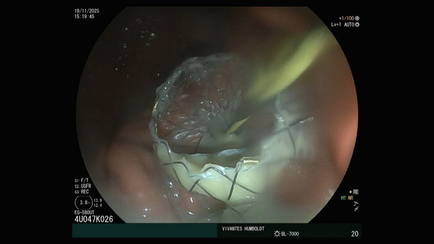
A lumen-apposing metal stent was inserted through the stomach wall into an infected pancreatic cyst.
Lumen-apposing metal stents (LAMS) are special metal stents used to connect two hollow organs or cavities. They are primarily used in interventional endoscopy, for example in cases of pancreatic necrosis or cholecystitis, and can be placed minimally invasively via an endoscope without the need for major surgery. With their help, infected fluid can be effectively drained or directed into an infected wound cavity using the endoscope to remove dead and infected tissue. Procedures that previously required traditional surgery involving an abdominal incision can now be performed from the inside with much less damage to healthy structures.
Prof. Dr. Weismüller explains: “With their help, for example, necrosis resulting from severe pancreatitis can now be treated gently. Under endosonographic guidance, the stent is inserted from the stomach into the necrotic cavity, allowing the dead tissue to be removed endoscopically from the inside. This allows the inflammation to heal—a procedure that previously often required risky surgery. These stents are also used in cases of acute cholecystitis, especially in patients who are too ill or too old for surgery.
In such cases, the gallbladder can be drained from the inside, allowing the pus to drain and the inflammation to subside. At the same time, endoscopes have become increasingly slender and capable. Modern devices, just 3 mm thick, now allow, for example, a direct view into the bile ducts or the pancreatic duct, making it possible to detect even the smallest changes and treat them precisely—a difference comparable to that between the camera on a modern smartphone and a model from 20 years ago.
Thanks to this improved image quality, we can now plan procedures with greater precision and provide more targeted treatment. This is increasingly supported by artificial intelligence, which serves as a tool to aid in the detection and classification of abnormalities. In addition, closure techniques such as clips and suture systems have been further developed in recent years, allowing the access site or wound surface to be closed safely and gently after the endoscopic procedure is complete.”
The use of interventional endoscopic methods has been proven, based on evidence, to be extremely effective for a wide range of conditions. These include, in particular, the treatment of precancerous and neoplastic lesions in the stomach, intestine, and esophagus, as well as functional disorders such as achalasia.
However, even these procedures have their limitations, particularly in oncology. As long as a tumor is confined to the outermost layer of the mucosa, it can be removed endoscopically—this is referred to as early-stage carcinoma. However, as soon as the tumor has invaded deeper into the submucosa (the layer of the gastrointestinal wall), there is a risk that it has metastasized to the lymph nodes. These are not accessible endoscopically, which is why surgical removal of the lymph nodes is necessary in such cases.
In practice, this means: After endoscopic submucosal dissection (ESD), the pathology department examines the removed tissue. If this reveals an increased risk of metastases, surgery is necessary as a second step—often performed today using minimally invasive robotic systems, which spare patients the trauma of major surgery. This collaboration between endoscopy and surgery exemplifies the modern concept of visceral medicine: both disciplines work hand in hand to find the gentlest yet most effective treatment for each patient.
In addition to medical limitations, there are also individual factors that influence the use of endoscopic procedures—such as age or pre-existing conditions. In very elderly patients, the risk of a procedure may outweigh the potential benefits. Nevertheless, there are also very elderly people who are still biologically fit and can benefit from minimally invasive treatment. “There is therefore no rigid age limit—what matters is always the overall condition and quality of life of the individual patient,” explains Prof. Dr. Weismüller.
The submucosa is a layer of the intestinal and stomach wall that lies directly beneath the mucosa. It consists of connective tissue, blood and lymph vessels, and nerves, and supplies the mucosa with nutrients. During endoscopic procedures, this layer is often distended by injecting saline solution so that surgeons can work within this boundary layer to separate tumor tissue from healthy tissue.
Today, gastroenterology encompasses much more than just gastroscopy and colonoscopy—it deals with the entire digestive tract, including the liver, bile ducts, and pancreas, and covers a broad spectrum of clinical conditions.
“In gastroenterology, many different areas benefit from minimally invasive endoscopic procedures. These include, among others, endoscopic mucosal resections, endoscopic submucosal dissection (see box), endoscopic full-thickness resection, in which all layers of the intestinal wall are precisely removed, as well as procedures on the esophagus, such as peroral endoscopic myotomy (POEM) for achalasia (a rare disorder of the lower esophageal sphincter). In addition, modern, thin endoscopes (cholangioscopes) now enable procedures in the bile ducts and the pancreatic duct, for example, to break up stones or treat tumors using minimally invasive techniques. Technological developments in recent years have led to enormous progress: optics have improved significantly, and image quality now allows for a precise assessment of whether lesions are benign or malignant without the need for biopsies in every case.
“Artificial intelligence is increasingly being used in endoscopy to support diagnosis and optimize treatment planning,” explains Prof. Dr. Weismüller.
Peroral endoscopic myotomy (POEM) is a minimally invasive procedure for treating achalasia. It involves creating a small, tunnel-like opening in the esophageal mucosa to precisely sever the thickened and spasmodic muscle tissue at the lower end of the esophagus. This allows the esophagus to open again, enabling food to pass more easily into the stomach. The advantage: No external incision is necessary; the treatment is performed entirely from the inside and is less invasive than traditional surgery.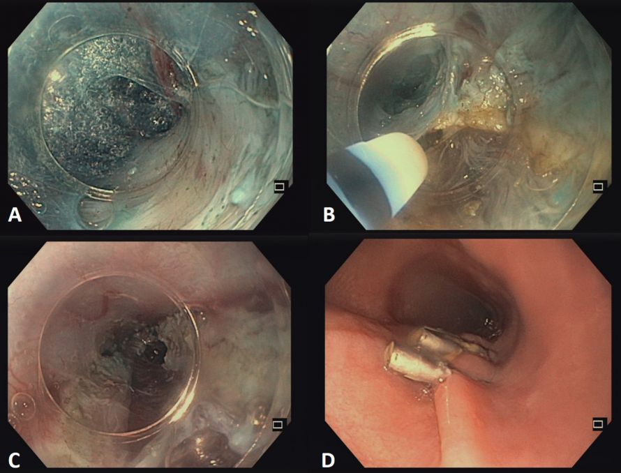
In the case of achalasia, an endoscopic tunnel is first created in the esophageal wall (A). The esophageal muscle layer is then severed beneath the intact mucosal layer using a tiny endoscopic knife (B). Once the muscle layer in the area of the lower esophageal sphincter has been completely severed (C), the tunnel entrance is closed with endoscopic clips (D).
Although advanced endoscopic procedures are generally safe and effective, they are associated with certain risks despite technical innovations. Although rare, complications such as bleeding, infections, perforations, and unintended damage to surrounding structures can occur in individual cases.
Prof. Dr. Weismüller explains: “Minimally invasive endoscopic and interventional procedures carry specific risks and complications that depend heavily on the particular procedure. In general, these procedures are less risky than major open surgeries; nevertheless, the risk of complications increases with the extent of the intervention. The risk of bleeding is particularly relevant, as many blood vessels are located in the surgical field during transvisceral procedures.
In addition, infections can occur if a procedure involves passing from non-sterile areas into sterile structures, such as the bile ducts; this risk is addressed prophylactically with antibiotic therapy. Added to this are the general risks of general anesthesia, which is necessary for longer procedures. However, minimally invasive procedures clearly offer the advantage of less surgical trauma, shorter hospital stays, and the ability to preserve affected organs.”
In general, a minimally invasive endoscopic procedure is significantly more advantageous for the postoperative period, particularly in terms of pain and recovery time.
“Since access is gained without an external incision, many patients hardly notice after the operation that a procedure has taken place, and there are usually only minor symptoms, depending on the area treated. To ensure safety and to be able to take swift action in an emergency, patients are typically monitored as inpatients for one to two days, undergo regular laboratory tests, and are often given prophylactic antibiotics.
The length of stay depends heavily on the procedure: For elective procedures such as endoscopic submucosal dissection (ESD), one to two days are often sufficient, while more complex procedures such as POEM require about four days. Acute emergencies—such as patients with inflamed necrotic lesions—remain in the hospital for a correspondingly longer period. “Overall, the length of hospital stay varies depending on the procedure, risk, and general health status, with planned procedures involving the esophagus or colon typically requiring a significantly shorter inpatient stay than complex emergency situations,” explains Prof. Dr. Weismüller.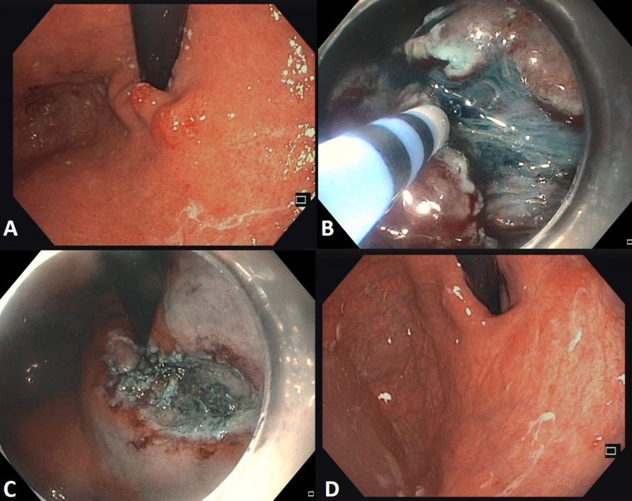
A tumor at the entrance to the stomach (A) is excised from the stomach wall during a gastroscopy using a millimeter-sized endoscopic knife (B). The resulting wound defect (C) heals completely within a few weeks (D).
Endoscopic submucosal dissection (ESD) is a minimally invasive procedure in which precancerous lesions or early-stage cancers in the stomach, colon, or esophagus are removed in one piece (en bloc) using an endoscope. In this procedure, a fine needle knife is used to separate the diseased mucosa from the underlying healthy muscle layer, allowing the wound to heal and the organ to be preserved.
Technological advancements—such as the integration of precise imaging systems and computer-assisted support systems, as well as modern anesthesiological techniques—further enhance safety. In addition, for complex procedures, careful patient counseling, a preoperative risk assessment, and close monitoring during and after the procedure are always carried out.
Prof. Dr. Weismüller emphasizes: “Endoscopic and minimally invasive procedures are now offered at many large hospitals in Germany, though not yet universally available everywhere. Developments in this field are very dynamic: At every conference, new innovations, techniques, and procedures are presented that continue to push the boundaries of minimally invasive treatment options and open up new possibilities year after year.
It is particularly important for patients to provide as many previous medical records as possible, including results from earlier endoscopic examinations or imaging studies, so that the team has a complete overview of the situation. At Vivantes, treatment is characterized by extremely close interdisciplinary collaboration between gastroenterology and surgery. This ensures that, should surgery be necessary, action can be taken immediately and patient care continues seamlessly.
Likewise, our surgical colleagues rely on endoscopic expertise when complex procedures must be performed using minimally invasive techniques. Close cooperation, the provision of comprehensive prior medical records, and clear communication enable efficient planning and execution of the procedures. “Patient inquiries should therefore be as detailed as possible—not just two or three sentences—to ensure a smooth process and the best possible individualized care,” he adds, concluding:
“There are many outstanding endoscopists throughout Germany, and it is not necessary to come to Berlin for every treatment. For northwest Berlin and the surrounding communities in Brandenburg, however, we are a very good point of contact, as we can offer the full spectrum of minimally invasive procedures.
Close interdisciplinary collaboration is particularly important here: In the past, internal medicine and surgery were viewed separately; today, we work closely with abdominal surgery and other specialties to provide patients with the best possible, holistic care. This collaboration is essential to ensuring optimal treatment.”
Thank you very much, Professor Dr. Weismüller, for your detailed explanation of endoscopic procedures!
- Prof. Dr. Tobias Weismüller is Chief of the Departments of Gastroenterology and Hepatology at Vivantes Humboldt Hospital and Vivantes Spandau Hospital in Berlin.
- Specialist in diseases of the digestive tract, liver, bile ducts, and pancreas.
- Many years of experience and extensive expertise in diagnostics, endoscopy, and interventional therapy.
- Areas of focus: Procedures on the bile ducts and pancreas, treatment of esophageal diseases such as achalasia or diverticula, and minimally invasive tumor removals in the esophagus, stomach, and intestines.
- Relies on innovative endoscopic procedures for particularly gentle and precise treatments.
- Is committed to interdisciplinary collaboration with oncology, surgery, and radiology.
- Member of several national and international professional societies.
- Conducts research at leading university hospitals such as Hannover Medical School and the University Hospital of Bonn.
- Committed to the training and continuing education of young physicians.
- Author of over 100 scientific articles, books, and presentations
- Committed to high-quality medical care, modern procedures, and empathetic patient care.
Share this article
Expert Interviews
Read next
- Expert interview
Expert interview with Professor Dr Elmar Herbst, PhD
Oct 15, 2026
Patient-centred and personalised knee surgery
Read more - Expert interview
Cardiogeriatrics: Significance and benefits in the context of hospital reform
Sep 21, 2026
Cardiogeriatrics: Significance and benefits in the context of hospital reform
Read more - Expert interview
Expert Interview with Professor Dr. Amadeus Hornemann, MPH
Sep 18, 2026
Prof. Amadeus Hornemann on Precision Medicine in Gynecology: Modern Surgery, New Techniques, Better Quality of Life
Read more