Expert Interviews
Aneurysm – When a Blood Vessel Wall Weakens

Alexandra Pfitzmann ·
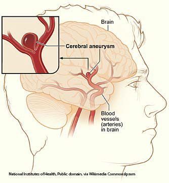
A brain aneurysm does not develop suddenly; rather, it is usually the result of a long, gradual process in which the blood vessel wall loses its stability. The arteries in the brain consist of several layers that work together to absorb blood pressure and distribute it evenly. If one of these layers is weakened—for example, due to structural changes, inflammation, or prolonged pressure—the vessel loses its strength.

“In an intracranial aneurysm, the tunica muscularis—the middle muscular layer—is missing from the three classic vascular layers. Yet it is precisely this layer that is crucial for the vessel’s stability. When only the inner and outer layers—which are less resistant to pressure—remain, blood pressure exerts a constant force on the weakened wall. At these points, the blood vessel can expand and bulge like a balloon—and this is precisely where the danger lies: that the pressure will eventually become so great that the thin wall ruptures and bursts.
This often raises the question of whether there is a specific “aneurysm gene.” The answer is no, even though there are families in which aneurysms occur more frequently and several first-degree relatives are affected. In such cases, the condition is referred to as aneurysmatosis, and a different approach to detection and monitoring is recommended compared to patients in whom an aneurysm occurs only sporadically as an incidental finding or following a rupture.
Despite these familial clusters, the absence of the middle vascular layer is not clearly linked to a specific genetic predisposition. Aneurysms are observed in both men and women, without a clear pathophysiological mechanism or a typical age peak being identifiable—they can occur in both younger and older individuals, either as an incidental finding or when symptoms are present. “In a way, we’re still ‘fishing in murky waters’ here,” explains Sascha Zink at the beginning of our conversation, and continues:
“An aneurysm can develop spontaneously; it doesn’t have to be present from the start—it simply grows. This also explains why, unlike with other conditions, there are no established preventive or screening programs, and why it’s not possible to make a blanket recommendation that everyone should undergo an MRI to screen for intracranial aneurysms.
There are no clear age groups or risk groups in which such changes occur more frequently. If an aneurysm is detected and treated, follow-up care remains important nonetheless—not only for the specific aneurysm that was treated, but also because of what’s known as ‘de novo synthesis’: New aneurysms can develop over time. To detect such changes in a timely manner, regular imaging follow-ups and structured aftercare are recommended.”
Some changes in the brain are preceded by subtle warning signs, while others do not appear until an aneurysm becomes unstable or is on the verge of rupturing. Since such symptoms can also occur with other conditions, it is important to seek medical evaluation if you experience any relevant symptoms.

“Aneurysms are often discovered by chance—frequently during investigations for headaches. Many patients seek medical evaluation due to persistent or new types of headaches, and it is only then that an aneurysm is detected. Such vascular bulges can reach a considerable size and press on nerves that run parallel to the blood vessels in the confined intracranial space. This can lead not only to localized pain but also to visual disturbances, double vision, or nonspecific symptoms such as neck pain. Some patients also present with autonomic symptoms such as nausea and vomiting.
This may be due to so-called “warning leaks”—small tears in the aneurysm wall through which blood escapes and irritates the meninges. In such cases, typical symptoms appear even if the aneurysm is not yet clearly visible on imaging. This is followed by MRI scans and a lumbar puncture to detect blood breakdown products in the cerebrospinal fluid. If the suspicion is confirmed, an invasive angiography is performed to confirm or rule out the finding. If the finding is confirmed, acute treatment is initiated. An unruptured aneurysm often remains asymptomatic. When it does cause symptoms, it is usually because it is pressing on surrounding structures.
Typical symptoms include one-sided headaches, visual disturbances, changes in pupil size, or sensory disturbances in the face. Some patients report new, unfamiliar types of headaches that differ from their previous symptoms. Recurring pain behind the eye or double vision can also be signs that an aneurysm is growing or irritating nerves. The situation becomes acutely dangerous when an aneurysm becomes unstable or ruptures. This often results in a sudden, extremely severe headache—often described as “the worst headache of my life.”
This pain may be accompanied by nausea, vomiting, neck stiffness, sensitivity to light, altered consciousness, or neurological deficits. Such symptoms constitute an absolute medical emergency, as bleeding in the brain can become life-threatening within a very short time. “Even though many of these symptoms may have other causes, the rule is: Sudden, unusually severe, or new neurological symptoms should always be evaluated by a doctor,” explains Sascha Zink.
Today, the diagnosis of an aneurysm relies on modern imaging techniques that visualize blood vessels in the brain in high resolution and with minimal effort. The key factor here is the specific clinical question at hand: Is an aneurysm suspected, does an incidental finding need to be evaluated, or is there suspicion of acute bleeding?
“Once a definitive diagnosis has been made, various treatment options are available, depending on the size, location, and individual risk profile of the aneurysm. The ‘watch-and-wait’ approach—that is, controlled observation under neurological supervision—which was once common, was used primarily in younger patients with very small incidental findings. For a long time, guidelines were based on size categories of less than 7 mm, 7–12 mm, and over 12 mm, up to so-called giant aneurysms (giant) starting at about 25 mm. Small aneurysms were generally considered to require monitoring, medium-sized ones were deemed to carry moderate risk, and large ones were considered particularly high-risk.
However, this classification is now only valid to a limited extent. More recent data—including studies from Düsseldorf and the Charité—show that even very small aneurysms measuring 3–5 mm in the anterior circulation can rupture. In addition, the risk of rupture increases along certain segments of the vessels: aneurysms in the posterior circulation are considered particularly high-risk. Against this backdrop, it is now recommended to treat all detected aneurysms as a matter of principle, rather than simply monitoring them over an extended period. This brings the decision between two active procedures into focus: clipping, i.e., neurosurgical intervention, and coiling, the endovascular treatment performed by neuroradiology. Each case is discussed individually in interdisciplinary neurovascular conferences—regardless of whether it is referred by neurology, neurosurgery, or radiology.
Numerous factors are taken into account in the decision: the size and location of the aneurysm, the vascular anatomy, comorbidities such as high blood pressure, lifestyle factors such as smoking, the patient’s age, and the available imaging data. Based on these parameters, a recommendation is made using a scoring system—such as the PHASES score. Whether surgery or endovascular treatment is performed depends on anatomical and technical criteria. A clip permanently seals the aneurysm; with coiling, small residual aneurysms may remain, which is why follow-up care is more frequent.
While imaging follow-up is eventually completed after clip placement (apart from screening for possible de novo aneurysms), patients undergo regular follow-up for a longer period after coiling, via CT or MRI depending on the center. A purely conservative approach remains the exception today. It is only considered if the aneurysm is very small, causes no symptoms, is anatomically non-critical, and the individual risk assessment clearly argues against intervention,” explains Sascha Zink.
Today, an aneurysm is usually detected using MRA (magnetic resonance angiography) or CTA (computed tomography angiography): MRA does not use radiation and is suitable for cases of unclear symptoms or a family history of risk, while CTA is very fast and is used primarily when acute bleeding is suspected. Both procedures reliably show the size, shape, and location of an aneurysm. If a subarachnoid hemorrhage is suspected, a cranial CT scan can determine within minutes whether there is blood in the brain; if no aneurysm is found, a CTA is usually performed to identify the cause. If the non-invasive methods do not provide a clear result or if treatment is planned, digital subtraction angiography is used—the gold standard for highly precise vascular imaging. Imaging is always necessary when new neurological symptoms, unusually severe headaches, incidental findings, or family history of risk factors are present, and today it enables very early and reliable detection.
Treatment of an aneurysm always depends on its size, shape, location, risk of rupture, and the patient’s individual health status.
Sascha Zink, a specialist in head and brain neurosurgery, explains the treatment methods: “After receiving a diagnosis, many patients feel a strong desire to ‘get rid of’ the aneurysm. The idea of carrying a potential danger inside their head leads to constant inner tension for many: Every rise in blood pressure, every nonspecific headache, could feel like a warning sign. That is precisely why, after receiving an explanation, most patients quickly opt for active treatment—clipping or coiling—rather than waiting for an extended period.
Both procedures are performed under general anesthesia and are accompanied by pre-hospitalization preparations, which are organized in advance from home. These include an MRI scan with contrast dye to visualize the blood vessels, blood draws, the provision of blood units for emergencies, and a pre-anesthesia consultation. “On the day of treatment, the patient arrives at the hospital, is anesthetized, and then treated either endovascularly or surgically,” he explains in detail:
“During coiling, the neuroradiologist punctures the femoral artery and guides fine catheters through the carotid artery into the cerebral vessels. There, he assesses the anatomy of the aneurysm and fills it with detachable titanium coils. These coils seal off the aneurysm from the inside, allowing it to thrombose while maintaining normal blood flow in the parent vessel. After the procedure, a pressure bandage remains in place on the groin for about 24 hours.
A follow-up CT scan is performed the next day; the patient is then mobilized and can usually go home after three days. Further follow-up care takes place on an outpatient basis. For clipping, the preparation is identical, but the procedure itself is performed by a neurosurgeon. After general anesthesia, an access point is created via a small incision along the hairline up to just in front of the ear—a partial shave is sufficient for this. The aneurysm is directly visualized and permanently closed with a titanium clip. The patient is extubated on the same day and initially monitored in the intensive care unit.
The next morning, a CT scan is performed, followed by recovery on the general ward. Here, too, the hospital stay is usually three to four days. The wound is checked on an outpatient basis after one week; the small shaved area can be easily concealed. Both procedures have their specific advantages and disadvantages. The endovascular approach is considered more elegant because it requires no incision or shaving. However, it often requires more frequent follow-up care, as small residual aneurysmal remnants may remain.
Clipping, on the other hand, permanently and definitively seals the aneurysm, so that follow-up visits can usually be concluded sooner—apart from monitoring for possible de novo aneurysms. In interdisciplinary neurovascular conferences, an individualized treatment plan is developed for each case, taking into account the location, size, vascular architecture, comorbidities, and lifestyle factors.”
The greatest challenge in surgery for an intracranial aneurysm is to safely and precisely navigate toward the aneurysm without damaging the surrounding healthy brain tissue or accidentally causing the aneurysm to rupture.
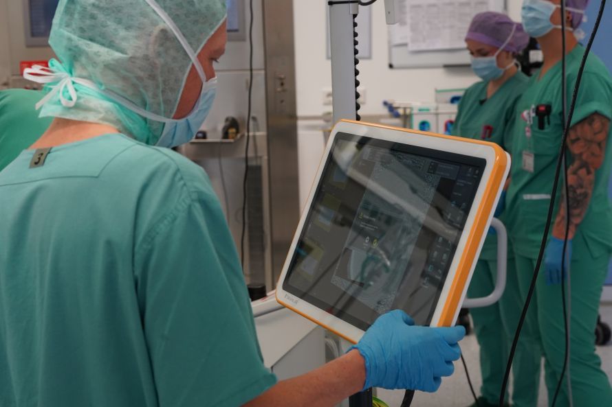
Sascha Zink comments on the challenges of the surgery: “Although the surgical approach is precisely planned—we know which anatomical spaces to open and how to approach the parent vessel—the dissection remains a highly delicate moment. To keep the access route clear, brain spatulas are used to gently hold the brain to the side. Even this necessary manipulation can be critical if the aneurysm wall is extremely thin, affected by atherosclerosis, or further weakened by small ‘baby aneurysms.’ During dissection, fine suction devices must also be used to gently remove a minimal amount of brain tissue in order to fully visualize the course of the feeding vessel. This is because when placing the clip, only the aneurysm itself must be occluded—never the feeding or draining vessel.
A mistake would cause a circulatory disturbance and thus a stroke. The most delicate phase is the moment when the aneurysm could rupture due to manipulation. At that point, the assistant must react in a flash and control the spurting blood with the suction device so that the surgeon can still place the clip safely. In an open skull, such a rupture is a serious but manageable situation—quite unlike in everyday life, where the rise in pressure within the closed cranial cavity would be life-threatening.
Another challenge lies in the selection and precise placement of the clip. There are numerous variants—long, short, curved, with a recess for the supporting vessel, or right-angled—and the choice must precisely match the individual vascular anatomy. Ideally, the clip should be placed in a single, precise step. Multiple attempts would mechanically irritate the aneurysm, dislodge thrombi, and, in the worst case, cause emboli that could lead to strokes in other areas of the brain. It is precisely this combination of delicate dissection, the potential risk of rupture, and the need for absolute precision that makes this operation one of the most challenging procedures in neurosurgery.”
Follow-up care after aneurysm treatment is a crucial part of the therapy because it determines how well the brain recovers and how stable the treatment outcome remains in the long term. It begins immediately after the procedure and continues for months or even years—depending on whether clipping or coiling was performed and how complex the initial findings were.
“Once a clip has been successfully placed, the affected aneurysm is permanently closed off. A new aneurysm cannot form at the same site. In these cases, follow-up care primarily serves to confirm the outcome and monitor the remaining brain vessels. So we’re not monitoring the clipped aneurysm itself, but rather checking to see if new changes could develop in other areas. With coiling, the situation is somewhat different. Here, too, the aneurysm is closed off, but in rare cases, a so-called ‘neck remnant’ may occur—a small remnant at the neck of the aneurysm through which a minimal amount of blood can flow. Some of these remnants disappear on their own over time, others remain stable and cause no problems, and still others slowly refill.
In such cases, repeat endovascular treatment may be advisable or—depending on the findings—clipping at a later date. Such decisions are always made on a case-by-case basis, as the course of the condition and anatomy vary from patient to patient. Regardless of the procedure, there is always the possibility that new aneurysms may develop elsewhere. Anyone who has ever had an aneurysm therefore remains under long-term care and is not discharged from follow-up care. The prognosis depends not only on the procedure but also on the patient’s willingness to consistently reduce risk factors: smoking and high blood pressure are considered the most significant risk factors.
Diet, exercise, and stable blood pressure control also play a role. Some patients additionally benefit from rehabilitation after the procedure, for example, if they experience psychological stress, concentration problems, or exhaustion. If there have already been several cases of aneurysms in the family, screening for relatives may also be advisable. “Treatment does not end with the hospital stay—it transitions into long-term care that combines medical monitoring, lifestyle optimization, and safety in everyday life,” emphasizes Sascha Zink.
A stable long-term outcome is achieved primarily through consistent management of risk factors: well-controlled blood pressure, quitting smoking, healthy cholesterol levels, and stable vascular function reduce the risk of new or growing aneurysms. After stent procedures, blood-thinning medication may be temporarily necessary. Following a rupture, neurological rehabilitation supports recovery. The prognosis depends on whether the aneurysm had already ruptured, how quickly treatment was administered, which procedure was chosen, and how stable the condition remains. Aneurysms detected early and safely clipped usually have a very good prognosis, whereas the course of the disease can be significantly more critical following a subarachnoid hemorrhage.
At the Knappschaftkliniken Bottrop, aneurysms have been treated continuously since the launch of neurosurgical and neuroradiological care in January 2025.
“Looking at the period up to June 2026—that is, about a year and a half—a total of approximately 18 patients were treated. About half of these procedures were surgical, and the other half were endovascular. These include both specifically referred cases and incidental findings that were discovered during other treatments—such as thrombectomies—and subsequently treated as well. For a hospital that did not have its own neurosurgery department until early 2025 and is only now on its way to becoming a neurovascular center, these figures are remarkable. They are on par with what even larger tertiary care providers outside the university medical system achieve.
University hospitals naturally treat significantly more cases—sometimes one aneurysm per week—but these are specialized centers with a supraregional catchment area. In Bottrop, on the other hand, this infrastructure is only just being established—and the case numbers reflect both the rarity of the condition and the successful development of the new care services. It is also essential that care is guaranteed around the clock.
Neurosurgery and neuroradiology provide a full on-call service every day, ensuring that emergencies from within the hospital, from partner hospitals, or from external sources can be handled at any time. “This means that Bottrop offers 24/7 care, which is by no means a given for a facility of this size and is becoming increasingly established in the region,” emphasizes Sascha Zink, speaking about the future Neurovascular Center in Bottrop:
“At the neurovascular center currently under construction in Bottrop, neurology, neurosurgery, neuroradiology, and neuroanesthesia are already working together as a functional unit. Diagnostics, electrophysiological procedures, conservative and surgical therapies, as well as care in monitoring, intermediate care, and intensive care units are fully covered. This enables vascular diseases such as aneurysms or arteriovenous malformations to be treated with the necessary personnel and technical resources.
The goal is to further develop this structure so that it is officially recognized as a neurovascular center—a status tied to defined case numbers, documented quality, and seamless 24/7 care. This requires that all participating departments systematically monitor and statistically record their treatments. The data is later incorporated into the evaluation by the German Society for Neurosurgery, which decides whether a center receives the corresponding certification based on case numbers, expertise, and quality of care.”
In neuro-oncology and spinal surgery, there are collaborations with the respective professional societies; senior physicians and chief physicians hold the appropriate certifications, and procedures are performed in accordance with guidelines.
At the conclusion of our conversation, Sascha Zink, Chief of Neurosurgery, explains: “The hospital has received funding from the state of North Rhine-Westphalia for further expansion—including the development of the pediatric clinic, the expansion of the neurovascular center, additional monitoringand rehabilitation beds, as well as possibly a second DSA system for diagnostics and therapy (digital subtraction angiography—the gold standard for high-resolution imaging of cerebral vessels).
Modern technology such as neuronavigation is also part of this, enabling the precise planning of surgical approaches. Another key component is intraoperative neuromonitoring: A dedicated electrophysiologist accompanies every operation, records SSEPs, MEPs (key procedures in intraoperative neuromonitoring), and other potentials, and monitors the function of the affected nerve pathways in real time. This significantly increases safety during tumor, spinal, and vascular procedures. In addition, the clinic is already a member of the Ruhr Region Neurovascular Network, which lists facilities that provide all three treatment modalities—conservative, surgical, and endovascular—around the clock.
Regular conferences and participation in regional case conferences underscore the clinic’s commitment to quality. Looking ahead, the clinic plans to use the data and experience it has gathered to officially position itself as a DGNC flagship clinic (an officially certified center of excellence for neurosurgical conditions) and a neurovascular center of excellence. Another aspect of quality assurance concerns intraoperative monitoring: During microsurgical procedures, angiography using indocyanine green dye is performed. Modern Leica microscopes or virtual reality visualizations immediately show whether the aneurysm has been completely excluded from the circulation and whether all feeding and draining vessels remain patent.
Together with neuromonitoring, this ensures a high level of safety—both for the surgical team and for the patients, who can see immediately that the “time bomb” has been reliably defused.”
- Chief of Neurosurgery at the Knappschaft Clinics in Bottrop, Specialist in Neurosurgery
- Specialist in brain and skull base surgery: cerebrovascular diseases, brain tumors, pituitary surgery, traumatic brain injury
- Spine expert: microsurgical procedures on the cervical, thoracic, and lumbar spine; endoscopic spine surgery; cervical spine prostheses
- Peripheral nerve surgery and pain management procedures
- Certified additional qualifications: specialized oncological neurosurgery (DGNC), endoscopic spine surgery, interventional radiology, and radiation protection
- Extensive clinical career, progressing from resident to a leadership position at the Evangelisches Klinikum Niederrhein
- Broad range of treatments: surgical precision combined with conservative therapeutic approaches
Share this article
Expert Interviews
Read next
- Expert interview
Expert interview with Professor Dr Elmar Herbst, PhD
Oct 15, 2026
Patient-centred and personalised knee surgery
Read more - Expert interview
Cardiogeriatrics: Significance and benefits in the context of hospital reform
Sep 21, 2026
Cardiogeriatrics: Significance and benefits in the context of hospital reform
Read more - Expert interview
Expert Interview with Professor Dr. Amadeus Hornemann, MPH
Sep 18, 2026
Prof. Amadeus Hornemann on Precision Medicine in Gynecology: Modern Surgery, New Techniques, Better Quality of Life
Read more