Expert Interviews
The Sarcoma Center in Hamburg – High Level of Expertise and Innovative Treatment Options Using Intraoperative Radiotherapy: An Expert Interview with Prof. Tonus

Alexandra Pfitzmann ·
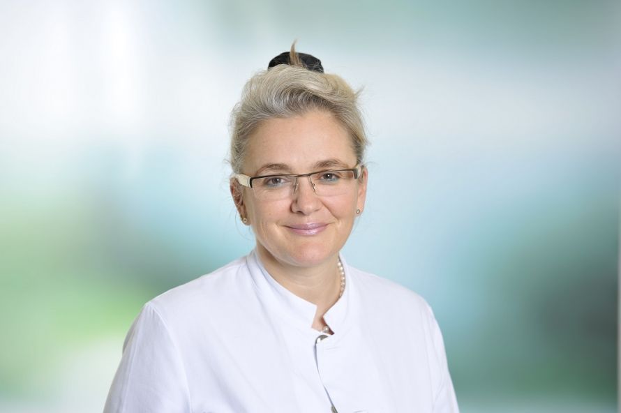
Professor Dr. med. habil. Carolin Tonus is a recognized specialist in visceral surgery, particularly in colorectal surgery, proctology, and oncological surgery. She is highly regarded internationally and specializes in the treatment of tumors of the gastrointestinal tract as well as rare soft-tissue tumors such as sarcomas. Her expertise encompasses complex procedures at both early and advanced stages of the disease. Particularly noteworthy is her introduction of intraoperative radiation therapy (IORT), which she successfully established at the Asklepios Klinik St. Georg. This procedure makes it possible to precisely irradiate residual tumor tissue during surgery to prevent recurrences and spare healthy tissue.
Since 2016, Prof. Dr. Tonus has headed the Department of General and Visceral Surgery and served as Medical Director at the Asklepios Clinic St. Georg in Hamburg. In this role, she plays a key role in optimizing clinical processes and quality assurance. Her clinical work focuses primarily on the treatment of colorectal cancer, covering both early and advanced stages. She demonstrates her high level of expertise particularly in complex procedures involving the gastrointestinal tract.
In 2017, Prof. Dr. Tonus founded the Central Sarcoma Center at the St. Georg Clinic, which is considered one of the leading centers for the treatment of rare soft tissue tumors in Germany. This center facilitates interdisciplinary collaboration among experts from various medical fields—including radiology, pathology, oncology, radiation therapy, and surgical subspecialties—to develop a personalized treatment plan for each patient. Prof. Dr. Carolin Tonus has established herself as a leading expert in the field of visceral surgery and is setting new standards in the treatment of tumors and complex abdominal diseases with innovative methods.
The editorial team of the Leading Medicine Guide was able to learn more about sarcomas and innovative treatment options, such as intraoperative radiation therapy (IORT), in a conversation with Prof. Dr. Tonus.
Sarcomas are a rare and particularly challenging group of tumors that can originate in bone, muscle, fat, cartilage, and blood vessels. Due to their rarity and the often difficult diagnosis, their treatment is one of the most complex in oncology. Many patients are faced with the question of how they can best defend themselves against these aggressive tumors. But thanks to innovative treatment options, there is new hope today. Intraoperative radiation therapy makes it possible to deliver radiation directly and precisely to the tumor site during surgery—a promising method that can significantly reduce the risk of recurrence and spare surrounding tissue. To ensure precise diagnosis and optimal treatment when sarcoma is suspected, the Asklepios Tumor Center Hamburg brings together the expertise of experienced specialists from across Hamburg at the Asklepios Clinic St. Georg and is the only clinic in northern Germany—and one of only three clinics nationwide—to offer intraoperative radiation therapy (IORT) using the brachytherapy technique. Prof. Dr. Tonus completed his habilitation on this procedure in 2002.
A sarcoma is a rare type of malignant tumor that originates in the body’s connective tissue, including bones, muscles, fat, cartilage, and blood vessels.
“Unlike carcinomas, which arise from epithelial tissue, sarcomas primarily affect the body’s supportive and soft tissue structures. A distinction is made between bone sarcomas, such as osteosarcoma, and soft tissue sarcomas, such as liposarcoma or leiomyosarcoma. The exact cause of sarcoma is not always known, but there are several risk factors that may contribute to its development. Sarcomas are malignant mesenchymal tumors, of which there are over 200 subtypes. Fortunately, they are very rare—affecting 3 to 5 patients per 100,000 people. Risk factors for developing a sarcoma include radiation exposure, asbestos exposure, and nonspecific causes. There are no symptoms specific to sarcomas as such. Hardened areas are sometimes felt in the extremities, and in the abdomen, displacement and compression can occur in advanced stages, which manifests as an increase in circumference. “About 80% of patients are affected by sarcomas in the extremities, while 20% develop sarcomas in the abdominal region, behind the peritoneum,” states Prof. Dr. Tonus.
Depending on their type and location, sarcomas can vary greatly in size. Some sarcomas reach sizes of several centimeters, while other tumors in advanced stages can grow to 20 cm or more and, in some cases, weigh 15 kilograms. Particularly large sarcomas can displace surrounding tissue and cause symptoms such as pain or functional impairment.
Diagnosis begins with a clinical examination, during which the doctor palpates for any swelling and assesses the symptoms. Imaging techniques such as ultrasound, X-ray, magnetic resonance imaging (MRI), or computed tomography (CT) are used for a more detailed evaluation. MRI is the gold standard for diagnosis. A fan-shaped biopsy, in which at least six tissue samples are taken and examined under a microscope, is crucial for confirming the diagnosis and classifying the tumor. In addition, molecular tests may be performed to identify specific genetic alterations that could be relevant for targeted therapy. Early diagnosis is crucial, as it can significantly improve the chances of successful treatment. “The patient’s subsequent treatment is discussed in an interdisciplinary tumor board to develop the best possible therapy. The board consists of colleagues from radiology, pathology, radiation oncology, medical oncology, and angiology, as well as the surgeon with the necessary specialized expertise—for example, a visceral surgeon, orthopedic surgeon, vascular surgeon, neurosurgeon, or colleagues from plastic surgery,” explains Prof. Dr. Tonus.
Sarcoma Support Group
With support from the “German Sarcoma Foundation” and “KISS Hamburg” (Contact Self-Help Referral Service), a self-help group was founded for this very rare form of tumor. Contact the support group through the General and Visceral Surgery Secretariat at the Asklepios Clinic St. Georg. Tel.: 040 1818-852144.
The treatment of sarcomas is generally multimodal, meaning it combines various therapeutic approaches to achieve the best possible results.
The exact selection and sequence of these treatments depend on factors such as the type, size, location, and stage of the tumor. “Complete surgical removal of the tumor—an R0 resection—is always the preferred option and the only curative approach. We must always strive to remove the tumor as radically as possible!” emphasizes Prof. Dr. Tonus, adding: “The procedure, which includes intraoperative radiation therapy, is initially performed surgically just as in any tumor surgery. The adjunctive radiation therapy, which was previously discussed in an interdisciplinary tumor board, is then an ‘add-on’ to a surgery that would be necessary anyway. This is because, after the tumor is resected, the wound is not immediately closed as is usually the case; instead, an intermediate step is performed—intraoperative radiation therapy, which is administered as brachytherapy at the Asklepios Klinik St. Georg. In this procedure, the desired radiation source is placed directly into the area of the resected tumor—specifically, the high-risk zone of the tumor bed. This allows the radiation to target the diseased tissue where microscopic tumor cells may still be present. The surrounding healthy tissue is largely spared in this process. This procedure is not a standard treatment but is used for large, locally advanced tumors as well as for recurrences, i.e., tumors that have returned. IORT is primarily used for rectal cancer and soft tissue tumors (sarcomas), but it is also used for other advanced tumors. IORT is only appropriate when, after tumor resection, only small residual tumor fragments remain in the tumor bed, since the radioactive rays do not penetrate deep into the tissue.” Because the radiation travels only a short distance within the body, the name “brachytherapy” is derived from the Greek word “brachys,” meaning “short.”
Compared to conventional external beam radiation therapy (EBRT), IORT offers several advantages and is specifically used in the treatment of sarcomas.
“The entire treatment is shaped by the surgical component. To ensure the therapy proceeds optimally, surgeons, radiation oncologists, and medical physicists work closely together. The great thing is that the computer system used understands what the patient’s body looks like after surgery, allowing it to precisely reconstruct the required flap so that the radiation—whose dose was calculated by the radiation physicist—can be delivered with pinpoint accuracy. Imagine the flap as a small ball-shaped mat with integrated hollow catheters, made of biocompatible materials. This size-customized flap, which functions as a radiation applicator, is used to deliver precise treatment. This method allows for high accuracy and efficiency. Through the thin, hollow tubes, the radiation source is delivered directly to the tumor bed—which, based on the prior calculation, remains exactly where it is needed—using a device called an afterloader, a computer-controlled device, ensuring that the radiation is delivered with extreme precision. The hollow catheters deliver the radiation at a customized dose directly into the tumor bed without unnecessarily exposing surrounding tissue or nearby organs to radiation. Radiation-sensitive organs, such as the small intestine or the ureters, can be kept out of the radiation field for the duration of treatment using retractors and abdominal drapes. The dose also depends on the type of tumor being treated, with sarcomas requiring a very high radiation dose. Once the radiation treatment is complete, the afterloader automatically retracts the radiation source, and the catheters can be removed. The duration of the procedure always depends on the size of the area to be irradiated, averaging between 30 and 60 minutes.
The vital signs of the patient, who is covered with a sterile drape, are closely monitored by the anesthesiologist via cameras and monitors throughout the entire treatment in the so-called brachytherapy suite, a separate, enclosed room where the patient lies. All stages of the IORT treatment can be viewed on a monitor by the surgeon, anesthesiologist, radiation oncologist, and physicists in an adjacent room. Using the software, it is possible to see exactly how the flap fits within the patient’s body, how the radioactive material flows through the catheters, and how much radiation is delivered and where. The surgeon is, of course, always nearby to intervene if necessary. This separate room must meet many criteria regarding statutory radiation protection; it is built on 60 metric tons of steel and shielded with 60 metric tons of lead. Otherwise, we would not be permitted to work with radioactive substances, as required by the Nuclear Weapons Act,” explains Prof. Dr. Tonus. “IORT is very manageable, safe, and has few side effects,” the passionate surgeon emphasizes.
In the days following surgery, patients are closely monitored to detect any potential side effects or complications early on. Depending on the type of tumor and the individual’s situation, treatment may be continued—for example, with adjuvant radiation therapy, chemotherapy, or rehabilitation measures.
“IORT does not replace adjuvant external beam radiation therapy, but conversely, IORT can effectively target areas that external beam radiation therapy would not reach—for example, because organs are in the way, which can be specifically moved out of the way during IORT. And if part of the required radiation dose has already been administered during surgery, the duration of the subsequent radiation therapy is shortened. This is, of course, also beneficial for the patient from a time perspective, as they need to visit their treating physician less frequently for radiation therapy,” explains Prof. Dr. Tonus.
Thank you very much, Prof. Dr. Tonus, for this fascinating insight into intraoperative radiation therapy!
Share this article
Expert Interviews
Read next
- Expert interview
Expert interview with Professor Dr Elmar Herbst, PhD
Oct 15, 2026
Patient-centred and personalised knee surgery
Read more - Expert interview
Cardiogeriatrics: Significance and benefits in the context of hospital reform
Sep 21, 2026
Cardiogeriatrics: Significance and benefits in the context of hospital reform
Read more - Expert interview
Expert Interview with Professor Dr. Amadeus Hornemann, MPH
Sep 18, 2026
Prof. Amadeus Hornemann on Precision Medicine in Gynecology: Modern Surgery, New Techniques, Better Quality of Life
Read more