A femoral hernia (also known as a thigh hernia, hernia femoralis, or femoral hernia) develops below the inguinal ligament at a weak spot next to the groin. Women are particularly affected—older women more frequently than men—and repeated pregnancies, weak connective tissue, heavy lifting, or previous surgeries (e.g., after inguinal hernia surgery) increase the risk. Typical symptoms include swelling or a bulge on the thigh and a pulling pain; the condition becomes dangerous if the hernia becomes incarcerated (incarceration = an acute complication accompanied by severe pain). Treatment for a femoral hernia is usually surgical—either open surgery or laparoscopic surgery (TEP/total extraperitoneal or TAPP/transabdominal preperitoneal technique)—often performed minimally invasively through small skin incisions using a synthetic mesh.
Symptoms of a femoral hernia: Swelling or a bulge below the inguinal ligament in the groin area, pulling pain in the thigh; if there is severe pain, a firm, non-reducible bulge, or nausea/vomiting, suspect an incarcerated hernia—in which case surgery is necessary.
Treatment: Femoral hernia surgery is performed as open surgery or laparoscopic surgery (TEP/TAPP, a minimally invasive technique)—often on an outpatient basis. The goal is to reduce the hernial sac back into the abdominal cavity and to repair the hernial orifice with minimal tension using a mesh (synthetic mesh). The choice of surgical technique in hernia surgery depends on various factors (size of the hernia, previous surgeries, general health). Can a femoral hernia go away without surgery? No—a femoral hernia does not heal on its own; surgical treatment reduces the risk of incarceration and recurrence.
Definition – What is an inguinal hernia?
A hernia is a rupture of the abdominal wall with tissue or a hernial sac protruding through a hernial orifice. In a femoral hernia, tissue protrudes below the inguinal ligament into the femoral canal—usually as a palpable swelling in the groin area or on the inner thigh. Femoral hernias are more likely to become incarcerated than inguinal hernias; therefore, surgical treatment is usually necessary. Women are more commonly affected; femoral hernias in men are less common. Diagnosis: physical examination, ultrasound; CT or MRI if findings are unclear.
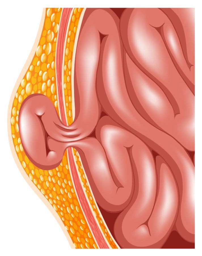
Causes and Development of a Femoral Hernia
A femoral hernia (also known as a thigh hernia, hernia femoralis, or femoral hernia) develops at a weak spot in the abdominal wall below the inguinal ligament in the groin region. Increased abdominal pressure and/or weakness of the connective tissue causes the hernial orifice in the femoral canal to widen. Initially, fatty tissue often protrudes; later, the hernial sac (a protrusion of the peritoneum) may fill with contents from the abdominal cavity—visible as a bulge or swelling on the inner side of the thigh near the groin.
Risk Factors—When Does a Femoral Hernia Develop?
- Women in particular (femoral hernias in women, especially older women) are anatomically more prone to femoral hernias than men.
- Repeated pregnancies, obesity, heavy lifting, chronic coughing or straining.
- Previous surgeries (e.g., after inguinal hernia surgery) resulting in scarring in the area of the connective tissue structures.
- General weakness of connective tissue associated with aging.
Incarceration as a complication:
If parts of the intestines pass through the hernial orifice, they can become trapped (incarceration)—often causing severe pain. Blood supply may be compromised, leading to complications that can include the necrosis of intestinal loops. Due to the proximity to the femoral vessels, the situation must be evaluated by a physician promptly.
Lateral distribution:
About 60% of inguinal hernias occur on the right side, and about 20% are bilateral.
If a femoral hernia occurs, a femoral hernia typically does not heal on its own—if symptoms or signs of incarceration are present, a diagnosis (e.g., physical examination, ultrasound) should be made promptly.
Diagnosis of a femoral hernia
How do you recognize a femoral hernia? A typical sign is swelling or a bulge below the inguinal ligament at the junction of the thigh and groin. The bulge may become more pronounced when coughing or straining. In cases of obesity, skin and tissue sometimes obscure the signs—making the inguinal hernia more difficult to detect (“recognizing an inguinal hernia” is not always clinically straightforward).
Diagnosis: First, a physical examination is performed with the patient standing and lying down, including a coughing or straining test. Ultrasound (sonography) then typically confirms the findings and visualizes the hernial orifice and hernial sac. If the physical examination findings are unclear or if differentiation from an inguinal hernia is necessary, CT or MRI can reliably visualize the hernia.
Warning Signs (Emergency): In cases of severe pain, a firm, non-reducible bulge, nausea/vomiting, or fever, a strangulated hernia must be suspected—in such cases, prompt medical evaluation is required, as blood flow may be compromised.
Surgery for Femoral Hernias
Surgery is usually necessary for a femoral hernia because the risk of incarceration is high—femoral hernias often cause acute symptoms. If symptoms of a femoral hernia (swelling or pain in the groin area) are present, or if the intestine is strangulated, surgery should be performed promptly; in an emergency, surgery is required immediately. Does a femoral hernia go away on its own? No—conservative treatments do not cure the hernia. Inguinal hernias are less common in men than in women.
Anesthesia & General Conditions: The procedure can be performed under local or general anesthesia; in cases of incarceration, general anesthesia is usually preferred to allow for enlargement of the incision if necessary. Many procedures are performed on an outpatient basis. Depending on the findings, the surgery often takes less than an hour.
Surgical Procedures / Techniques
: Open surgical procedures and minimally invasive/laparoscopic techniques are available. The goal is to reduce the hernial sac (return it to the abdominal cavity) and to close the hernial orifice with minimal tension, usually using a mesh implant.
- Open Surgery: Through a targeted skin incision in the groin area (below the inguinal ligament), the hernial sac is exposed, its contents are returned to the abdominal cavity, and the hernial orifice is closed. For very small defects, a suture in the connective tissue may be sufficient; more commonly, a synthetic mesh is inserted for stabilization to prevent recurrence.
- Laparoscopic Surgery (TEP/TAPP):
TEP (total extraperitoneal) and TAPP (transabdominal preperitoneal) are preperitoneal procedures performed through small skin incisions. The surgeon works gently using a camera and instruments; the hernia is repaired from the inside, and the mesh is placed behind the hernial orifice without tension. These procedures are particularly gentle and often allow for a quick recovery.
Why use a mesh? A mesh implant relieves pressure on the connective tissue and reduces the risk of recurrence. The choice of technique (open vs. TEP/TAPP) depends on various factors: the size and location of the hernia, previous surgeries, comorbidities, and the center’s experience.
Important: If there are signs of incarceration (severe pain, a firm, non-reducible swelling, nausea/vomiting), prompt action is required—surgery is necessary in these cases.
Preparatory Measures Before Inguinal Hernia Surgery
Many inguinal hernia surgeries (femoral hernia) can be performed on an outpatient basis—especially when using laparoscopic, minimally invasive techniques (TEP/TAPP). On the day of surgery, you may go home accompanied by someone after the usual monitoring. In cases of a strangulated hernia or planned open surgery, a short inpatient stay is often advisable.
Information & Planning
Before the procedure, the surgeon and anesthesiologist will discuss the surgical technique (open vs. TEP/TAPP), the planned mesh implant (synthetic mesh), and the type of anesthesia (local anesthesia or general anesthesia). A history of previous surgeries (e.g., for inguinal hernia), comorbidities, and medications is taken; blood thinners are adjusted as needed.
Preparation on the Day of Surgery
- Fasting as instructed (no food or drink).
- Shaving and disinfection of the groin area; marking of the planned incision.
- Laparoscopic surgery involves small skin incisions; open surgery involves a targeted incision below the inguinal ligament.
- Depending on the procedure, surgery often takes less than 60 minutes.
After the Surgery
After a brief period of monitoring, you will receive instructions on wound care and mobility. A wound check is usually performed after 2–3 days; sutures are removed later—depending on the material—or dissolve on their own. If there are signs of incarceration (acute pain, a firm, non-reducible swelling, nausea/vomiting), the surgery cannot be postponed.
Surgical Methods for Inguinal Hernia Repair
The goal of surgical treatment is to reduce the hernial sac (return it to the abdominal cavity) and to close the hernial orifice with minimal tension—usually using a mesh implant (synthetic mesh). Two surgical techniques are available:
Open femoral hernia surgery via an abdominal incision
Through a precise skin incision in the groin area (usually below the inguinal ligament), the hernial sac is exposed, its contents are returned to the abdominal cavity, and the hernial sac is shortened if necessary. The hernial ring is then closed:
- Direct suture (for very small hernias) in the connective tissue.
- Fascial duplication (Shouldice): overlapping sutures of the abdominal wall fasciae for mesh-free stabilization.
- Lichtenstein method: A synthetic mesh placed as an onlay on the fascia transversalis (between the abdominal wall fascia and the muscles).
The mesh implant generously overlaps the stable tissue so that connective tissue can heal reliably and recurrences are less common.
Closed inguinal hernia surgery via laparoscopy
This laparoscopic, minimally invasive procedure is performed through small skin incisions (approx. 2–12 mm) using a camera and instruments. The hernia is exposed at its origin, the hernial sac is reduced, and the hernial orifice is secured prepereitoneally with a non-absorbable polypropylene mesh; the peritoneum is then closed. Two established procedures:
- TEP (total extraperitoneal mesh repair): extraperitoneal approach; the peritoneum remains closed.
- TAPP (transabdominal preperitoneal mesh repair, “TAPP”): Transabdominal approach, preperitoneal mesh placement, closure of the peritoneum. Advantages: minimally invasive, rapid return to activity, often performed on an outpatient basis; the surgery typically takes less than 60 minutes.
Risks and Complications of Inguinal Hernia Surgery
In addition to the possible general complications common to all surgeries, such as
- bleeding,
- infections,
- thrombosis, and
- the risk of embolism
, swelling in the surgical area is possible following inguinal hernia surgery. This swelling is caused by bruising or fluid accumulation in the tissue and usually subsides after a short time.
Mild pain in the surgical area or sensory disturbances may also occur after inguinal hernia surgery. While the pain usually subsides quickly, sensory disturbances in small areas of the skin may persist for a longer period.
Surgery in close proximity to the intestines, bladder, blood vessels, and nerves always carries a potential risk to these structures. However, injury to major blood vessels during femoral hernia surgery is extremely rare.
In particular, blood flow in the large vein that runs through the femoral canal must remain unimpeded. Otherwise, there is a risk of thrombosis.
Furthermore, there is a risk that a femoral hernia will recur at the same site. This risk exists with all femoral hernia surgery procedures; however, it is lower when synthetic meshes are used.
Hardening and shrinkage in the area of the implanted synthetic meshes are extremely rare in femoral hernia surgery. Allergic reactions or rejection of the synthetic meshes are virtually nonexistent.
What to Expect After Inguinal Hernia Surgery
Immediately after the procedure:
- After inguinal hernia surgery, depending on the type of anesthesia and procedure, you’ll usually be able to get out of bed on the day of the surgery. Prolonged bed rest is not necessary and can actually be harmful due to the risk of thrombosis.
- For outpatient procedures, you’ll go home with an escort after a brief monitoring period; for inpatient procedures, you’ll also be encouraged to get moving early.
Wound Care & Follow-Up Visits:
- The first dressing change usually takes place after 2–3 days; from then on, showering is generally possible again (just briefly wet the wound and pat it dry).
- Sutures are removed around day 10—this is not necessary if absorbable sutures were used.
- Watch for signs of infection in the groin area (increasing swelling, redness, warmth, fever) or secondary bleeding. If you notice any of these symptoms, please see a doctor.
Physical Activity & Daily Life:
- Work: Depending on your job, you can return to work after 2–3 weeks; physically demanding work may require a longer recovery period.
- Sports: Light activity (walking) immediately; more intense sports after 3–4 weeks—depending on whether minimally invasive or open surgery was performed.
- Lifting: Avoid lifting loads >10 kg for about 6–8 weeks to allow the hernia opening to heal properly (especially after mesh placement).
- Recovery is often faster after laparoscopic surgery with small skin incisions than after open surgery with a larger skin incision.
When should you seek immediate medical attention?
- Increasing pain or a firm, non-reducible bulge in the groin area, fever, nausea/vomiting, or significant postoperative bleeding. These symptoms may indicate a complication and must be evaluated promptly.

About the medical author
Prof. Dr. med. Ferdinand Köckerling
Medical Author · Director
Prof. Dr. med. Ferdinand Koeckerling – medical author: expert articles, professional insights and medical knowledge in the Leading Medicine Guide.
More about the medical author →