Definition: Liver Resection
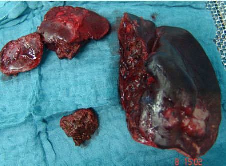
Liver resection is a surgical procedure in which a portion of the liver is removed or resected. Possible reasons for this removal or resection include benign or malignant conditions, as well as injuries resulting from accidents.
There are various surgical options for liver resection. Depending on the extent of the liver disease,
- a small piece of the liver (atypical liver resection),
- a portion of the liver (segmental resection), or
- half of the liver (hemihepatektomy)
is surgically removed.
Anatomy of the Liver
Visually, the liver is defined by
- a strong ligamentous structure (the falciform ligament and the origin of the round ligament of the liver) and
- an indentation on the side facing the other organs (the sagittal fissure)
into a larger right lobe and a smaller left lobe.
However, this does not correspond to the functional structure of the liver. The functional structure is based on the branching of the hepatic veins (portal branching) into individual, independent subunits. These are called liver segments. The hepatic veins supply the liver with blood.
According to Couinaud (a French anatomist and surgeon), eight liver segments are distinguished. These are numbered clockwise, beginning on the underside of the liver with the so-called lobus caudatus (“caudate” liver segment) as Segment I.
What makes the liver unique is its high regenerative capacity. Even if more than fifty percent of its total mass is damaged, it can regenerate almost completely. The causes of this damage are often poisoning or alcohol.
Even after surgical removal (resection) of up to 75 percent of the liver’s total mass, the liver can “regrow” completely. However, this depends on liver function.
The liver accounts for a total of 20 to 30 percent of the human body’s cardiac output. Cardiac output is a measure of the heart’s pumping function. It is calculated using the heart rate and the heart’s stroke volume per minute.
Blood is supplied via
- arterial (10 to 20 percent of the blood supply) and
- portal venous (inflows from the vena cava—portal vein)
vessels (80 to 90 percent of the blood supply) in a three-dimensional network.
Liver veins drain blood from the liver. Other vessels draining the liver are the bile ducts. The duct systems have a higher content of collagen and elastin. These are components of the walls of the individual structures. As a result, they differ significantly in their structure and resilience from the organ tissue (parenchyma) of the liver. The bile ducts are the most resilient of these structures.
Resection Techniques in Liver Resection
These characteristics can be utilized during liver resection. Resection techniques (dissection methods) that take advantage of these differences in tissue composition are referred to as selective.
These primarily include
- so-called blunt dissection,
- the so-called ultrasonic aspirator (CUSA®), and
- the so-called water-jet dissector (Water-Jet).
These should be distinguished from non-selective surgical techniques, which do not differentiate between liver parenchyma and bile duct structures.
Examples include
- mechanical instruments such as the scalpel, scissors, and—to a limited extent—the stapler, as well as
- thermal instruments such as the so-called radiofrequency coagulator, the laser, or the scissors of the so-called UltraCision®, which operate both thermally and mechanically.
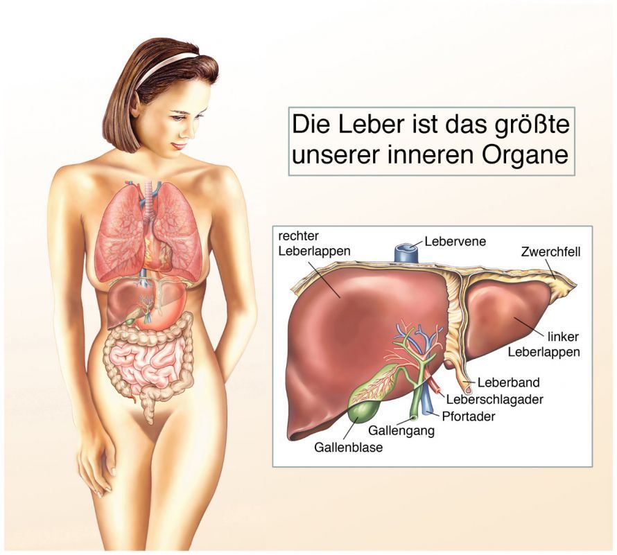
Appearance and anatomy of the liver © Henrie | AdobeStock
Outcomes of Liver Resection
Key factors influencing the postoperative outcome and the patient’s survival are
- the amount of intraoperative blood loss and
- the extent of the transfusion requirement. This refers to the amount of blood products required per operation—primarily red blood cell concentrates and blood plasma.
In modern liver surgery, therefore, surgical techniques should be used that
- minimize damage to liver tissue and
- minimize bleeding.
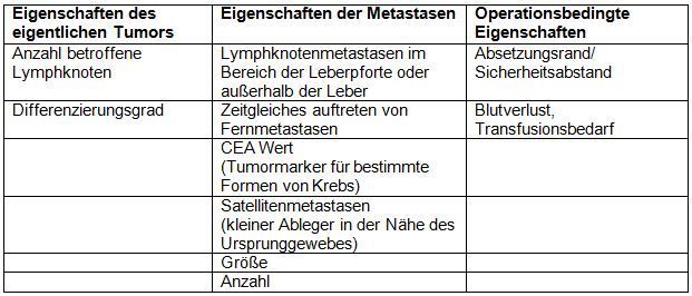
Thanks to continuous improvements in surgical techniques, the mortality rate for liver resections currently stands at 2 to 4 percent. The most important prognostic factors for survival after liver resection are listed in Table 1.
Procedure for Liver Resection
The following section discusses selected surgical approaches to liver resection. The technique used to dissect liver tissue depends heavily on the surgeon’s habits and training.
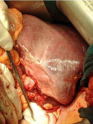
Liver Resection Using Blunt Dissection
The finger fragmentation technique was first described in 1958. In this method, the liver parenchyma is crushed between the fingers. This allows larger vessels to be isolated and subsequently ligated.
This technique is very archaic and unsuitable for modern, blood-sparing, parenchyma-sparing, segment-oriented liver surgery. This primitive form of dissection is still mentioned in a few textbooks but is no longer used in everyday clinical practice today.
Blunt dissection using a clamp is a further development of this method. In this technique, the liver tissue is compressed between clamps. At the same time, more resistant blood vessels and bile ducts are mechanically isolated from the parenchyma. The clamp technique is still used, although blood loss and operative time remain unsatisfactory. In principle, however, all variants of liver resection are possible using clamp dissection.
A modification of this clamp technique is blunt scissor dissection. In this procedure, the liver parenchyma is carefully pushed apart using closed scissors, thereby isolating the ductal structures.
The smaller ductal structures are subsequently closed with metal clips. The surgeon ligates (sutures or ties off) the larger vessels using surgical suture and a needle.
Blunt scissor dissection is a commonly used method that can be performed quickly and cost-effectively. In numerous centers, this type of dissection is
- non-nodular (cirrhotic) and
- non-enzymatically altered (cholestatic)
livers.
Liver Resection with an Ultrasonic Aspirator (CUSA®)
The principle of the ultrasonic aspirator is based on the conversion of electrical energy into mechanical energy via ultrasound. CUSA® stands for Cavitron Ultrasonic Surgical Aspirator. It uses ultrasound to cut through the liver tissue and then aspirates the resulting suspension. The suspension is a mixture of fluid and liver tissue. The energy generated by the ultrasound triggers the cutting of the liver tissue.
Due to the different tissue compositions, selective cutting of the various structures within the liver tissue is possible. Tissue with a high water content (parenchyma) is cut more quickly than tissue with a higher solid content (vessels, bile ducts).
Irrigation with saline cools the device, dissolves the severed tissue, and then aspirates it. The aspirated fluid, along with the severed tissue, can subsequently be sent for histological examination.
Another advantage of the simultaneous suction function is the reduced risk of intraoperative tumor cell seeding during tumor removal.
Studies have shown that the use of an ultrasonic aspirator during liver resections results in a significant reduction
- in intraoperative blood loss,
- the need for transfusions,
- operative time,
- morbidity and mortality, as well as
- the length of the hospital stay
. However, the use of this technique requires a relatively long period during the operation in which the liver is deprived of blood flow (known as the Pringle time).
Liver Resection with a Water-Jet
The water-jet dissector uses a high-pressure water jet to cut through liver tissue. The high-pressure liquid jet operates at a pressure of 20 to 50 bar and has a nozzle diameter of 0.1 to 0.2 mm.
This allows the liver parenchyma to be “washed away” from the blood vessels and bile ducts according to their tissue composition (hardness gradients).
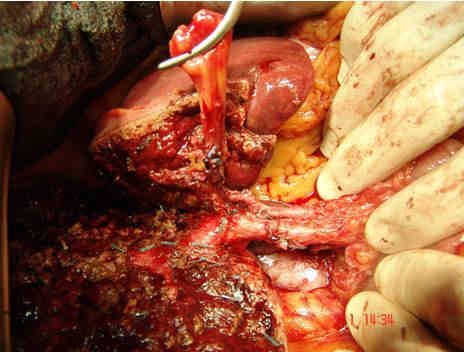
Liver resection (dissection) using the water jet can also be performed using the “keyhole technique” (laparoscopically). Studies show that, using the water jet,
- reduces intraoperative blood loss,
- liver resection time, and
- the ischemia time (the time during which the liver is not perfused)
can be significantly reduced.
The additional application of radiofrequency energy or laser energy can significantly increase the speed of liver resection. Larger vessels can thus be preserved, and smaller ones (up to 1 mm in diameter) can be cauterized with radiofrequency energy.
Due to the advantages mentioned above, water-jet-assisted liver resection is the standard procedure at our center for both open and laparoscopic liver resections.
Conclusion on Liver Resection
Surgical techniques for the resection of liver tissue have been continuously improved. Consequently, the procedure is a safe and standardized operation, particularly when performed at specialized centers.
Currently, open surgery remains the method of choice, particularly for extensive oncological liver resections (in accordance with tumor standards). However, the development of suitable instruments for efficient and safe liver surgery has led to significant advances in laparoscopic liver surgery.
The current literature shows low rates of postoperative complications for both laparoscopic and open liver resections.
Laparoscopic liver resection is associated with a shorter hospital stay and a lower complication rate. Therefore, the method should be used in patients who meet the appropriate criteria, namely
- benign liver lesions and
- smaller carcinomas located in the peripheral region
be the preferred choice.
Extensive liver resections are currently still more frequently performed using open surgery. However, these procedures are associated with higher mortality rates and longer hospital stays.
The literature lacks meaningful, large-scale studies on the oncological value of extensive liver resections performed using laparoscopic and open techniques. These studies should also include a comparison of mortality, morbidity, and length of hospital stay. Smaller studies have already demonstrated that even the removal of half the liver can be safely performed laparoscopically.
Currently, the performance of extensive laparoscopic and laparoscopically assisted liver resections is still the subject of critical debate. In laparoscopic liver resections, particularly with extensive, central lesions, there are disadvantages regarding the surgeon’s precise three-dimensional orientation, for example during dissection of the major vessels.
Bleeding complications are the most common reason for conversion to open liver resection. Other disadvantages of laparoscopic procedures include
- the often greater time required,
- the higher costs, and
- greater dependence on the individual surgeon.
Nevertheless, laparoscopic liver resections performed by experienced surgeons will increasingly become the gold standard in liver surgery in the future.