Percutaneous transhepatic cholangiography, or PTC for short, is a diagnostic procedure for the bile ducts. PTC is typically used to diagnose and, if necessary, treat bile flow disorders.
Here you will find further information as well as a selection of specialists and centers for percutaneous transhepatic cholangiography.
Definition: What is percutaneous transhepatic cholangiography (PTC)?
Percutaneous transhepatic cholangiography, or PTC for short, is an interventional imaging procedure. It is an X-ray examination that allows the bile ducts inside and outside the liver to be visualized.
Before the examination, the patient is injected with a contrast agent that is visible on X-ray images. The X-ray image clearly shows the path the contrast agent takes through the bile ducts.
PTC enables physicians to diagnose an obstruction of bile flow. If bile accumulates in the bile ducts, it can lead to various diseases. Gallstones or tumors are common causes of such obstructions.
Through the access point created for a PTC, a diagnosed bile obstruction can also be resolved in the same procedure. This is done using what is known as percutaneous transhepatic cholangiodrainage, or PTCD for short.
Indications for a PTC
A PTC is generally used to diagnose and, if necessary, treat obstructions of bile flow. However, PTC is usually performed only when a diagnosis and resolution of the bile stasis cannot be achieved using the less invasive procedure known as endoscopic retrograde cholangiopancreatography (ERCP). In these cases, the bile duct opening cannot be reached endoscopically.
Bile stasis can have both benign and malignant causes. Benign conditions include, for example, gallstones, which block the bile ducts and thus lead to bile stasis. Benign tumors and inflammatory changes can also cause an obstruction and necessitate a PTC.
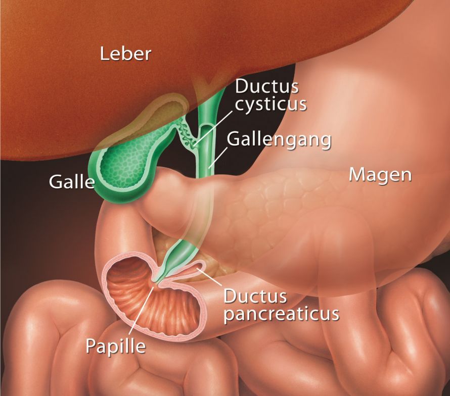
Possible malignant conditions that can lead to obstruction or compression of the bile ducts include, among others
- hepatocellular carcinomas,
- liver metastases, or
- other malignant (cancerous) tumors.
Another indication for performing a PTC is so-called leakage in the biliary drainage system. This can occur, for example, after liver surgery, after a Whipple procedure, or after a liver transplant.
Under certain circumstances, a PTC is not possible. These include, among others, severe coagulation disorders and marked ascites (accumulation of fluid in the free abdominal cavity).
Preparation for Percutaneous Transhepatic Cholangiography
A PTC is always performed on an inpatient basis. On the evening before the procedure, the doctor provides the patient with comprehensive information about the procedure and the necessary preparations.
Medications that inhibit blood clotting must be discontinued in advance. Extensive blood tests and, in some cases, comprehensive preoperative evaluations may be necessary before the PTC.
Procedure for a PTC
First, the medical team establishes sterile conditions. This includes disinfecting the patient’s skin. Next, local anesthesia is administered to the skin and the liver capsule in the area of the right flank or upper abdomen.
Next, a thin hollow needle is inserted through the skin into the biliary tract under radiographic and/or ultrasound guidance. An X-ray contrast agent is then injected into the bile ducts through this hollow needle, allowing the biliary tract system to be visualized on an X-ray image. The goal is to clearly visualize the biliary tract system.
Next, a soft wire is inserted into the bile ducts through the hollow needle, and a drainage tube is passed over it. The accumulated bile is drained outward (or, if necessary, inward) through this drainage tube (PTCD).
It is also possible to use this access to place a stent in the biliary tract system, which keeps narrowings or blockages open. The stent restores the bile drainage pathway.
Possible Complications of PTC
PTC is a safe procedure. It has relatively low complication rates and an overall mortality rate of about 1.7 percent.
However, since complication rates for ERCP are even lower than for PTC, an ERCP is usually performed—whenever possible—in cases of biliary obstruction.
Possible complications that may occur in rare cases during PTC include:
- minor complications such as bruising and hematomas, which do not require further treatment
- Hypersensitivity reactions caused by the contrast agent used
- Bleeding due to injury to venous vessels or the formation of vascular fistulas
- Bleeding from the puncture site
- Injury to the large blood vessels in the liver, resulting in bleeding into the abdominal cavity
- Injuries to the pleura if the puncture site is selected too high. This can lead to air entering the space between the parietal and visceral pleura, resulting in a pneumothorax
- Infections, localized inflammation, or blood poisoning (sepsis)
Complications such as bleeding may require further therapeutic measures such as
- blood transfusions,
- intensive care,
- surgery, or
- a chest tube
.