Angiography is an imaging technique used to diagnose vascular diseases and related injuries. This radiological procedure can be used to diagnose diseases of the veins and arteries, such as an aneurysm or peripheral arterial disease.
Below you will find additional information as well as a selection of angiography specialists.
What is an angiogram?
Angiography is an imaging procedure that provides information about the condition of blood vessels. To perform the procedure, a blood vessel is punctured (that is, a small opening is made through the skin) and visualized using X-rays. To improve the image quality, a contrast agent is usually injected. This makes the blood vessels easier to see on the X-ray image.
Doctors can then analyze the image of the inside of the blood vessel—called an angiogram—to make a diagnosis. An angiography can
- arteries (arteriography) and
- veins (phlebography), as well as
- the lymphatic system (lymphography)
.
When is an angiography performed?
Vascular diseases are the leading cause of death in Germany. For this reason, it is advisable to take preventive measures. This generally includes regular blood tests and blood pressure checks. Depending on the situation, angiography can also help examine the blood vessels using imaging techniques.
This allows for the detection of an aneurysm—a dangerous dilation of an artery, also known as an arterial bulge. If, for example, an aortic aneurysm ruptures, it poses an acute threat to life.
Peripheral arterial disease (PAD) can also be diagnosed using angiography. It is commonly known as “intermittent claudication.” In this condition, an artery is narrowed or blocked.
To facilitate diagnosis, a contrast agent is injected during the examination. The agent then spreads throughout the vascular system, making it highly visible.
Angiography has proven to be an effective method for examining vascular abnormalities and the course of blood flow.
However, many abnormalities in the blood vessels can also be detected using ultrasound examinations. An ultrasound examination carries fewer risks and is also very accurate when used on large vessels. For this reason, ultrasound is often performed before an angiography is conducted.
If the ultrasound results are inconclusive and questions remain, an angiography may follow.
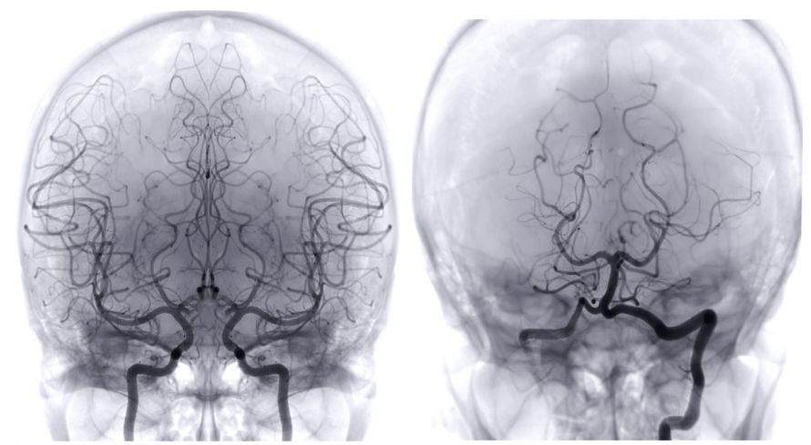
How is conventional digital subtraction angiography (DSA) performed?
In conventional digital subtraction angiography (DSA), a vessel—usually in the groin—is punctured. The patient generally feels little or nothing during the procedure. Only the injection of the contrast agent may occasionally cause a general sensation of warmth.
The physician then inserts a catheter and advances it to the target region. Next, the contrast agent is injected; it appears white on the subsequent images. The angiography machine digitally subtracts the images taken before and after the contrast agent is administered (=subtraction). Afterward, it displays only the contrast agent within the vessels, thereby revealing their precise location and course in detail.
This allows physicians to clearly identify narrowings and blockages.
If necessary, narrowings or blockages can then be treated during the same procedure. This is done using a balloon catheter, which is stabilized from the inside with a stent.
After the catheter is removed from the femoral artery, pressure must be applied to the puncture site for approximately 10–15 minutes. The patient is then given a pressure bandage to wear until the next day to prevent bleeding.
What is CT or MR angiography?
In CT or MR angiography, the contrast agent is injected through a vein in the arm. It is not necessary to puncture an artery.
The CT or MRI scanner then takes cross-sectional images, which are digitally reconstructed into three-dimensional images.
Computed tomography (CT) is a form of X-ray examination, meaning it uses X-rays. A CT angiography takes only a few seconds.
In MR angiography, a magnetic resonance imaging (MRI) scanner is used for imaging. An MRI uses magnetic fields instead of radioactive radiation. Therefore, there is no radiation exposure. An MR angiography takes about 20 minutes.
A major advantage of these two specialized procedures is that the risk of bleeding is very low, since no artery needs to be punctured.
However, if narrowings are detected, they cannot be treated simultaneously as they would be with conventional DSA.
When is which angiography technique preferred?
The three methods cannot be used equally for every patient.
For example, an MRI scan is not possible if the patient has metal implants or devices in their body, such as
- an insulin pump,
- a neurostimulator
- or a cardioverter-defibrillator.
In such cases, the physician may opt for CT angiography instead. Local anesthesia is usually sufficient.
An MRI angiography is recommended in cases of
- a known allergy to iodine-containing contrast agents or
- hyperthyroidism.
A different contrast agent is used here than in CT angiography or DSA. This agent does not contain iodine, which significantly reduces the risk of hyperthyroidism or allergic reactions.
What are the risks of angiography?
Complications from angiography are generally rare, but a residual risk cannot be ruled out.
The four most common complications are:
- Bleeding and post-procedural bleeding
- Allergic reaction to the contrast agent
- Kidney damage
- Hyperthyroidism
How can bleeding be prevented?
The puncture site is usually in the groin. Bleeding into the surrounding tissue can occur here. In extreme cases, this may require surgical suturing, but it can often be treated or prevented with compression.
Therefore, the patient must remain lying quietly in bed after the examination. A pressure bandage is applied to the groin and left in place for 24 hours.
If you regularly take blood-thinning medications, you must inform your doctor. Under certain circumstances, you may need to stop taking these medications before the examination.
In addition, it is essential to have your current lab values checked. This ensures that your body has adequate blood clotting ability.
How can the risk of a contrast agent allergy be reduced?
Before using an iodine-containing contrast agent, the patient must be screened for intolerance. Allergic reactions to the contrast agent used are among the most common complications associated with angiography.
If an allergy is detected in advance, taking antiallergic medications is essential. Alternatively, an MR angiography can be performed, which does not use an iodine-containing contrast agent.
What should be considered in cases of impaired kidney function?
The kidneys are responsible for excreting the contrast agent. To do so, they must be functioning properly. If problems with excretion are anticipated, medications can help improve kidney function for the procedure.
Therefore, it is essential to check kidney function through a blood test. You should also drink plenty of fluids before and after the exam—ideally mineral water. If necessary, intravenous fluid therapy may also be required.
Why is hyperthyroidism dangerous?
Iodine stimulates thyroid function. If the patient has hyperthyroidism, thyroid function may need to be reduced before the examination. Otherwise, a shock reaction may occur during and after the angiography.
In such cases, MR angiography can be used as an alternative. Therefore, it is very important that any known functional disorders be reported prior to the examination.
What test results should be brought to the angiography appointment?
Essential tests before the procedure at a glance:
- Thyroid function test
- Kidney function test
- Test for an allergy to the contrast agent
- Blood tests (blood clotting)
- Allergy card (if available)