Early detection of breast cancer is essential for successful treatment. The earlier the diagnosis is made, the better the chances of recovery. Therefore, every woman should take advantage of the screening exams offered and consult her gynecologist if she feels a lump. However, not every lump is malignant. Breast cancer diagnostics therefore aim not only to detect tumors but also to determine whether they are benign or malignant.
Here you will find information on breast cancer diagnostic procedures as well as selected breast cancer diagnostic specialists and centers.
At what tumor size can breast cancer be diagnosed?
Many factors are important for the diagnosis of breast cancer, such as
- the size of the breast,
- the density of the glandular tissue, and
- the patient’s age.
Therefore, there is no one-size-fits-all answer to the question of exactly when breast cancer can be diagnosed. Depending on the size of the breast, inexperienced individuals can usually feel a lump starting at two centimeters in size. An experienced gynecologist can also feel lumps measuring one to 1.5 centimeters.
Earlier detection is possible with the help of imaging techniques. Mammography can detect calcifications in the breast. Calcifications are often found in what is known as carcinoma in situ. This allows lumps to be detected even before they form.
Under optimal conditions, physicians can detect tumors as small as five to ten millimeters using ultrasound and magnetic resonance imaging (MRI).
Below is an overview of the procedures used in breast cancer diagnosis.
Medical History and Physical Examination by a Gynecologist
Your gynecologist is your first point of contact if breast cancer is suspected. The diagnostic process always begins with a medical history. During a detailed consultation, your gynecologist will ask you about any possible
- symptoms,
- previous medical conditions, and
- your family medical history
.
As part of the subsequent physical examination, they will palpate your breast. They will check for any lumps and assess their location and size. The doctor will also palpate your armpits to detect any enlarged lymph nodes.
When detecting breast cancer through a physical examination, the size of the breast is the most important factor. The location of the lump also plays a significant role. The deeper the lump is located, the later it is detected and the larger it is at the time of diagnosis. Superficial lumps are usually detectable earlier.
You can also perform physical examinations yourself. At the gynecologist’s office, this is done during the annual preventive exam.
In many women, the glandular tissue is naturally somewhat lumpy. Unfortunately, this makes it more difficult to feel tumorous changes. Furthermore, a physical examination provides only an indication of tissue changes; it does not reveal whether the changes are benign or malignant.
Imaging Techniques for the Diagnosis of Breast Cancer
Imaging techniques are essential for the early detection and comprehensive diagnosis of breast cancer. Typical procedures include:
- Mammography (X-ray examination)
- Breast ultrasound
- Magnetic resonance imaging (MRI)
Mammography and breast ultrasound are the classic standard procedures in breast cancer diagnosis. Magnetic resonance imaging is used when the standard examinations have not answered all questions regarding the diagnosis.
Mammography
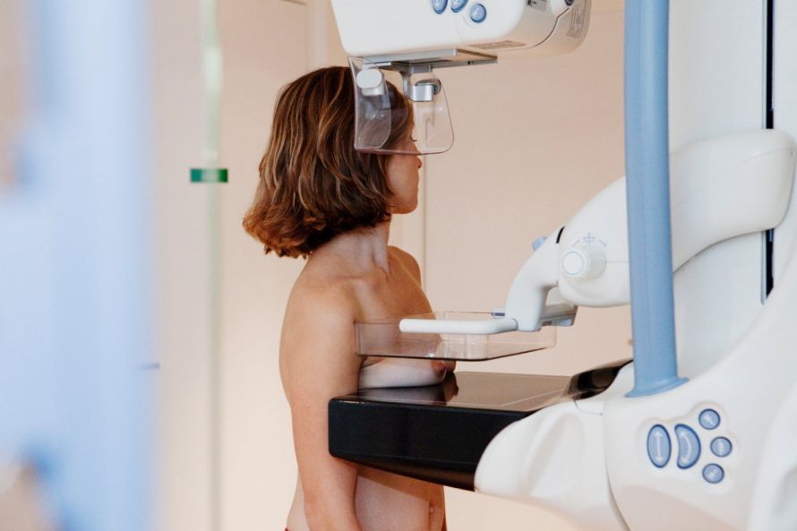
Mammography is an X-ray examination. The procedure provides images of the breast from various angles. Standard views in a mammogram are
- the top-to-bottom view,
- from the center outward, and
- from the front center diagonally toward the back and out.
If necessary, special images (e.g., of the armpit or décolletage) are also taken to better visualize specific areas.
Breast Sonography
The X-ray examination is supplemented by ultrasound examinations of the breast. However, breast ultrasound cannot replace a mammogram.
It is particularly useful in cases where mammography reaches its limits: in patients with very dense breast tissue. Even if a mammogram already provides good diagnostic information, breast ultrasound can further confirm the diagnosis.
The results of the ultrasound examination depend heavily on
- the quality of the equipment and
- the experience of the examining physician
. Therefore, an experienced specialist should perform both the mammography and the breast ultrasound.
Magnetic Resonance Imaging (MRI)
If the results of the mammogram and breast ultrasound are inconclusive, a magnetic resonance imaging (MRI) scan is also performed. Its results are very accurate. They help determine whether the carcinoma is present in other areas of the breast or has already metastasized.
Biopsy and histological examination of the breast
After the imaging procedures have been performed, it must be determined whether the lesion is benign or malignant. This is where a histological examination comes into play.
For this purpose, a so-called punch biopsy is performed. This is an outpatient procedure.
This makes it easier to plan surgical access to tumors, as the results of the core biopsy allow the doctor to visualize the lesion. As a result, safety margins around the tumor can be precisely maintained, and the procedure also yields better cosmetic results.
Frequently Asked Questions About Biopsies:
Is it possible for cancer cells to spread throughout the body as a result of the biopsy?
While the spread of cancer cells can never be completely ruled out, the risk is very low. Studies have shown that cancer cells are more likely to die off in a different location than to grow.
Is the biopsy painful?
The moment the needle pierces the skin is somewhat uncomfortable. For this reason, a local anesthetic is administered, and pain is barely noticeable.
Will the appearance of the breast change after the procedure?
Fine-needle biopsies generally do not leave visible scars. However, since punch biopsies use thicker needles, minor scarring cannot be completely ruled out.
Additional Tests in Breast Cancer Diagnosis
There are other procedures available for breast cancer diagnosis.
Galactography
Galactography can be helpful when mammography and breast ultrasound findings are unclear. In this procedure, the milk ducts are visualized on an X-ray image using a contrast agent that is injected beforehand.
This makes it easier to detect possible changes in the milk ducts.
Thermography
Breast thermography can also play a role as a diagnostic tool in breast cancer screening. In this procedure, the doctor uses infrared analysis to measure the heat emitted by the tissue.
Tumorous tissue has a higher blood supply and therefore generates more heat. This heat emission should be clearly visible in a thermogram.
However, benign tissue changes can also generate heat. Therefore, thermography cannot provide definitive results regarding whether a lump is benign or malignant. For this reason, it is not covered by health insurance.
Laboratory Tests
For laboratory testing, a blood sample is taken and analyzed for specific characteristics. These characteristics indicate how the liver, kidneys, and other organ systems are functioning.
Confirming a Breast Cancer Diagnosis
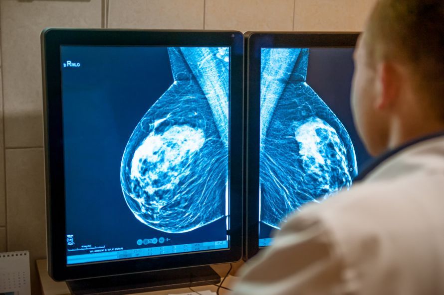
In the case of breast cancer, confirming the diagnosis is essential for further treatment. To do this, the diagnostician and pathologist collaborate. They review the results of imaging studies and histological examinations.
In practice, this means the following: If a mammogram suggests a malignant finding but the core needle biopsy results are unremarkable, a quality control check of the biopsy must be performed.
The pathologist and the physician who performed the core needle biopsy jointly review the test results for plausibility. If the results are consistent, a definitive diagnosis can be made. Appropriate treatment measures can then be initiated.
Breast cancer diagnosis involves a tremendous amount of organizational and logistical effort. For this reason, diagnoses are typically performed at certified breast centers. Treatment also takes place at specialized breast cancer centers.
Conclusion: The Goals of Breast Cancer Diagnostics
Breast cancer develops over a period of about five to ten years. Only then does the lump reach a size that can be felt. However, there are also types of cancer that grow faster or slower.
Cancers that closely resemble breast cells grow significantly slower than those that bear little resemblance to breast cells. Yet even fast-growing breast cancers can sometimes take several years to become detectable.
For this reason, breast cancer diagnostics pursue the following goals:
- early detection of previously undetected breast cancer cells
- confirming the diagnosis on an outpatient basis
- Completion of all diagnostic steps, ideally in a single appointment
- Improving the quality of treatment and patient satisfaction, while simultaneously improving quality of life
- Reducing the risk of mortality and improving the recovery rate
In addition, thorough and high-quality breast cancer diagnostics help ensure that women are better informed about breast health and breast diseases.
You can also find more information on the advice pages of the Breast Center at the University Women’s Hospital in Tübingen.

About the medical author
Prof. Diethelm Wallwiener
Medical Author · MedicalDirector – University Women’s Clinic · Tuebingen
More about the medical author →Sources
- https://www.krebsinformationsdienst.de/tumorarten/brustkrebs/diagnostik.php
- https://www.krebsgesellschaft.de/onko-internetportal/basis-informationen-krebs/krebsarten/brustkrebs/diagnose.html
- http://www.brustkrebs-studien.de/ratgeberbrustkrebs.php