Cardiology is the study of heart diseases and their treatments. This includes both surgical procedures and medication. Heart diseases can have many different causes. Coronary heart disease is common; it is characterized by a gradual “hardening” of the coronary arteries and can lead to angina pectoris or a heart attack.
Here you will find further information as well as a selection of doctors specializing in heart disease.
Common Symptoms of Heart Disease
The symptoms of heart disease can vary widely. Symptoms do not always appear at all. High blood pressure, for example, is often detected only through a blood pressure measurement.
The most common symptoms associated with heart disease are
- chest pain that may radiate to the back or arms,
- palpitations,
- chest tightness (“angina pectoris”),
- shortness of breath during physical exertion,
- decreased stamina accompanied by fatigue and a feeling of weakness, as well as
- serious heart rhythm disturbances.
Some heart conditions are acutely life-threatening and require treatment by an emergency physician. These include, above all, heart attack and cardiogenic shock.
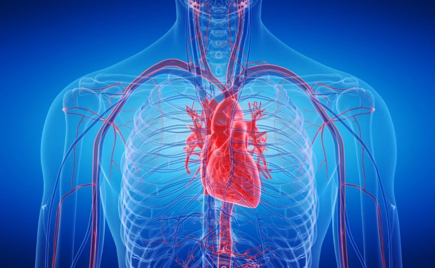
High blood pressure (hypertension)
High blood pressure (hypertension) does not cause pain and is otherwise very difficult to detect. In most cases, high blood pressure is a classic incidental finding during a medical examination.
However, consistently elevated pressure in the blood vessels can lead to damage to the vessel walls or even heart disease. If the coronary arteries are affected, this can ultimately lead to a heart attack.
With high blood pressure, the heart must work against very high resistance in the blood vessels as it pumps blood. The heart muscle must therefore work harder and harder to maintain the same pumping capacity. As a result, the muscle—and thus the heart itself—enlarges, particularly in the left ventricle.
At the same time, this enlargement impairs the oxygen supply to the muscle cells. The heart can become “tired,” resulting in heart failure.
On the other hand, high blood pressure is also a consequence of vascular and heart diseases. For example, pressure in the blood vessels can rise as a result of narrowing of the arteries (atherosclerosis).

Functional Heart Disorders (Cardiac Neuroses)
Functional heart disorders are often not organic heart diseases. Very often, psychological factors play a major role. Symptoms of functional heart disorders include, for example
- palpitations,
- chest pain,
- shortness of breath, and
- a feeling of tightness in the chest.
The associated anxiety can escalate to the point of fear of death. This led to these cardiac symptoms being labeled “cardiac neurosis.” In about 15 percent of patients with heart conditions, the symptoms cannot be explained by organic causes.
Nevertheless, organic heart disease should first be ruled out by a doctor. Only then can one speak of functional heart complaints.
In cases of functional heart conditions, common heart medications such as beta-blockers and sedatives are often ineffective. They can even make the symptoms worse.
Cardiac neuroses stem from psychological imbalance and states of anxiety. In some cases, they respond very well to psychotherapy and relaxation techniques.
Heart diseases of the inner lining (pericarditis, endocardial diseases, endocarditis)
The inner lining of the heart (the endocardium) is a very thin layer of epithelium that lines the inside of the heart. The heart valves also originate from the endocardium. After surgery or prolonged colds, bacteria can colonize the endocardium.
A very common cause of heart diseases affecting the endocardium is rheumatic fever. It occurs as a result of a streptococcal infection in childhood. The resulting inflammation leads to endocardial defects (e.g., scarring) and can also impair heart valve function. Heart diseases involving the endocardium therefore often result in heart valve defects.
The symptoms of endocardial heart disease can be very severe. These include
- chills,
- high fever,
- anemia,
- heart failure, and
- embolisms.
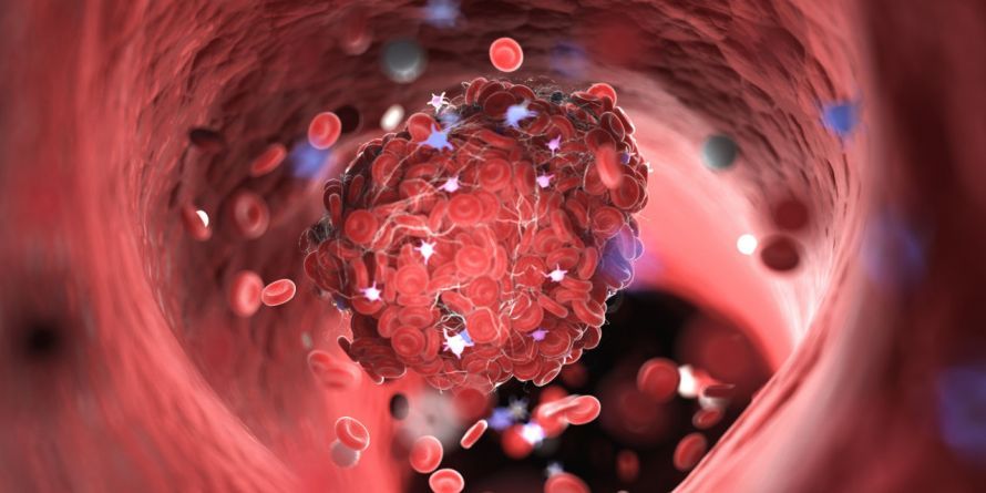
Embolisms are caused by blood clots forming in the body’s small blood vessels. Once such a blood clot breaks loose, it can travel, for example, to the brain and block small capillaries there. This results in a stroke.
Heart Valve Diseases
Heart valve defects can be congenital or develop over the course of a person’s life as a result of an infection of the pericardium.
Inflammation of the pericardium (pericarditis) initially heals with scarring. If this occurs on the heart valves, they can shrink, become distorted, or even partially fuse together. As a result, the function of these heart valves is impaired.
Heart valve defects can manifest as stenosis or insufficiency.
In valve regurgitation, the heart valves no longer close properly. With each heartbeat, blood is still pushed out through the affected valve. However, it then flows back into the ventricle because the valve does not close completely. This so-called regurgitant flow causes a murmur that a doctor can hear with a stethoscope.
Valvular stenosis, on the other hand, prevents the affected heart valve from opening fully. As a result, the opening through which blood flows is narrowed. This means the heart can pump less blood with each heartbeat.
Blood backs up in the heart in front of the valve. Over time, this blood congestion can persist and, depending on which heart valve is affected, lead to further heart conditions, including
- venous congestion or
- pulmonary edema.
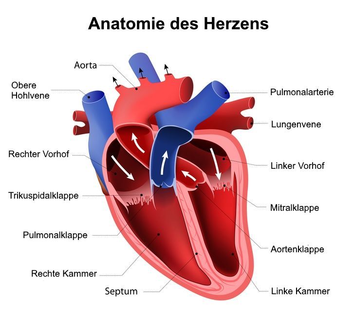
The Anatomy of the Heart © designua | AdobeStock
Diseases of the pericardium
The pericardium is also known as the heart sac. Diseases of the pericardium are therefore also referred to as pericardial diseases or pericarditis.
Heart diseases involving the pericardium often result from infections caused by
- bacteria,
- fungi, or
- viruses.
Also,
- infections in neighboring organs, such as the lungs,
- heart attacks,
- tumors in the chest cavity, and
- open-heart surgery
can lead to pericardial diseases.
Essentially, a distinction is made between acute and chronic pericarditis.
Both forms are characterized by severe pain behind the breastbone and breathing difficulties. These two main symptoms are caused by the occurrence of what is known as dry or wet pericarditis.
Dry pericarditis often occurs in the early stages of the condition. As the disease progresses, it transitions to wet pericarditis. Both epithelial surfaces of the pericardium (the inner and outer layers) are affected.
In dry pericarditis, the two epithelial surfaces of the pericardium rub dry against each other. This causes severe pain as well as friction sounds reminiscent of creaking leather.
In wet pericarditis, a fluid-filled space—known as an effusion—forms between the two layers of the pericardium. The heart rate increases to counteract the pressure. At the same time, breathing difficulties become more pronounced, as the effusion also restricts the space available for the lungs to expand during breathing.
If left untreated, pericarditis leads to overt heart diseases such as heart failure (cardiac insufficiency). Blood flow to the heart becomes increasingly restricted. As a result, there are severe impairments in heart function, and edema develops throughout the body.
Heart Muscle Diseases
The heart muscle is medically referred to as the myocardium. Inflammation of the heart muscle is therefore called myocarditis. Myocarditis is a life-threatening heart condition, as the inflammation leads to
- impaired heart function as well as
- cardiac arrhythmias
.
Bacterial or viral infections are very common triggers of myocarditis. In rare cases, however, medications (e.g., penicillin) and allergies can also trigger this heart condition.
Symptoms of myocarditis include
- shortness of breath,
- chest pain,
- rapid pulse, and
- extrasystoles (heart rhythm disturbances)
.
A rare complication of myocarditis is cardiogenic shock. This is the result of impending heart failure.
Due to myocarditis, the heart is no longer able to pump a sufficient amount of blood throughout the body. The person feels anxious and suffers from acute oxygen deprivation due to reduced blood flow.
Cardiogenic shock is classified as a cardiac emergency. The patient must be positioned with the upper body elevated until emergency medical services arrive.
A characteristic feature of cardiogenic shock is a gradual mottling of the skin on the neck, extremities, and chest.
Heart Diseases with Rhythm Disorders (Arrhythmias)
Heart conditions involving disturbances in the heart’s rhythm can be divided into three main groups:
- Tachycardia (> 100 beats per minute),
- bradycardia (< 60 beats per minute), and
- premature beats (extrasystoles).
These disorders are usually caused by problems with the conduction of electrical impulses or with the generation of electrical impulses that trigger the heartbeat. Metabolic processes, such as hyperthyroidism, can also cause cardiac arrhythmias.
Cardiac arrhythmias are dangerous whenever they significantly impair the heart’s pumping capacity. Heart rates above 160 or below 40 beats per minute lead to a noticeable reduction in the volume of blood pumped. As a result, the brain is deprived of oxygen, leading to
- dizziness,
- visual disturbances, and
- loss of consciousness
may occur.
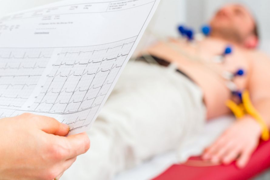
In cases of severe heart disease or a heart attack,
- atrial flutter (220 to 350 beats per minute) and
- atrial fibrillation (350 to 600 beats per minute)
may occur, leading to complete ventricular arrhythmias. These are life-threatening.
Coronary Heart Disease (CHD) and Heart Attack
Coronary heart disease is a complex of various heart conditions. Together, they claim the lives of about one in five people. These conditions include
- atherosclerosis of the heart’s coronary arteries,
- angina pectoris,
- heart attack, and
- arrhythmias, and
- heart failure.
The main risk factors for these heart diseases are
smoking,
- high blood pressure,
- high blood lipid levels,
- diabetes,
- severe obesity,
- lack of exercise, and
- advancing age.
Atherosclerosis involves the buildup of substances on the inner walls of blood vessels, which progressively narrows the vessel’s cross-section. This is also colloquially referred to as vascular calcification. The cross-sectional area of the blood vessels decreases until, in the worst-case scenario, the vessel is completely blocked.
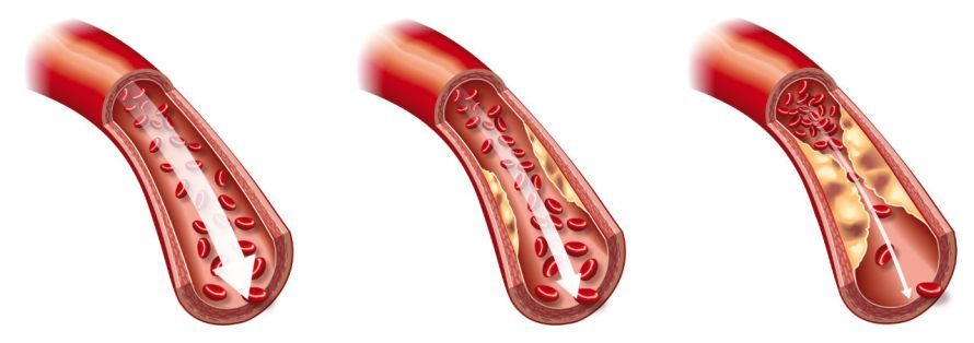
The insufficient supply of oxygen to the heart muscle leads to angina pectoris and, in the worst-case scenario, to a heart attack. It is characterized by a feeling of “tightness in the chest.” This occurs when the coronary arteries (blood vessels that supply the heart muscle) are already severely narrowed due to atherosclerosis. Especially during physical exertion (e.g., climbing stairs, walking briskly), the blood flow through these vessels is no longer sufficient to supply the heart muscle with enough oxygen.
As a result, parts of the heart muscle are deprived of blood flow for three to 20 minutes. The heart muscle reacts to this with pain, which is experienced as “chest tightness” accompanied by a stabbing pain in the heart and shortness of breath.
Rest and nitroglycerin spray provide rapid relief of symptoms. During rest, the blood flow through the narrowed vessels once again meets the muscle’s oxygen demand.
Angina pectoris attacks are always considered warning signs of an impending heart attack, so regular medical monitoring is essential.
In the case of a heart attack, the lack of blood flow to the heart muscle persists for an even longer period. This results in irreversible tissue death in the heart muscle.
In most cases, a branch of the coronary arteries is completely blocked, so that the area it supplies no longer receives blood. As a result, heart muscle cells die.
The symptoms do not differ significantly from those of an angina pectoris attack, but they usually last for more than 30 minutes. The pain associated with a heart attack is typically felt in the left side of the chest and radiates down the left arm. In women, however, a heart attack very often manifests as pain in the upper abdomen. For this reason, heart attacks in women are too often unrecognized.
A heart attack is a life-threatening emergency and requires immediate emergency medical treatment. Coronary heart disease is the most common cause of sudden cardiac death.
Heart Failure
Heart failure (cardiac insufficiency) is a heart condition characterized by the heart’s inability to pump blood effectively. As a result, less and less blood is pumped into the circulatory system.
The causes of heart failure are numerous and include
- valve defects,
- arrhythmias, and
- a heart attack.
A distinction is made between left-sided heart failure and right-sided heart failure.
Left-sided heart failure affects the left side of the heart. In this condition, blood backs up in front of the left ventricle. This accumulation of blood causes it to back up into the lungs, leading to pulmonary hypertension and the formation of edema.
The main symptoms of left heart failure therefore include, among others,
- shortness of breath,
- cardiac asthma,
- pulmonary edema,
- oxygen deficiency in the body, and
- rapid breathing.
Right-sided heart failure usually develops as a result of prior left-sided heart failure. The backflow of blood continues through the lungs and into the right heart. There, it leads to the full-blown picture of “congestive” right-sided heart failure.
Typical symptoms of these heart conditions include
Congenital heart defects
Congenital heart diseases include all conditions that develop before birth,
- valve and septal defects as well as
- vascular abnormalities of the heart.
Congenital heart defects occur in about one percent of newborns.
In an atrial septal defect, the wall between the right and left atria of the heart is not closed. Due to the increased pressure in the left atrium, oxygen-rich blood flows into the right atrium.
A naturally occurring atrial septal defect is the ductus arteriosus, which all unborn children have. It functions as a shunt to bypass the lungs, which are not yet fully functional. Thus, the ductus arteriosus is not considered a heart disease in newborns.
The second most common defect among congenital heart defects is a ventricular septal defect. In this condition, the wall separating the right and left ventricles is not closed. As a result, blood flows from the left ventricle into the right ventricle.
Symptoms depend on the size of the defect and can range from signs of oxygen deprivation to shortness of breath.
Other congenital heart defects usually affect the large blood vessels that branch off from the heart. The aorta and the pulmonary artery may be transposed at their origins. This leads to oxygen-poor blood in the systemic circulation and is life-threatening for the newborn.
Narrowing (stenosis) can also occur in the area of the pulmonary valves or the aortic arch. In what is known as tetralogy of Fallot, four heart defects occur simultaneously:
- a ventricular septal defect,
- pulmonary valve stenosis,
- enlargement of the right side of the heart, and
- aortic anomalies.
The more severe the heart defect, the more likely it is that only heart surgery can help.
FAQ
What is an acute heart attack, and how can you recognize it?
In an acute heart attack, a branch of the coronary arteries is blocked by a clot; typical symptoms include persistent chest pain that radiates into the left arm—in women, this is often felt as upper abdominal pain.
What risk factors contribute to heart disease?
The most important risk factors include smoking, high blood pressure, diabetes, high triglycerides, obesity caused by an unhealthy diet, and lack of physical activity—many of which can be reduced by adopting a healthy lifestyle.
How are heart diseases diagnosed?
Diagnostic tests include an ECG, ultrasound, blood tests, and imaging procedures; based on the findings, the cardiologist decides on further medical or surgical steps.
What does the German Heart Foundation do?
The German Heart Foundation educates those affected about common heart diseases, promotes early detection, and supports research projects aimed at improving heart health in Germany.
Share this article

About the medical author
Sabine Schneider
Editor-in-Chief
Sabine Schneider – medical author: Explore expert articles and medical expertise in the Leading Medicine Guide.
View full expert profile →