A rectal prolapse occurs when portions of the rectum protrude through the anus, forming a visible or palpable hernia. This condition is usually caused by weakness of the pelvic floor, loosening of the rectal suspensory structures, or a long-standing pattern of straining due to chronic constipation. Depending on the severity, the rectum may prolapse inward or outward, leading to mucus discharge, pain, functional disturbances, and, in some cases, fecal incontinence.
Rectal prolapse results from structural and functional changes in the rectum, the rectal wall, the pelvic floor muscles, and the connective tissue supporting structures. It is often caused by a combination of overstretching, straining during bowel movements, chronic constipation, age-related changes, scarring from difficult childbirth, or anatomical weaknesses.
Symptoms range from protrusion, mucus discharge, pain, and the urge to defecate to impaired bowel movements, incontinence, and episodic bleeding.
Diagnosis includes a physical examination, visual examination, proctoscopy, rectoscopy, colonoscopy, dynamic defecography, and imaging studies of the abdominal cavity.
Treatment depends on the severity of the condition and ranges from dietary changes and pelvic floor exercises as conservative therapy to surgical intervention. The goal of all procedures is to reposition the prolapsed tissue, restore stability and continence, and alleviate functional symptoms.
What is a rectal prolapse?
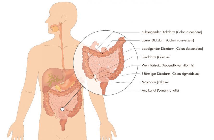
The rectum is a part of the large intestine.
A rectal prolapse refers to the invagination or protrusion of the rectum through the anal canal. A distinction is made between:
- internal prolapse, in which the intestine appears gathered inside,
- and external anal prolapse, in which a circular protrusion or a complete protrusion of the anal canal occurs from the opening of the anus.
In many cases, the condition goes unnoticed at first and occurs only during defecation; however, as it becomes more severe, the prolapse may become permanent and significantly impair quality of life.
The condition is often associated with impaired function of the anal sphincter system, leading to incontinence. Anatomical factors such as changes in the rectal wall, the positional relationship of the rectum, or age-related weakness of the supporting tissue can contribute to the progression from functional dysfunction to a visible rectal prolapse.
Epidemiology
Rectal prolapse occurs particularly frequently in older women, as the structure of the pelvic floor loses stability over the course of a woman’s life due to hormonal and childbirth-related factors (= pelvic floor descent).
Difficult childbirth is also considered a risk factor, as it can cause persistent dysfunction of the pelvic floor muscles.
Repeated straining, chronic constipation, or chronic strain on the sphincter muscle also play a role in its development.
The incidence increases with age due to a general weakening of connective tissue and muscle structures.
Diagnosis
Diagnosis of rectal prolapse begins with a thorough physical examination, during which a visual assessment can already provide clues as to the type of prolapse. If necessary, a proctoscopy is performed to assess changes in the anal region and inside the anal canal. A rectoscopy or colonoscopy is performed to evaluate the entire rectum and rule out other causes, such as hemorrhoids.
Dynamic imaging plays a central role, particularly defecography, which visualizes the mechanism of defecation and reveals whether an internal prolapse, intussusception, or external anal prolapse is present. In unclear cases, imaging of the abdominal cavity or a laparoscopy can provide additional information, particularly in the presence of comorbidities and complications.
Causes
The causes of rectal prolapse are diverse and often interrelated. Typically, there is a combination of structural weakness, functional strain, and mechanical factors. These include, above all, increasing overstretching of the rectal wall, impaired function of the pelvic floor muscles, and disorders affecting the sphincter and anal sphincter system.
Many affected individuals report long-standing constipation or regular, forceful straining, which leads to a gradual loosening and descent of the supporting structures. Following difficult childbirth, there may also be undiagnosed injuries to the pelvic floor that, over time, contribute to the development of a rectal prolapse. Anatomical variations or an unfavorable position of the rectum can also increase the risk of prolapse.
Symptoms:
Symptoms usually develop gradually. Initially, patients report occasional pressure in the anal area, a sensation of a foreign body, or difficulty passing stool. As the condition progresses, mucosal protrusion, pain, and a visible bulge of tissue may occur. Many patients experience a strong urge to have a bowel movement without being able to fully empty their bowels.
In advanced stages, functional disorders such as incontinence or fecal incontinence may occur. Occasional bleeding is often caused by friction against the sensitive mucous membrane.
Conservative Treatment
In the early stages, conservative therapy is used. This includes managing constipation, strengthening the pelvic floor muscles through physical therapy or biofeedback, a high-fiber diet, adequate fluid intake, and consciously avoiding excessive straining, all of which can alleviate symptoms. These measures improve the course of the condition but cannot completely halt the progression of a severe rectal prolapse.
Various Surgical Techniques for Rectal Prolapse
Over the past few decades, more than 100 variations of surgical procedures have been performed to treat rectal prolapse.
The different surgical techniques can be divided into the following broad categories:
- Surgery approaching the rectum from the pelvic floor or anus (perineal surgery) or surgery through the abdomen (abdominal surgery)
- Open, conventional surgery or keyhole procedures (laparoscopic / minimally invasive)
- Surgeries with or without fixation of the rectum (with or without pessary)
- Surgeries with or without removal of excess colon (resection / non-resection surgeries)
Based on the current body of research, no general guidelines for choosing a procedure have yet been established.
Nevertheless, it can be noted that surgeries performed via the pelvic floor or anus are somewhat less invasive. They are also associated with slightly lower complication rates. However, compared to abdominal surgeries, they are not as effective in treating symptoms in the long term.
We therefore recommend perineal surgery for
- patients of very advanced age, as well as
- patients with significant comorbidities that pose a high risk for more extensive surgery.
However, the principle of minimally invasive surgery is increasingly being taken into account, particularly in minimally invasive surgery.
Centers specializing in minimally invasive surgery can thus offer laparoscopic resection-rectopexy to the majority of patients. In this procedure, the excess bowel is removed using a minimally invasive technique through the abdomen
- the excess bowel is removed,
- the continuity of the bowel is restored using a stapler,
- the pelvic floor is elevated, and
- the rectum is secured in its original position using a suture.
Other surgical procedures involve fixation using synthetic meshes. However, this carries some additional risks and does not promote optimal functional outcomes.
Possible Complications and Risks of Laparoscopic Resection Rectopexy
Every surgery generally carries risks, such as impaired wound healing and others. In addition, the (very rare) leakage at the intestinal anastomosis (anastomotic insufficiency) is particularly worth mentioning. This can lead to the complication of peritonitis.

Before and after surgery
The surgery is performed under general anesthesia. It lasts between 90 and 150 minutes.
Patients can and should get out of bed on the day of the surgery, and they can leave the hospital after five to six days.
A significant improvement in a pre-existing bowel movement disorder usually occurs very quickly and in about 70% of cases. However, pre-existing incontinence usually improves only over the course of the following months. (Biofeedback)
The success rate for incontinence is also approximately 70%.
Course and Prognosis
The course of the condition depends on the severity and anatomical factors. While early stages can still be effectively stabilized with conservative therapies, a pronounced anal prolapse leads to persistent symptoms and a significant impairment of continence in the long term. Surgical repair has shown very good results in studies. Abdominal surgical procedures, in particular, lead to high stability, a significant improvement in anal canal function, and a marked reduction in pain and constipation. Most patients report an improved quality of life just a few weeks after the procedure.
Frequently Asked Questions
Can a rectal prolapse resolve on its own?
A complete rectal prolapse does not resolve spontaneously. Conservative treatment can alleviate symptoms but does not replace surgery if the prolapse is severe.
How is a rectal prolapse diagnosed?
Typical symptoms include protrusion, mucus discharge, pain in the anal area, urgency to defecate, and difficulty passing stool. The diagnosis is confirmed through a physical examination, rectoscopy, proctoscopy, colonoscopy, and defecography.
What happens during a rectopexy?
An excess segment of the intestine is resected, and the rectum is anatomically correctly fixed and stabilized so that the prolapsed portion cannot protrude again.
Share this article