
A retinal detachment (medical term: ablatio retinae) is an ophthalmic emergency that must be taken very seriously: Without prompt treatment, it can lead to blindness in the affected eye. Therefore, if you experience any of the following symptoms, see an ophthalmologist immediately. Possible symptoms of retinal detachment include seeing flashes of light and the appearance of dark spots or shapes in your field of vision.
Here you will find all the important information as well as recommended specialists for the treatment of retinal detachment.
Definition: What is a retinal detachment?
This leads to visual impairment—ranging from loss of peripheral vision to complete blindness. However, if the condition is treated early, vision in the affected eye can usually be preserved.

A retinal detachment viewed under a slit lamp
Prevalence and Causes of Retinal Detachment
Generally, physicians distinguish between three different forms of the condition based on the cause: rhegmatogenous, exudative, and tractional retinal detachment.
Rhegmatogenous retinal detachment
If retinal detachment occurs due to a tear in the retina, doctors refer to it as rhegmatogenous or tear-induced retinal detachment. It is most often caused by the shrinkage of the vitreous body that occurs with age. The vitreous body is a gel-like mass inside the eyeball.
As the vitreous shrinks, it “pulls” on the retina, causing it to tear. Holes form, allowing fluid from the vitreous to escape. This fluid seeps between the retina and the underlying pigment epithelium, leading to retinal detachment.
The primary risk factor for rhegmatogenous retinal detachment is the patient’s age. Also,
can also contribute to retinal detachment.

Older people, in particular, have an increased risk of a retinal detachment caused by a tear © Wira SHK / AdobeStock
Tractive retinal detachment
Tractive retinal detachment occurs when tissue in the vitreous body and/or layers of the retina scar. This causes the area around the scar to shorten, creating tension on the retina. In many cases, diabetic retinopathy—that is, damage to the retina caused by long-term diabetes—is the trigger for tractional detachment. Sometimes, however, it is also a late complication of what is known as retinopathy of prematurity.
Tractive retinal detachment is more commonly observed among relatives. The risk of this condition is therefore higher if it has already occurred in family members.
Exudative retinal detachment
Exudative retinal detachment is caused by fluid accumulation and is diagnosed only very rarely. In this condition, fluid leaks from the choroidal vessels and accumulates between the retina and the pigment epithelium.
The causes are often inflammation or tumors in the eye. Inflammation occurs in association with severe systemic diseases—such as AIDS. In the case of tumors, such as choroidal melanoma, the newly formed blood vessels “leak,” leading to fluid accumulation under the retina.

The Structure of the Human Eye
Symptoms of a retinal detachment
Flashes of light, specks of soot, loss of visual field
Typical symptoms of retinal detachment include flashes of light, as well as spots, lines, and patches of fog. In addition, some patients report seeing specks of soot—or even a “rain of soot”—that is, what appears to be a swarm of tiny black dots.
Others perceive a dark curtain or shadow in their visual field as a disturbance: In the areas of the eye affected by retinal detachment, light perception is not possible. This results in a loss of visual field, which the affected person perceives as a curtain or veil.
If you notice the symptoms described, you should see an ophthalmologist immediately!
Floaters
In contrast, so-called “mouches volantes,” or flying gnats, are usually harmless—they are caused by vitreous opacities. Characteristic features include nearly transparent streaks, specks, or “floaters.” They are particularly noticeable against a bright background or while reading.Nevertheless, caution is advised: If you notice “mouches volantes” for the first time or observe that they are changing, you should definitely consult an ophthalmologist as a precaution—if only because similar symptoms can also occur with a hole-related retinal detachment.

“Floaters” in the Eyes
Diagnosis of a retinal detachment
An ophthalmologist makes the diagnosis by examining the back of the eye. If the ophthalmologist observes only tears in the retina, these can be treated with a laser. However, if the retina has already detached, surgery is usually the only effective treatment. Just like retinal detachment, retinal holes cannot be treated with medication.
The doctor usually examines both eyes, even if the patient only notices symptoms in one eye. In many cases, changes are also present in the retina of the seemingly healthy eye. These often turn out to be a precursor to retinal detachment. The doctor can treat them preventively with a laser.
Ophthalmoscopy & More
The actual examination is usually performed using an ophthalmoscope with the pupil dilated using medication. This procedure is necessary because the retina, located at the back of the eye, cannot be seen with the naked eye.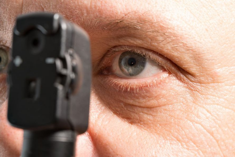
Eye examination using ophthalmoscopy © Henrik Dolle / AdobeStock
For this purpose, the patient is given eye drops beforehand containing a medication that promotes pupil dilation. In this case, the patient should not drive themselves to the ophthalmologist’s office on the day of the examination: The permanently dilated pupils render the patient unfit to drive immediately after the examination.
Using a light source, the ophthalmologist then illuminates the back of the eye to reveal any potential retinal detachment.
It is not uncommon for a so-called contact lens to be used to diagnose a retinal detachment. This is a magnifying-glass-like device that the ophthalmologist places on the eyeball. This allows the doctor to diagnose a retinal detachment and, in some cases, its cause—such as a tear—in the retina. If a retinal detachment is already present, it often manifests as gray, raised folds.
If bleeding in the vitreous humor makes it impossible to see the retina, the ophthalmologist will instead perform an ultrasound examination. This allows changes on the retina to be identified.
Treatment of Retinal Detachment
Surgery is almost always necessary to treat a retinal detachment. There are several surgical procedures. The two most important are
- hump surgery and
- vitrectomy.
Laser therapy is also used to treat retinal detachments.
Laser Treatment
A retinal detachment or retinal tear cannot be treated with medication. In the case of a retinal tear—that is, before a retinal detachment begins—laser treatment of the affected eye is helpful.
The laser beam can be used to trigger an inflammatory response at the site of the injury. As a result, the tissue scars over and the hole in the retina closes. In this way, retinal detachment can often be prevented.
Eye Surgery
However, if retinal detachment is diagnosed, laser treatment proves ineffective: in such cases, eye surgery is the only option. The surgical method depends largely on the type and stage of the retinal detachment.
The primary focus here is on reattaching the detached retina and addressing the underlying causes—such as changes in the vitreous humor. Other forms of treatment
If retinal detachment is caused by inflammation, treating the underlying condition is essential.
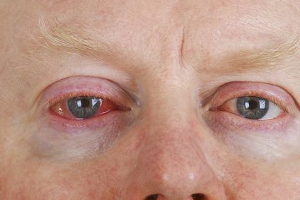
Eye inflammation can cause retinal detachment © Birgit Reitz-Hofmann / Fotolia
If, on the other hand, tumors are responsible, a specialized cancer center is responsible for treatment. There are various treatment options available here: On the one hand, treatment can involve surgical removal of the tumors or radiation therapy. The latter can involve irradiating the entire eyeball, or it can be performed as localized radiation therapy using radiation sources placed directly on the eye.
Surgery for Retinal Detachment
The following surgical procedures are available for treating a retinal detachment: Buckle surgery with a foam seal
If there is a single hole or tear, a procedure known as buckling surgery is used.
In this procedure, the eyeball is indented in the area of the defect using a foam tamponade, which the surgeon sutures to the sclera from the outside. The application of cold via a cryoprobe helps to maintain this adhesion. The surgeon drains the fluid that has accumulated under the detached retina from the outside via a puncture.
A special form of this indentation procedure involves applying a band that presses against the sclera, known as a cerclage.

A retinal detachment almost always requires surgical treatment © DragonImages / AdobeStock
Circular Band Surgery
If there are multiple holes, a cerclage is indicated: The surgeon wraps a band around the entire eyeball and tightens it until a circular constriction is created. A puncture in the fluid-filled space between the retina and the choroid is used to remove fluid. The surgeon then seals the holes or tears using a plug or band.
Intraocular Surgery
The vitreous humor is removed from the eye to detach the adhesions and eliminate the strands of connective tissue.
The surgeon replaces the vitreous humor: this can be done using either gas or silicone oil. Both allow the retina to be pressed against the choroid from the inside. The silicone oil usually needs to be removed in a subsequent surgery.
Chances of Recovery and Prognosis for Retinal Detachment
In nine out of ten patients, the eye surgery is successful the first time. This means that retinal detachment can be cured with surgery. In some cases, however, retinal detachment recurs later. Another procedure is then unavoidable.
The treatment of retinal detachment places high demands on the surgeon. Patients should be aware of this. Accordingly, you should choose an eye center that specializes in this type of surgery.
Even after successful retinal surgery, vision is usually reduced compared to before the detachment, or there may be a loss of visual field.
How a retinal detachment ultimately progresses depends on various factors: On the one hand, the extent of the detachment—that is, how far it has already progressed—is crucial. On the other hand, time is a very significant factor—specifically, how long the detachment had been present before surgery.
This means that the course and prognosis of a retinal detachment—both for the eye and for vision—are all the better and more favorable
- the less retinal tissue is affected and
- the shorter the duration of the retinal detachment, and
- the less complicated the initial findings are.
Follow-up Care After a Retinal Detachment
Anyone who has undergone retinal surgery should avoid physical exertion, at least for the next few weeks. This includes sports and lifting heavy objects. In the first few days, you should avoid anything that requires your eyes to move rapidly back and forth. This includes, above all, reading. Watching television, on the other hand (where you are looking steadily in one direction), is permitted.
Follow-up eye exams should be scheduled at intervals of several weeks, then several months.
If the symptoms mentioned—such as flashes of light, dark spots, or blurred vision—recur, you should immediately consult an ophthalmologist or visit an eye clinic.
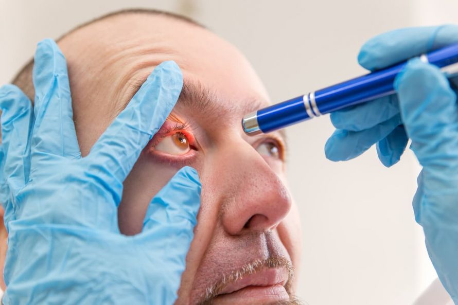
Regular eye exams are necessary after surgery © M. Dörr & M. Frommherz / AdobeStock
Prevention of Retinal Detachment
Examination Based on Symptoms
If you notice the first signs of retinal detachment, see an ophthalmologist immediately. If you notice flashes of light or sudden black spots, an immediate visit to the ophthalmologist is essential. If you’re experiencing visual field defects, you can assume that the retina is already beginning to detach.
Regular Examinations
- you have severe nearsightedness,
- you recently underwent surgery for cataracts, or
- you have had a retinal detachment in the past.
An examination of the back of the eye can also reveal potential degenerative changes that could lead to more serious conditions.
Laser treatment of such retinal changes serves as a preventive measure. The scars that form later at the treated site fuse the retina to the choroid.
It should be expressly noted: This preventive measure is only possible as long as a retinal detachment has not yet occurred.
FAQ
How is a retinal detachment treated?
A retinal detachment is almost always treated surgically. Depending on the findings, a scleral buckling procedure or intraocular surgery may be performed—in cases of a simple retinal tear, laser treatment may sometimes be sufficient.
2. What are the symptoms of retinal detachment? In addition to flashes of light, other symptoms include floaters, dark veils, and visual field defects. If you experience these symptoms, you should see an ophthalmologist immediately.
What is traction retinal detachment?
In traction retinal detachment, scar tissue forms in the vitreous humor or within the retinal layers, creating a pulling force on the retina. The most common cause is diabetic retinopathy.
Will full vision be restored after surgery?
Even after a successful surgery, vision may remain impaired. In some cases, repeat surgery may be necessary if a detachment occurs again.
Share this article

About the medical author
Sabine Schneider
Editor-in-Chief
Sabine Schneider – medical author: Explore expert articles and medical expertise in the Leading Medicine Guide.
View full expert profile →