A cataract is a common eye condition in which the lens of the eye becomes progressively cloudy. This clouding of the lens prevents light from reaching the retina clearly, impairing vision. Early symptoms often include blurred vision or a haze that makes images appear less clear.
Cataracts usually develop gradually and primarily affect older adults. In many cases, the natural lens of the eye is affected, causing visual acuity to decline continuously. A timely diagnosis by an ophthalmologist is important to determine the right time for surgery. Cataracts are usually treated with surgery, in which the clouded lens is replaced with an artificial lens. Today, this procedure is usually performed on an outpatient basis and is painless.
Definition: What is a cataract?
A distinctive feature of cataracts (medical term: cataract) in the advanced stages of the disease is a grayish discoloration behind the pupil. This is caused by the gradual clouding of the eye’s lens.
The term “cataract” is derived from this discoloration. It primarily affects older adults over the age of 60, which is why cataracts are often referred to as age-related cataracts.
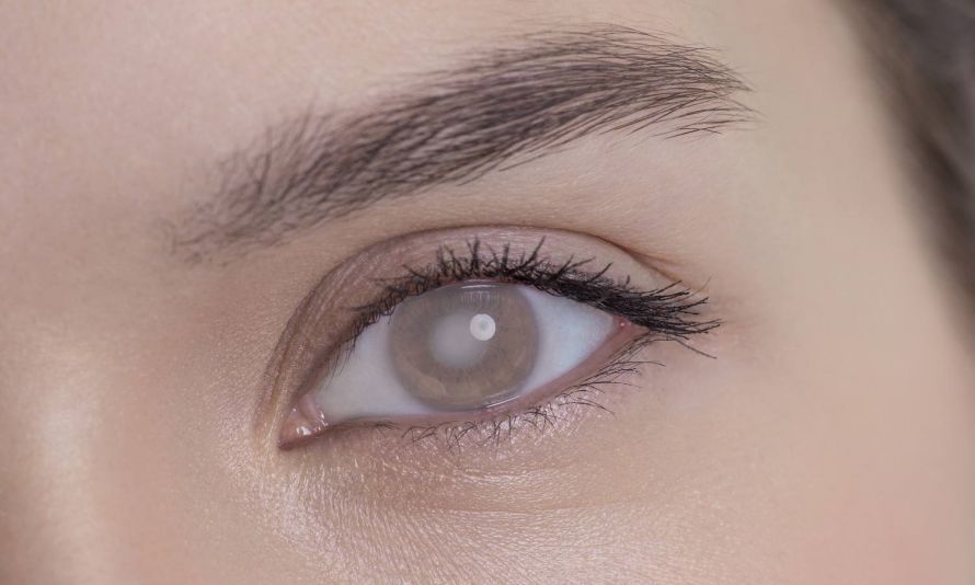
In the advanced stages of cataracts, a grayish discoloration appears behind the pupil
Prevalence of the Disease
Cataracts are considered the leading cause of blindness worldwide. They are particularly common in developing countries, mainly due to a lack of medical care.
Every year, more than 600,000 people in Germany undergo surgery to treat cataracts. The chances of a cure for this eye disease are very good. As many as 90 percent of those who undergo surgery subsequently achieve visual acuity between 50 and 100 percent.
Among people aged 52 to 64, the probability of developing the condition is just over 50 percent. Between the ages of 65 and 75, nearly everyone develops cataracts. However, 50 percent of those affected do not notice the condition until they reach the age of 75.

Cataracts develop in almost everyone as they grow older
Causes of the condition
Cataracts can have many causes. In most people, cataracts develop around the age of 60 (age-related cataracts), and the causes are usually hereditary. Only in very rare cases do children and adolescents suffer from this condition.
Furthermore, cataracts can also be triggered by medications such as cortisone. In addition, various types of radiation are considered a cause of cataracts:
- UV light from the sun
- Infrared light
- radioactive radiation
- X-rays
- Heat rays
Therefore, wearing protective eyewear is essential to protect your eyes. Especially in the tropics, where there is direct sunlight, eye diseases can develop very early on. Here, cataracts often appear as early as between the ages of 40 and 50.

Sunlight damages the eyes. Protect yourself from UV rays!
Smoking and other eye injuries also contribute to the development of the disease. Eye injuries include:
- Contusion
- open wounds
- Foreign objects entering the eye
- Injuries caused by electric shock or lightning strikes
Another risk factor is diabetes. The form of cataract caused by diabetes, known as diabetic cataract, primarily affects younger people with type 1 diabetes. With type II diabetes, on the other hand, age-related cataracts can develop prematurely. This is because these individuals only have insulin resistance in the tissues that process glucose; insulin deficiency does not occur until later.
A deficiency in important nutrients or skin conditions such as atopic dermatitis can also trigger the development of this eye disease. Cataracts can also be congenital. This is either due to hereditary factors or results from a complication during pregnancy, such as the mother contracting rubella.
Symptoms: What are the signs of cataracts?
Cataracts cause the lens of the eye to become cloudy. Incoming light is diffused by this cloudiness. This also causes a sharp increase in sensitivity to glare in those affected: The effect is comparable to a fogged-up car windshield that glows brightly when sunlight hits it head-on.
People often first notice cataracts while driving in poor weather conditions. Symptoms vary greatly depending on the severity of the condition. As a result, the extent of visual impairment differs from person to person.
The symptoms of cataracts usually become apparent later in the course of the disease:
- Blurred vision
- Double vision
- Loss of contrast
- Halos around objects
- The eyes can no longer adjust properly to light and dark
- Depth perception is severely impaired
Another major problem for many affected individuals is the change in visual acuity at both near and far distances. In the case of a posterior subcapsular cortical opacity, near vision is impaired. In the case of a so-called nuclear cataract in the affected person’s eye, an impact on distance vision can be observed.

Cataracts can also progress to what is known as a “total cataract,” which can lead to blindness. In this case, the entire lens of the eye is affected. In such cases, it is particularly important to plan cataract treatment early on.
Diagnosis of Cataracts
To make a diagnosis, the doctor uses a slit-lamp microscope. This microscope provides a magnification of six to forty times for the lens inside the eye. The cornea, the posterior surface, and the anterior chamber of the eye can also be examined using this microscope.
During the examination, eye drops are used to ensure that the pupil of the eye dilates widely for a short time.
 Slit-lamp examination
During this examination, the patient sits in front of the microscope. The patient’s chin and forehead are secured in place. First, the doctor examines the cornea, as vision problems caused by cataracts may originate there. One possible cause could be an irregular curvature of the cornea.
 Cataract under a slit lamp
If a cataract is suspected, videokeratoscopy can be used for further examination. This procedure creates a computer-generated image of the potentially affected cornea.
In addition, the thickness of the cornea and the endothelial cell density are usually measured. Using these methods, the doctor can rule out other eye conditions and make a definitive diagnosis.
As with many diseases, early detection plays an important role in cataracts. It is therefore generally recommended to undergo regular preventive eye exams with an ophthalmologist starting at age 40. Unfortunately, health insurance companies do not currently cover this exam.
Cataracts: Disease Progression and Prognosis
If left untreated, cataracts are associated with a gradual loss of vision that can lead to blindness. Initially, those affected become nearsighted. Since the disease often occurs in older age, existing age-related farsightedness is initially compensated for. This means that those affected can suddenly see again without glasses for a short time. However, vision continues to deteriorate. The image becomes cloudy and increasingly blurry. Without surgery, cataracts eventually lead to blindness.
Typically, both eyes are affected by cataracts. However, the disease can progress at different rates. The loss of vision depends, among other factors, on where the clouding of the lens originates. In younger people, the process often progresses more rapidly.
Cataract Surgery—A Common Procedure to Restore Good Vision
Cataract Treatment: Cataract Surgery
Cataracts are treated through cataract surgery, which involves lens replacement. During the procedure, the clouded lens is replaced with an artificial lens. The surgery is usually performed on an outpatient basis under local anesthesia.
There are essentially two methods of lens replacement: intracapsular and extracapsular. In the extracapsular procedure, the capsule remains in the eye. The most common procedure today is extracapsular phacoemulsification.
.jpg) Artificial Eye Lens
Artificial Eye Lens
After a consultation with the doctor, the decision to undergo cataract surgery may be easier to make. The timing of the procedure is usually determined based on the patient’s current life situation. The progression of the cataract and the resulting visual impairment play a particularly decisive role here.
Cataract Surgery Procedure: Phacoemulsification
Either eye drops or an injection are used to anesthetize the eye. The incision is only two to three millimeters long and heals on its own afterward.
Through this small incision, the doctor uses ultrasound to liquefy the nucleus and cortex of the lens. The resulting fluid is then suctioned out of the eye.
During cataract surgery, the surgeon leaves the lateral and posterior lens capsules intact. The artificial lens is first inserted into the capsular bag in a folded or collapsed state. Only once inside the eye does the soft artificial lens unfold and take its shape. The doctor then secures the lens to the remaining capsular bag using its fine retaining loops.
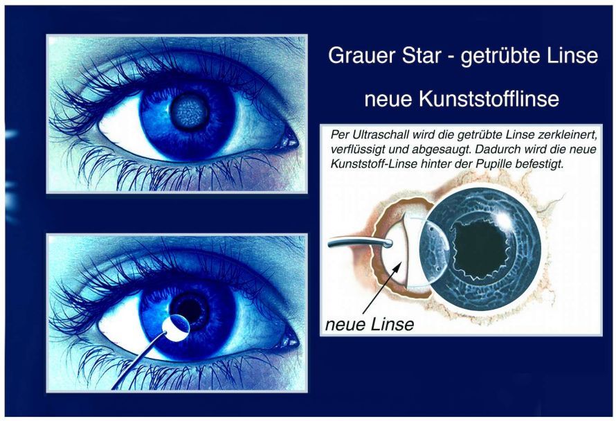
Non-folding lenses are used only very rarely today. In addition, it is also possible to implant the artificial lens behind the pupil. Here you will find detailed information about the cataract surgery procedure.
Postoperative Care
The surgery is followed by postoperative care involving an ointment dressing. This dressing generally remains on the eye until the next day.
During the first four weeks after surgery, antibiotic and anti-inflammatory eye drops must be applied at regular intervals. In addition, the treated eye must be examined by an ophthalmologist at regular intervals. After approximately six to eight weeks, the eye and the cataract can finally be considered healed.
If the second eye also requires surgery, this procedure is usually postponed for about one month. This is the only way to account for any potential complications that may arise during the healing process of the cataract in the first eye.
Preventing Cataracts
The development of cataracts cannot be prevented, but surgery can largely alleviate the symptoms. Furthermore, measures intended to slow the progression of cataracts have not yet been medically proven. As of the current state of medical knowledge, there are no measures available to prevent cataracts.
It is generally important to protect the eyes from UV radiation, for example by wearing sunglasses with certified UV protection.
To prevent congenital cataracts, expectant mothers should definitely get vaccinated against rubella.
Conclusion
Cataracts are an eye condition that usually develops gradually and is one of the most common eye diseases in older adults. Cataracts typically become noticeable around age 60, with initial symptoms such as blurred vision or difficulty seeing at a distance. Cataracts often affect both eyes, although the severity may vary from one eye to the other. Cataract symptoms are often underestimated at first, even though they progressively impair vision.
The cause of cataracts is usually an age-related change in the eye’s natural lens, although congenital cataracts are also possible. Over time, the lens in the eye becomes cloudy, requiring the cloudy lens to be replaced with an artificial one. Cataracts are an eye condition that, if left untreated, can lead to severe vision loss. However, cataracts can be treated, and treatment is highly effective today.
Cataract surgery is considered the standard treatment and is usually performed painlessly under local anesthesia. During the procedure, the cloudy lens is removed and replaced, allowing many patients to see better as early as the day after surgery. Typically, both eyes are operated on one after the other, and vision improves significantly within weeks of the surgery. The right time for surgery depends on how much daily life is affected.
Although cataracts can only be prevented to a limited extent, early diagnosis is crucial. A cataract diagnosis no longer automatically means severe impairment, as modern procedures deliver very good results. It is important to take the symptoms of cataracts seriously and act promptly. This helps prevent the cataract from progressing further; in rare cases, cataracts can lead to more serious complications. Overall, it is clear that symptoms and treatment are closely linked—those who act early can preserve their vision in the long term.
FAQ
What is cataract?
A cataract is an eye condition in which the lens of the eye becomes cloudy. This cloudiness causes the image to appear blurry and reduces visual acuity.
What are the symptoms of cataracts?
Typical symptoms of cataracts include blurred vision, a haze over the eye, increased sensitivity to glare, and deteriorating vision, especially while driving or in low-light conditions.
How are cataracts treated?
Cataracts are treated with surgery. During the procedure, the clouded lens is removed and replaced with an artificial lens to restore vision.
How is cataract surgery performed?
Cataract surgery is an outpatient procedure that is usually performed under local anesthesia. The clouded lens is removed and an artificial lens is implanted through a small incision.
When should you have surgery?
Surgery is recommended when vision is so severely impaired that it interferes with daily life. The ophthalmologist will determine the optimal time for surgery based on the diagnosis.
Share this article