
A fall onto an outstretched arm or a direct impact to the shoulder often results in a painful humeral head fracture. In this type of fracture, the upper part of the humerus breaks near the shoulder joint. Older adults, in whom osteoporosis weakens bone strength, are particularly susceptible to this injury. However, even in younger patients, an accident involving high-impact force can lead to a complicated humeral head fracture in which bone fragments are displaced. Treatment—whether surgical, using plates and screws, or conservative, involving immobilization—depends largely on whether the arm’s mobility is compromised and how many bone fragments are present. Consistent follow-up care is essential to restore shoulder function.
Definition, Prevalence, and Causes of a Humeral Head Fracture
Humeral head fractures refer to bone injuries of the head of the humerus. This type of injury occurs primarily after falls onto an outstretched arm or onto the shoulder.
They account for approximately 4 to 5 percent of all fractures. Furthermore, at 47 percent, humeral head fractures are the most common type of fracture in the shoulder girdle.
Risk factors for humeral head fractures include advanced age and the presence of osteoporosis. As the population continues to age and the prevalence of osteoporosis increases, the number of humeral head fractures is also rising steadily.
Women are affected 2 to 3 times more often than men, on average.
Anatomy of the Upper Arm
The humerus, together with the glenoid cavity, forms the main, movable part of the shoulder joint.
The bone of the humeral head is significantly larger than the shoulder socket. This allows for the greatest possible range of motion in the shoulder joint. However, as a result, the joint does not offer as much stability as, for example, the hip joint, where the femoral head is almost completely enclosed by the acetabulum.
The shoulder joint is therefore primarily stabilized by the attached soft tissues, namely
- tendons,
- muscles, and
- ligaments
. This knowledge is essential for understanding the following descriptions of humeral head fractures (fractures of the upper arm head) and their treatments.
The following diagram shows the basics of anatomy in a highly simplified form:
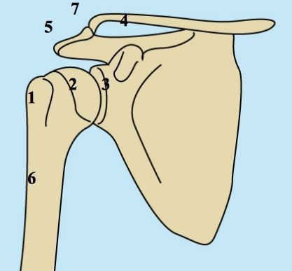
- Humerus head/tuberculum majus
- Humerus head: Articular cartilage
- Shoulder socket
- Clavicle
- Acromion
- Humerus shaft
- Acromioclavicular joint
In an accident, various structures of the humeral head can be injured. These injuries differ significantly in terms of diagnosis, treatment, and long-term outcome. Therefore, it is not possible to consider all humeral head fractures as a single category.
Different Types of Humeral Head Fractures
Depending on
- the number of fragments resulting from the humeral head fracture,
- the extent of fragment displacement, and
- the level of the fracture
the injury is classified into different types. Each type requires a different treatment approach.
Type 0 fractures—non-displaced “single-piece fractures”—are distinguished from Type A fractures. The latter are two-fragment fractures involving avulsion of the greater tubercle (tuberculum majus) or the lesser tubercle (tuberculum minus).
In addition, there are Type B fractures, which occur in the surgical neck and may involve 2 to 4 fracture fragments. Type C fractures occur in the anatomical neck. Here, too, 2 to 4 fracture fragments may be present.
Type X fractures refer to anterior or posterior dislocation fractures (dislocation of the shoulder joint accompanied by a bone fracture). After shoulder joint reduction (realignment), these fractures are further classified according to Types A through C.
In addition, we distinguish between comminuted fractures of the humeral head (so-called “head-splitting” and “impression” fractures).
Symptoms of a humeral head fracture
The main symptoms of a humeral head fracture are pain, as well as limited function and range of motion.
In the case of a humeral head fracture, the affected person holds the upper arm in a protective position to minimize pain. In most cases, there is swelling of the shoulder joint that varies in severity.
This is followed by the development of a bruise (hematoma) in the area of the affected shoulder. The hematoma may extend as far as the elbow.
Diagnosis of a humeral head fracture
An X-ray is used to confirm a suspected humeral head fracture.
Computed tomography (CT) is used as a follow-up examination for complex fractures of the humeral head. A CT scan also helps determine whether surgical treatment of the humeral head fracture is necessary.
Treatment of a humeral head fracture
The goal of treatment is to maintain a pain-free range of motion in the shoulder joint, taking into account the patient’s age and functional needs.
Therefore, the treatment plan must be individually tailored to the patient’s needs.
Treatment Principles for a Fracture of the Humerus Head (Humerus Head Fracture)
60 to 85 percent of humeral head fractures are Type 0 fractures. A non-displaced or only slightly displaced humeral head fracture (Type 0 fracture) does not require surgery.
This type of fracture is “splinted” by the periosteum, the joint capsule, and the muscles. Therefore, immobilization in a tubular bandage for 7 to 14 days until the pain subsides is sufficient.
Early functional exercise therapy is important to prevent stiffness in the shoulder joint. After about 6 to 8 weeks, the fracture has fully healed.
Joint Replacement
Fracture types A through C, Type X, and comminuted fractures of the humeral head result in
- severe malalignment,
- restricted movement, and
- pain.
Surgery is necessary to restore the humeral head to its original condition as much as possible. Depending on the type of fracture, various materials are used to stabilize the fracture. For example, a fracture can be stabilized using
- wires,
- screws, or
- plates
.
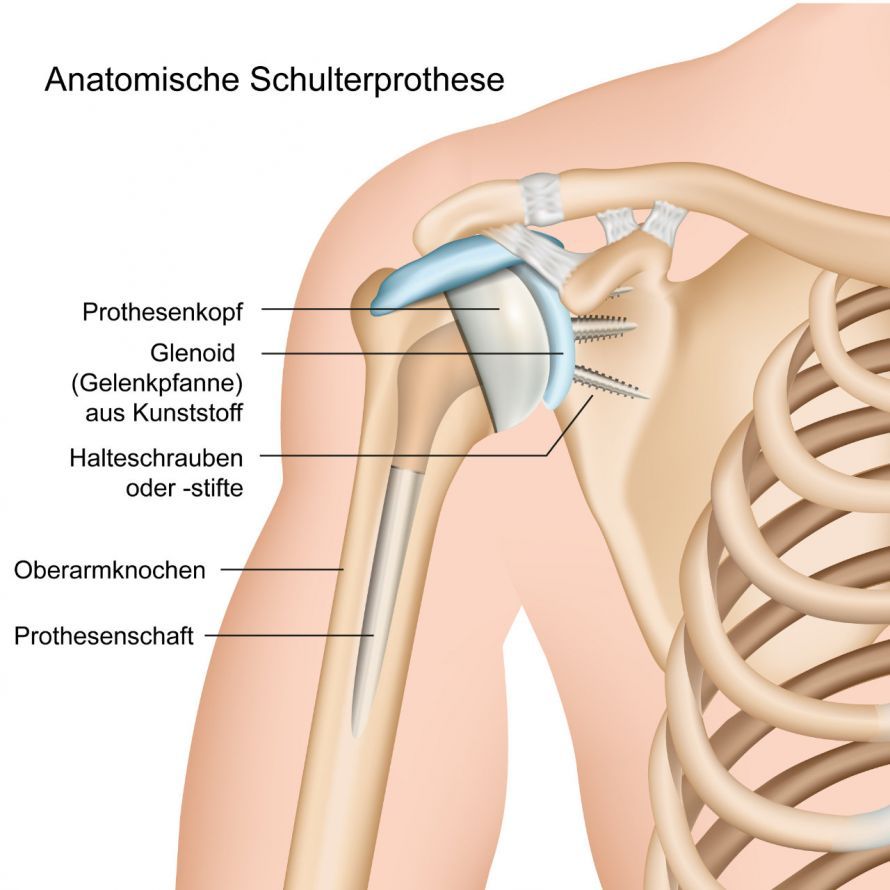
In some cases, replacement of the humeral head with a shoulder prosthesis (artificial joint replacement) is also advisable. A prerequisite for this is that
- the severity of the injury makes it impossible to perform surgery that preserves shoulder joint function, or
- the blood vessels supplying the humeral head are destroyed, making the likelihood of humeral head necrosis very high.
In recent years, special fracture prostheses have been developed for this type of joint replacement. They can be individually tailored to the fracture.
For example, the height of the prosthetic head can be adjusted to compensate for any height loss caused by the fracture. Furthermore, the fracture prosthesis has special features that allow the avulsed rolling hills (tubercle major and minor) to be reattached to the prosthesis in their anatomical position. This is important because the tendons of the rotator cuff attach to the rolling hills.
The humeral head comminuted fracture is a special case. If more than 40 percent of the articular surface is destroyed in this fracture, replacement of the articular surface with a humeral head prosthesis is also indicated.
Treatment of the Different Types of Humeral Head Fractures
One problem with fractures in the shoulder region is the rapid stiffening of the shoulder. Therefore, prolonged immobilization in a cast or bandage should be avoided in cases of a humeral head fracture.
Treatment for the various types of humeral head fractures varies greatly depending on the specific type. Therefore, the individual types of humeral head fractures and their treatments are discussed below.
Humeral head fracture with avulsion fracture of the tuberculum majus
In a humeral head fracture with avulsion of the tuberculum majus, the large bony protrusion to which the rotator cuff is attached is torn off.
The mechanism of injury is usually direct impact trauma or a dislocation of the shoulder. Typically, the avulsed portion of the tuberculum majus remains in place (see Figure 1).
After the shoulder is reduced, the bone either fits well back into its original position or remains in a malposition (see Figure 2).
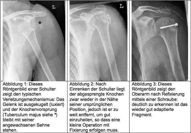
In the latter case, a minor surgical procedure is necessary. During this procedure, the bone fragment is repositioned to its original location. It is then fixed in place using screws or strong sutures until it has healed back into place. This usually takes about six weeks.
Follow-up care during this period involves guided movements without the patient applying their own force. A final X-ray examination can then confirm the success of the healing process.
Following this, active muscle and range-of-motion rehabilitation is necessary.
Subcapital Humerus Head Fracture
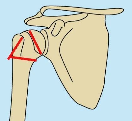
A subcapital humeral head fracture is defined as a fracture that extends across the entire circumference of the humeral head. The red lines in the diagram below illustrate the various fracture lines that can run through the humeral head.
In common parlance, these injuries are usually referred to as a humeral head fracture or a fracture of the humeral head.
Treatment of these injuries depends on the extent of displacement of the bone fragments.
If the bone fragments are only slightly displaced, treatment can be performed without surgery. In this case, immobilization in a Gilchrist splint for 3 weeks is sufficient. This is followed by physical therapy involving guided movements while the splint is still in place for 6 weeks. This treatment is also called “conservative” because it does not involve surgery.
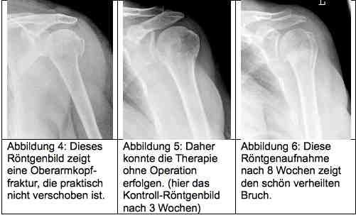
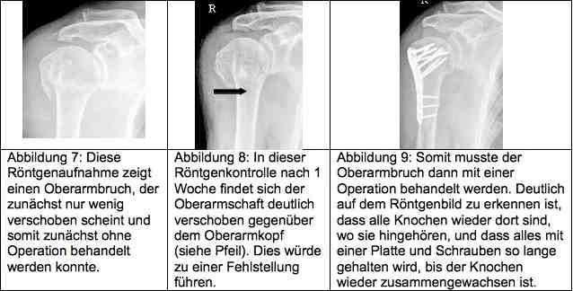
The healing process must be monitored at regular intervals using X-rays. Fractures can shift during this time. This is illustrated in the following example:
Comminuted fracture of the humeral head (head-split fracture)
Sometimes the humeral head is so severely comminuted that blood flow to the individual bone fragments is disrupted. In such cases, the bone can no longer heal and must be replaced with an artificial joint.
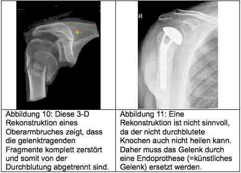
Whether the humeral head can still be saved after a comminuted fracture of the humeral head depends on a number of factors. The basis of bone healing is an adequate blood supply to the bone fragments. Without blood, healing cannot occur. Thus, the decision is significantly influenced by all factors that affect blood flow.
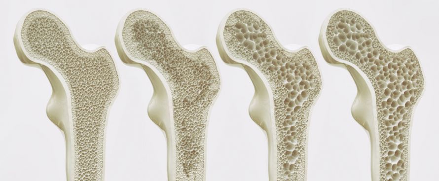
Another key factor is the degree of osteoporosis. This bone loss causes the bone to become thinner internally and less able to bear weight. Consequently, anchoring screws in severely osteoporotic bone is also considerably more difficult.
The decision as to whether the humeral head can be preserved or must be replaced also depends on the following factors:
- The patient’s age, as the severity of osteoporosis is strongly influenced by it
- The patient’s overall health
- Heavy smokers have poorer blood circulation from the outset.
- Chronic conditions such as dialysis, corticosteroid therapy, and diabetes impair blood flow.
After careful consideration, reconstruction can be attempted in young, healthy, and athletic patients. However, there is a higher risk that the reconstruction will not be successful and that a prosthesis will still be necessary. Patients must be aware of this risk.
If the procedure is successful, the joint can heal again.
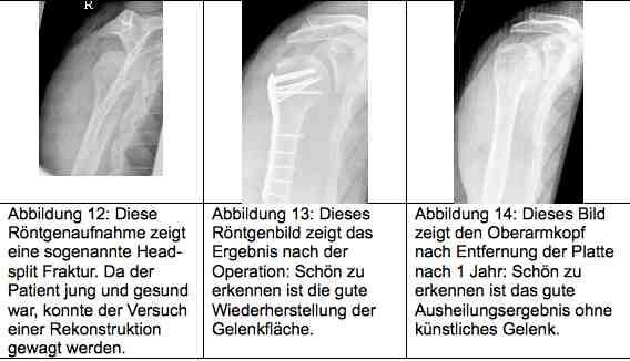
Surgical techniques for humeral head fractures have undergone a revolutionary improvement in recent years thanks to new, so-called angle-stable implants. As a result, it is now possible to reconstruct even the most severe fractures and treat them without an artificial joint. The surgeon’s experience is crucial in this regard.
Follow-up Care for a Fracture of the Humeral Head (Humeral Head Fracture)
Follow-up care must be tailored to the individual. The
- the duration of work disability and
- the timing of resuming sport-specific training
depend on the injury and the surgical procedure chosen.
Prognosis for humeral head fractures
The prognosis for humeral head fractures depends primarily on their complexity. As the number of fragments increases, the likelihood of fully restoring shoulder function decreases.
Other key factors affecting the prognosis for a humeral head fracture are
- on the one hand, the patient’s age,
- and, on the other hand, pre-existing, age-related, bony, or muscular limitations.
Risks of Surgery for Humeral Head Fractures
The risks of surgery include:
- Risk of infection and impaired wound healing
- Nerve damage
- Failure of the bone to heal, leading to the formation of a pseudarthrosis
- Implant failure (fracture of a screw or plate)
- Limited shoulder function
Conclusion on humeral head fractures
In summary, it can be stated that the number of humeral head fractures has increased significantly in recent years. The treatment of a humeral head fracture depends on the degree of displacement and the patient’s overall health.
If surgery is performed, the hospital should have as much experience as possible with cases of this type. The success of treatment depends significantly on the surgical procedure.
Images: Radiological images were kindly provided by Dr. Stefan Wirth.
Graphics: by Ms. Hella Thun
FAQ: The 8 Most Important Questions About Humeral Head Fractures
What are the typical symptoms and signs of a humeral head fracture?
The primary symptom is immediate, severe pain in the shoulder and upper arm, which makes any movement impossible. The arm is usually held close to the chest in a protective position. Within a short time, significant swelling and bruising (hematoma) develop, often extending down to the elbow and the chest wall. If a humeral head fracture is suspected, a doctor should be consulted immediately to rule out nerve damage.
How is a humeral head fracture diagnosed?
The diagnosis of a humeral head fracture begins with a physical examination to assess blood flow, motor function, and sensation. To reliably assess the fracture, an X-ray in two planes is standard. For complex humeral head fractures or to plan surgery, a computed tomography (CT) scan is necessary. It shows in detail whether a fragment is displaced or whether the fracture runs through the collum chirurgicum (predetermined fracture site) or the collum anatomicum.
When is conservative treatment used, and when is surgery performed?
The goal of treatment is always to restore mobility to the shoulder joint. About 80 percent of fractures are stable and show minimal displacement; these are treated conservatively (immobilization in a Gilchrist splint for two to three weeks). Surgery is necessary if parts of the humeral head are severely displaced (dislocated), the humeral head is comminuted, or bone fragments such as the tuberculum majus have been avulsed, as this would otherwise compromise the function of the rotator cuff and the muscle.
How does conservative treatment work?
Conservative treatment of a humeral head fracture begins with a short period of immobilization, usually in a shoulder sling. Early physical therapy is important: pendulum exercises begin after just a few days to prevent adhesions in the shoulder joint. After about two to three weeks, once initial bony union has been established, the load is gradually increased. Regular X-ray follow-ups ensure that no displacement occurs.
What complications can occur?
Possible complications include shoulder stiffness (frozen shoulder), which often requires intensive follow-up treatment. In severe fractures, blood flow to the humeral head may be impaired, leading to the death of bone tissue (humeral head necrosis). Injuries to nerves (axillary nerve) or blood vessels are also possible. Improperly healed fractures can cause chronic pain and osteoarthritis.
Why is osteoporosis a risk factor?
Osteoporosis reduces bone density, making the bone porous and brittle. In older adults, even a minor trauma—such as a fall from a standing position onto the shoulder—is often enough to cause a proximal humerus fracture. Because the bone is softer, surgically anchoring screws and plates is also more challenging.
How long does it take for a humerus fracture to heal?
Bone healing usually takes 6 to 12 weeks. However, functional recovery of arm mobility takes significantly longer. It often takes 3 to 6 months or longer for full strength and mobility to return. Special exercises as part of physical therapy are crucial for success.
What is a proximal humerus fracture?
The terms “humeral head fracture,” “fracture of the humeral head,” and “proximal humerus fracture” are often used interchangeably. They all refer to a fracture of the humeral head or the neck region at the upper end of the humerus. Classification is often based on the Neer system, depending on how many of the four main segments (head, shaft, greater tubercle, and lesser tubercle) are affected and displaced.
Share this article